


Abstract
1203
Objectives: Position emission tomography (PET) is increasingly being used to guide radiation therapy treatment planning processes. Best possible outcomes however, hinges on accurate target volumes derived from these PET images. This work aims to establish an optimal method for deriving target volumes with PET including 1) PET image reconstruction algorithms and reconstruction parameters, and 2) image volume delineation techniques.
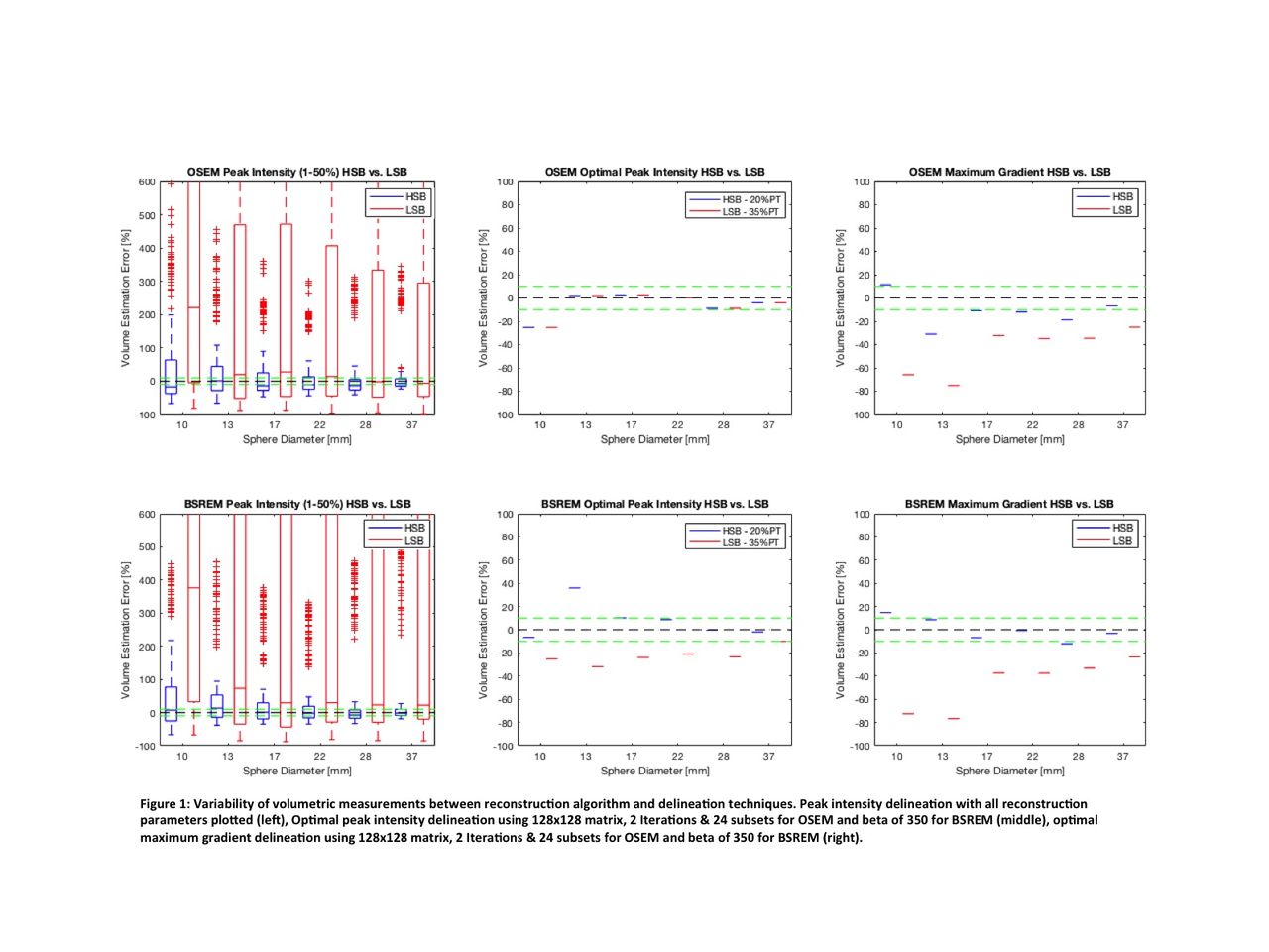
Methods: The NEMA hot-spheres phantom (10, 13, 17, 22, 28, 37 mm sphere diameters) was imaged twice using high (HSB) and low (LSB) sphere-to-background activity concentration ratios using ~40 MBq of 18F activity in each (87:1 and 4:1 ratios achieved, respectively). Images were acquired over 2.5 min using typical image acquisition parameters on a GE Discovery 710 PET/CT. PET images were reconstructed using two vendor provided reconstruction algorithms: Ordered subset (OSEM) and Bayesian penalized likelihood (BSREM) expectation maximization. Reconstructed image matrix size was varied (1282, 1922, 2562 and 3202 pixels per slice), as was the number of iterations (NoI) - from 2 to 10 in increments of 2, the number of subsets (NoS) - from 12 and 24, and the noise penalization variable (β) in BRSEM reconstructions - from 50 to 500 in increments of 50. Sphere volume delineation was achieved using both peak intensity relative thresholding (%PT) with varying percent thresholds and using maximum gradient (MG) segmentation. PET-image derived volumes were compared to known phantom volumes to calculate percent errors.
Results: ANOVA analysis indicated significant dependence of volume error on sphere size, spheres-to-background ratio, image matrix size, %PT and NoI/NoS (OSEM) (p<0.01 for each), but not on β (BSREM)(p>0.05). Small spheres were associated with the largest errors. With OSEM reconstruction, volumes defined using the %PT method had errors ranging between -98% to 809% [907%], but were optimized with small matrix size (128x128) and moderate number of iteration (e.g. 2 iteration, 24 subsets). Using this optimal reconstruction, volume errors ranged -25% to 3% [28%] for HSB data using 20%PT and -25% to 3% [28%] for LSB using 35%PT. Delineation using MG had greater errors ranging -31% to 11% [42%] and -75% to -25% [50%] for HSB and LSB scans respectively. Likewise, with BSREM, optimal reconstruction was 128x128 and little dependence on β value. Using β=350 the PT method was optimized to have errors ranging -7% to 36% [43%] for HSB data using 20%PT and -31% to -10% [21%] for LSB data using 35%PT, and MG method also had similar errors as with OSEM, -12 to 15% [27%] (HSB) and -77% to 23% [100%] (LSB).
Conclusions: While optimal volume accuracy can be achieved with OSEM, reconstruction parameter optimization is required. BSREM achieves slightly lower volume accuracy, but is consistent across a large range of reconstruction parameter values. Accurate volume measurements are easily achieved with high target-to-background contrast, but are challenging in low contrast scenarios. Threshold based segmentation is highly dependent on threshold values, while automated gradient based segmentation can perform accurately with sufficient contrast. The use of anatomical imaging alongside PET may further increase volume accuracy in low contrast scenarios.

In this issue


Jump to section
Related Articles
Cited By...
- No citing articles found.