
Main menu

  • Home
  • Content
    • Current
    • Ahead of print
    • Past Issues
    • JNM Supplement
    • SNMMI Annual Meeting Abstracts
    • Continuing Education
    • JNM Podcasts
  • Subscriptions
    • Subscribers
    • Institutional and Non-member
    • Rates
    • Journal Claims
    • Corporate & Special Sales
  • Authors
    • Submit to JNM
    • Information for Authors
    • Assignment of Copyright
    • AQARA requirements
  • Info
    • Reviewers
    • Permissions
    • Advertisers
  • About
    • About Us
    • Editorial Board
    • Contact Information
  • More
    • Alerts
    • Feedback
    • Help
    • SNMMI Journals
  • SNMMI
    • JNM
    • JNMT
    • SNMMI Journals
    • SNMMI

User menu

  • Subscribe
  • My alerts
  • Log in
  • Log out
  • My Cart

Search

  • Advanced search
Journal of Nuclear Medicine
  • SNMMI
    • JNM
    • JNMT
    • SNMMI Journals
    • SNMMI
  • Subscribe
  • My alerts
  • Log in
  • Log out
  • My Cart
Journal of Nuclear Medicine

Advanced Search

  • Home
  • Content
    • Current
    • Ahead of print
    • Past Issues
    • JNM Supplement
    • SNMMI Annual Meeting Abstracts
    • Continuing Education
    • JNM Podcasts
  • Subscriptions
    • Subscribers
    • Institutional and Non-member
    • Rates
    • Journal Claims
    • Corporate & Special Sales
  • Authors
    • Submit to JNM
    • Information for Authors
    • Assignment of Copyright
    • AQARA requirements
  • Info
    • Reviewers
    • Permissions
    • Advertisers
  • About
    • About Us
    • Editorial Board
    • Contact Information
  • More
    • Alerts
    • Feedback
    • Help
    • SNMMI Journals
  • View or Listen to JNM Podcast
  • Visit JNM on Facebook
  • Join JNM on LinkedIn
  • Follow JNM on Twitter
  • Subscribe to our RSS feeds
Meeting ReportNeurosciences

PET imaging of pre-synaptic protein SV2A in Rett syndrome mouse model

Alexandra DiFilippo, Carolina Ferreira, Jonathan Engle, Todd Barnhart, Qiping Dong, Qiang Chang and Bradley Christian
Journal of Nuclear Medicine May 2019, 60 (supplement 1) 117;
Alexandra DiFilippo
1Medical Physics Madison WI United States
  • Find this author on Google Scholar
  • Find this author on PubMed
  • Search for this author on this site
Carolina Ferreira
4University of Wisconsin-Madison Madison WI United States
  • Find this author on Google Scholar
  • Find this author on PubMed
  • Search for this author on this site
Jonathan Engle
3Medical Physics University of Wisconsin - Madison Madison WI United States
  • Find this author on Google Scholar
  • Find this author on PubMed
  • Search for this author on this site
Todd Barnhart
2University of Wisconsin Madison WI United States
  • Find this author on Google Scholar
  • Find this author on PubMed
  • Search for this author on this site
Qiping Dong
1Medical Physics Madison WI United States
  • Find this author on Google Scholar
  • Find this author on PubMed
  • Search for this author on this site
Qiang Chang
1Medical Physics Madison WI United States
  • Find this author on Google Scholar
  • Find this author on PubMed
  • Search for this author on this site
Bradley Christian
4University of Wisconsin-Madison Madison WI United States
  • Find this author on Google Scholar
  • Find this author on PubMed
  • Search for this author on this site
  • Article
  • Figures & Data
  • Info & Metrics
Loading

Abstract

117

Objectives: Rett syndrome is a rare neurological disorder resulting in intellectual disability caused by mutations in the X-linked methyl-CpG-binding protein 2 (MECP2) gene. Altered neuronal morphology and reduced dendritic complexity have been observed in cell and animal models of Rett syndrome. The goal of this study was to use [11C]UCB-J, a PET radioligand binding to the pre-synaptic protein SV2A, to assess changes in synaptic density in the Mecp2 knockout (Mecp2 KO) mice, a mouse model of Rett syndrome.

Methods: Two Mecp2 KO and two wild type littermate (WT) mice underwent [11C]UCB-J scans using a Sophie G4 PET scanner. Dynamic images were acquired during 45 minutes after injection of the radiotracer (57.5±14.4μCi, 1.07±0.004 pg ) and reconstructed with 15 dynamic frames (six-1min, two-2min, seven-5min). Using FSL FLIRT the integrated PET image was normalized to the mouse MR brain template (Paxinos, 2001). The whole cortex volume of interest was created using the Paxinos mouse brain atlas. The body weight SUV was calculated using the cortical values from the last 15 minutes of the scan for the Mecp2 KO and the WT mice.

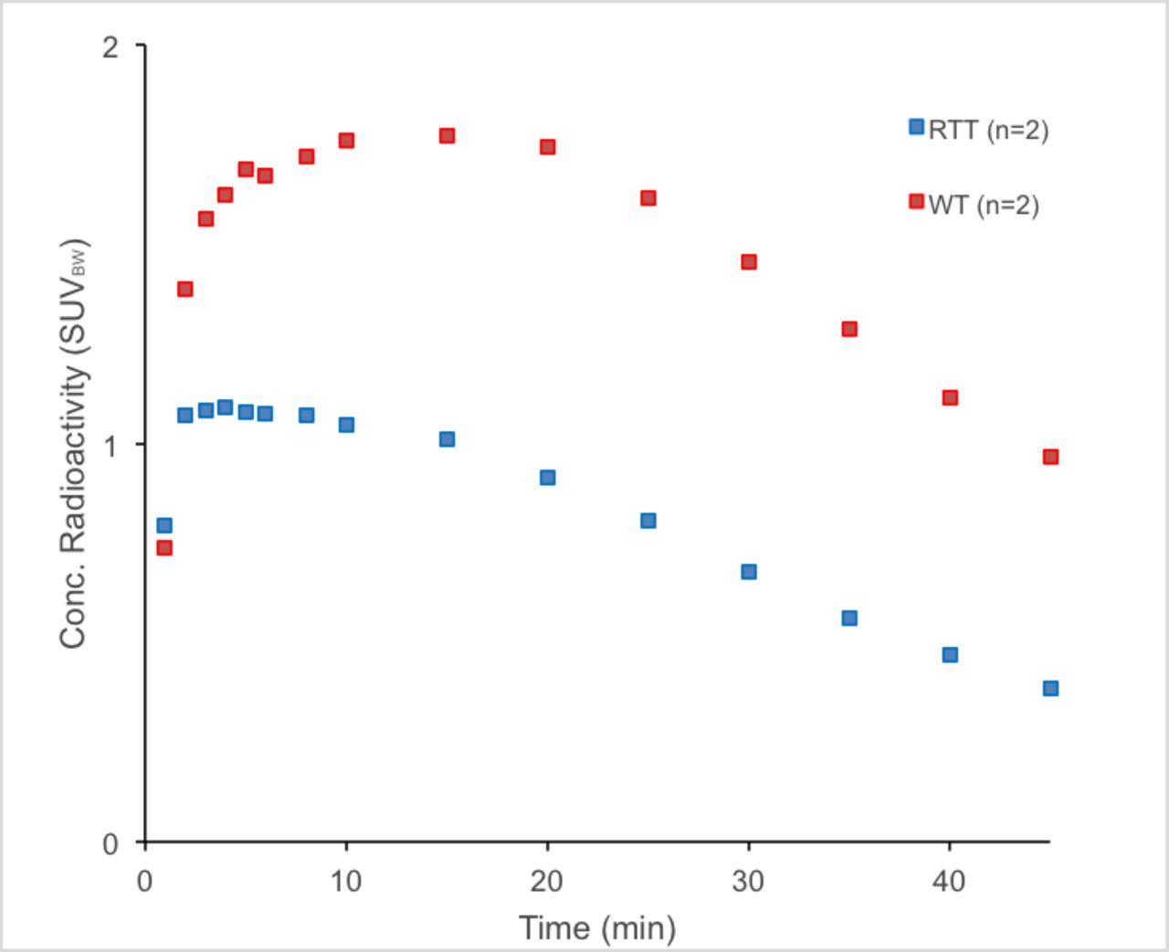
Results: SUVBW data calculated using the cerebral cortex region from 30 to 45 minutes of the scan gave values of 1.21±0.21 for the WT mice and 0.52±0.13 for the mutant mice.

Conclusions: These preliminary results reveal decreased uptake of [11C]UCB-J in the Rett syndrome mouse model are consistent with the hypothesis that the loss of Mecp2 gene may result in decreased synaptic density in the cerebral cortex. Studies are ongoing to increase the sample size that may inform future investigation into whether [11C]UCB-J imaging can serve as a valid biomarker in individuals with Rett syndrome.

Figure
  • Download figure
  • Open in new tab
  • Download powerpoint
Previous
Back to top

In this issue

Journal of Nuclear Medicine
Vol. 60, Issue supplement 1
May 1, 2019
  • Table of Contents
  • Index by author
Article Alerts
Sign In to Email Alerts with your Email Address
Email Article

Thank you for your interest in spreading the word on Journal of Nuclear Medicine.

NOTE: We only request your email address so that the person you are recommending the page to knows that you wanted them to see it, and that it is not junk mail. We do not capture any email address.

Enter multiple addresses on separate lines or separate them with commas.
PET imaging of pre-synaptic protein SV2A in Rett syndrome mouse model
(Your Name) has sent you a message from Journal of Nuclear Medicine
(Your Name) thought you would like to see the Journal of Nuclear Medicine web site.
Citation Tools
PET imaging of pre-synaptic protein SV2A in Rett syndrome mouse model
Alexandra DiFilippo, Carolina Ferreira, Jonathan Engle, Todd Barnhart, Qiping Dong, Qiang Chang, Bradley Christian
Journal of Nuclear Medicine May 2019, 60 (supplement 1) 117;

Citation Manager Formats

  • BibTeX
  • Bookends
  • EasyBib
  • EndNote (tagged)
  • EndNote 8 (xml)
  • Medlars
  • Mendeley
  • Papers
  • RefWorks Tagged
  • Ref Manager
  • RIS
  • Zotero
Share
PET imaging of pre-synaptic protein SV2A in Rett syndrome mouse model
Alexandra DiFilippo, Carolina Ferreira, Jonathan Engle, Todd Barnhart, Qiping Dong, Qiang Chang, Bradley Christian
Journal of Nuclear Medicine May 2019, 60 (supplement 1) 117;
Twitter logo Facebook logo LinkedIn logo Mendeley logo
  • Tweet Widget
  • Facebook Like
  • Google Plus One
Bookmark this article

Jump to section

  • Article
  • Figures & Data
  • Info & Metrics

Related Articles

  • No related articles found.
  • Google Scholar

Cited By...

  • No citing articles found.
  • Google Scholar

More in this TOC Section

Neurosciences

  • PET CT Quantification to direct Alzheimer’s patients to anti-amyloid therapy
  • Multicenter trial study for usefulness of physical parameters toward the standardization of brain SPECT image: relation to visual analysis
  • Assessment of Brain Perfusion 123I-IMP SPECT imaging Using Low Energy High Resolution Collimator image reconstructed by Flash 3D
Show more Neurosciences

Basic Science I (Neurosciences)

  • PET imaging of [11C](R)(-)NR2B-Me, a novel radioligand for the NR2B subunit of the NMDA receptor, in rhesus macaques
  • Antioxidative neuroprotective role of iron-gallic acid coordination nanoparticles and PET/MR usage in an ischemia stroke model
Show more Basic Science I (Neurosciences)

Similar Articles

SNMMI

© 2025 SNMMI

Powered by HighWire