
Main menu

  • Home
  • Content
    • Current
    • Ahead of print
    • Past Issues
    • JNM Supplement
    • SNMMI Annual Meeting Abstracts
    • Continuing Education
    • JNM Podcasts
  • Subscriptions
    • Subscribers
    • Institutional and Non-member
    • Rates
    • Journal Claims
    • Corporate & Special Sales
  • Authors
    • Submit to JNM
    • Information for Authors
    • Assignment of Copyright
    • AQARA requirements
  • Info
    • Reviewers
    • Permissions
    • Advertisers
  • About
    • About Us
    • Editorial Board
    • Contact Information
  • More
    • Alerts
    • Feedback
    • Help
    • SNMMI Journals
  • SNMMI
    • JNM
    • JNMT
    • SNMMI Journals
    • SNMMI

User menu

  • Subscribe
  • My alerts
  • Log in
  • Log out
  • My Cart

Search

  • Advanced search
Journal of Nuclear Medicine
  • SNMMI
    • JNM
    • JNMT
    • SNMMI Journals
    • SNMMI
  • Subscribe
  • My alerts
  • Log in
  • Log out
  • My Cart
Journal of Nuclear Medicine

Advanced Search

  • Home
  • Content
    • Current
    • Ahead of print
    • Past Issues
    • JNM Supplement
    • SNMMI Annual Meeting Abstracts
    • Continuing Education
    • JNM Podcasts
  • Subscriptions
    • Subscribers
    • Institutional and Non-member
    • Rates
    • Journal Claims
    • Corporate & Special Sales
  • Authors
    • Submit to JNM
    • Information for Authors
    • Assignment of Copyright
    • AQARA requirements
  • Info
    • Reviewers
    • Permissions
    • Advertisers
  • About
    • About Us
    • Editorial Board
    • Contact Information
  • More
    • Alerts
    • Feedback
    • Help
    • SNMMI Journals
  • View or Listen to JNM Podcast
  • Visit JNM on Facebook
  • Join JNM on LinkedIn
  • Follow JNM on Twitter
  • Subscribe to our RSS feeds
Research ArticleTheranostics

Parameters to Predict Progression-Free and Overall Survival After Peptide Receptor Radionuclide Therapy: A Multivariate Analysis in 782 Patients

Else A. Aalbersberg, Daphne M.V. Huizing, Iris Walraven, Berlinda J. de Wit-van der Veen, Harshad R. Kulkarni, Aviral Singh, Marcel P.M. Stokkel and Richard P. Baum
Journal of Nuclear Medicine September 2019, 60 (9) 1259-1265; DOI: https://doi.org/10.2967/jnumed.118.224386
Else A. Aalbersberg
1Department of Nuclear Medicine, ENETS Centre of Excellence, Netherlands Cancer Institute, Amsterdam, The Netherlands
  • Find this author on Google Scholar
  • Find this author on PubMed
  • Search for this author on this site
Daphne M.V. Huizing
1Department of Nuclear Medicine, ENETS Centre of Excellence, Netherlands Cancer Institute, Amsterdam, The Netherlands
  • Find this author on Google Scholar
  • Find this author on PubMed
  • Search for this author on this site
Iris Walraven
2Department of Radiation Oncology, Netherlands Cancer Institute, Amsterdam, The Netherlands
  • Find this author on Google Scholar
  • Find this author on PubMed
  • Search for this author on this site
Berlinda J. de Wit-van der Veen
1Department of Nuclear Medicine, ENETS Centre of Excellence, Netherlands Cancer Institute, Amsterdam, The Netherlands
  • Find this author on Google Scholar
  • Find this author on PubMed
  • Search for this author on this site
Harshad R. Kulkarni
3Theranostics Centre for Molecular Radiotherapy and Precision Oncology, ENETS Centre of Excellence, Zentralklinik Bad Berka, Bad Berka, Germany; and
  • Find this author on Google Scholar
  • Find this author on PubMed
  • Search for this author on this site
Aviral Singh
3Theranostics Centre for Molecular Radiotherapy and Precision Oncology, ENETS Centre of Excellence, Zentralklinik Bad Berka, Bad Berka, Germany; and
4GROW–School for Oncology and Developmental Biology, Maastricht University, Maastricht, The Netherlands
  • Find this author on Google Scholar
  • Find this author on PubMed
  • Search for this author on this site
Marcel P.M. Stokkel
1Department of Nuclear Medicine, ENETS Centre of Excellence, Netherlands Cancer Institute, Amsterdam, The Netherlands
  • Find this author on Google Scholar
  • Find this author on PubMed
  • Search for this author on this site
Richard P. Baum
3Theranostics Centre for Molecular Radiotherapy and Precision Oncology, ENETS Centre of Excellence, Zentralklinik Bad Berka, Bad Berka, Germany; and
  • Find this author on Google Scholar
  • Find this author on PubMed
  • Search for this author on this site
  • Article
  • Figures & Data
  • Supplemental
  • Info & Metrics
  • PDF
Loading

Article Figures & Data

Figures

  • Tables
  • Additional Files
  • FIGURE 1.
    • Download figure
    • Open in new tab
    • Download powerpoint
    FIGURE 1.

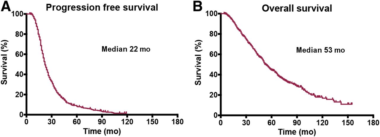
    Kaplan–Meier curves of PFS (A) and OS (B).

  • FIGURE 2.
    • Download figure
    • Open in new tab
    • Download powerpoint
    FIGURE 2.

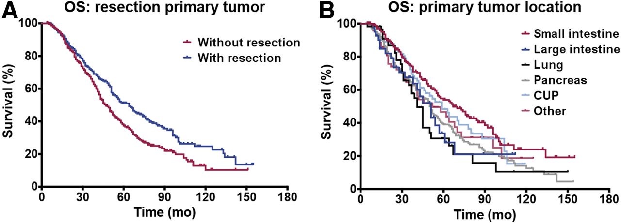
    Kaplan–Meier curves of OS after resection of primary tumor and OS per primary tumor location. Median OS is 43 and 65 mo without and with resection, respectively. Median OS is 67 mo for small-intestine NETs, 51 mo for large intestine, 41 mo for lung, 50 mo for pancreas, 59 mo for cancer of unknown primary (CUP), and 52 mo for other NETs.

  • FIGURE 3.
    • Download figure
    • Open in new tab
    • Download powerpoint
    FIGURE 3.

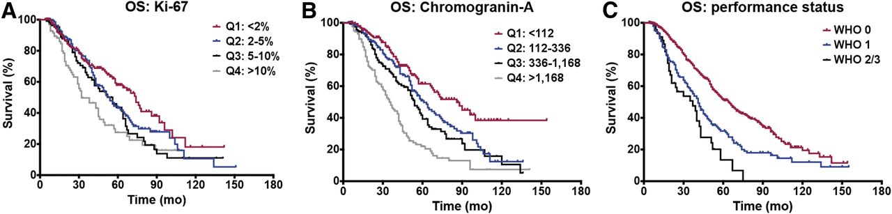
    Kaplan–Meier curves of OS per Ki-67 and CgA quartiles and performance status classes. Median OS is 74 mo for Ki-67 < 2%, 54 mo for 2%–5%, 56 mo for 5%–10%, and 32 mo for >10%. Median OS is 86 mo for CgA < 112, 61 mo for 112–336, 54 mo for 336–1,168, and 35 mo for >1,168. Median OS is 64 mo for WHO 0, 41 mo for WHO 1, and 37 mo for WHO 2/3.

Tables

  • Figures
  • Additional Files
    • View popup
    TABLE 1

    Patient, Tumor, and Treatment Characteristics

    VariableCharacteristicData
    SexMale444 (56.8%)
    Female338 (43.2%)
    Age (y)60.3 ± 11.0 (22–83)
    Location of primary tumor (n = 782)Small intestine221 (28.3%)
    Large intestine42 (5.4%)
    Lung60 (7.7%)
    Pancreas277 (35.4%)
    Unknown primary106 (13.6%)
    Other76 (9.7%)
    Tumor grade (n = 570)Grade 1182 (31.9%)
    Grade 2345 (60.5%)
    Grade 343 (7.5%)
    Ki-67 (n = 503)Q1≤2%
    Q23%–5%
    Q36%–10%
    Q4>10%
    Functional tumor (n = 555)Yes280 (50.5%)
    No275 (49.5%)
    Performance status (n = 782)WHO 0547 (69.9%)
    WHO 1190 (24.3%)
    WHO 2–345 (5.8%)
    ComorbiditiesHypertension (n = 239)99 (41.4%)
    Diabetes (n = 782)46 (5.9%)
    Prior treatmentInterferon-α (n = 727)36 (5.0%)
    Chemotherapy (n = 727)159 (21.9%)
    Resection, primary tumor (n = 710)336 (47.3%)
    Ablation (n = 727)100 (13.8%)
    Radiotherapy (n = 727)35 (4.8%)
    Somatostatin analog (n = 727)297 (40.9%)
    Isotope (n = 782)177Lu295 (37.7%)
    90Y96 (12.3%)
    177Lu and 90Y391 (50.0%)
    PRRT administrations (n = 782)3395 (50.4%)
    4329 (42.1%)
    5–659 (7.5%)
    Cumulative activity (GBq) (n = 782)177Lu21.7 (13.3–36.7)
    90Y11.5 (4.8–23.6)
    177Lu and 90Y18.2 (5.0–33.7)
    Blood count (n = 782)Normal546 (69.8%)
    CgA (n = 661)Q1<112
    Q2112–333
    Q3336–1,168
    Q4>1,168
    Serotonin (n = 627)273 (10–14,200)
    eGFR (n = 778)81.2 (30.56–259.08)
    Creatinine (n = 778)75.0 (27–186)
    • Continuous data are mean ± SD followed by range. Categoric data are number followed by percentage. When variables are divided into quartiles, range of quartile is given. Percentages may not add up to 100% due to rounding.

    • View popup
    TABLE 2

    PFS and OS per Primary Tumor Location

    PFS (mo)OS (mo)
    Primary tumor locationMedianIQRMedianIQR
    Small intestine2417–396736–111
    Large intestine2214–3351*24–68
    Lung18†14–2641*30–67
    Pancreas2316–3550*28–88
    Cancer of unknown primary2317–355936–104
    Other1914–315296–22
    Overall2216–355330–98
    • ↵* Significant in univariate analysis.

    • View popup
    TABLE 3

    PFS Analysis

    UnivariateMultivariate
    ParameterGroupHR95% CIPHR95% CIP
    Ki-67Q1: <2%10.00210.044
    Q2: 2%–5%1.1650.898–1.5131.1710.880–1.557
    Q3: 5%–10%1.4661.111–1.9341.4191.041–1.935
    Q4: >10%1.6311.230–2.1631.4931.090–2.045
    ComorbiditiesDiabetes1.3730.989–1.9070.058†1.7061.117–2.6050.013
    Prior treatmentChemotherapy1.3621.123–1.6520.002†1.3751.039–1.8200.026
    Interferon-α1.4871.046–2.1140.0272.0541.211–3.4850.008
    CgA (μg/L)Q1: <1121<0.0011<0.001
    Q2: 112–3331.1890.932–1.5171.2670.922–1.741
    Q3: 336–1,1681.5801.241–2.0111.4691.084–1.992
    Q4: >1,1682.1481.679–2.7472.0391.488–2.794
    • HR > 1 indicates greater risk for disease progression compared with reference value (HR = 1).

    • View popup
    TABLE 4

    Resection of Primary per Tumor Location

    Median OS (mo)Univariate Cox regression analysis
    Location of primaryResectedUnresectedHR95% CIP
    Small intestine77 (37–134)48 (32–90)0.6490.423–0.9950.047*
    Large intestine51 (28–∞)45 (24–61)0.6250.257–1.5220.301
    Lung51 (26–67)41 (31–45)0.7550.374–1.5220.432
    Pancreas65 (35–112)43 (24–69)0.5950.426–0.8320.002*
    Overall64 (32–112)47 (29–80)0.7150.584–0.8770.001*
    • ↵* Significant.

    • Data in parentheses are IQR.

    • View popup
    TABLE 5

    OS Analysis

    UnivariateMultivariate
    ParameterGroupHR95% CIPHR95% CIP
    Ki-67Q1: <2%10.00110.014
    Q2: 2%–5%1.3400.960–1.8711.3630.946–1.965
    Q3: 5%–10%1.5191.051–2.1941.2030.798–1.814
    Q4: >10%2.1431.488–3.0871.9301.285–2.899
    Performance statusWHO 01<0.00110.010
    WHO 11.7811.432–2.2141.5511.109–2.169
    WHO 2–32.8171.941–4.0892.3051.432–3.713
    Prior treatmentAblation1.4581.103–1.9280.0081.5191.041–2.2150.030
    Chemotherapy1.7921.434–2.239<0.0011.9791.412–2.773<0.001
    CgA (μg/L)Q1: <1121<0.0011<0.001
    Q2: 112–3361.5311.100–2.1311.6901.095–2.608
    Q3: 336–1,1681.8871.354–2.6311.8161.718–2.799
    Q4: >1,1683.3572.434–4.6312.6711.717–4.155
    • HR > 1 indicates greater risk for death of any cause and HR < 1 lower risk.

Additional Files

  • Figures
  • Tables
  • Supplemental Data

    Files in this Data Supplement:

    • Supplemental Data
PreviousNext
Back to top

In this issue

Journal of Nuclear Medicine: 60 (9)
Journal of Nuclear Medicine
Vol. 60, Issue 9
September 1, 2019
  • Table of Contents
  • Table of Contents (PDF)
  • About the Cover
  • Index by author
Print
Download PDF
Article Alerts
Sign In to Email Alerts with your Email Address
Email Article

Thank you for your interest in spreading the word on Journal of Nuclear Medicine.

NOTE: We only request your email address so that the person you are recommending the page to knows that you wanted them to see it, and that it is not junk mail. We do not capture any email address.

Enter multiple addresses on separate lines or separate them with commas.
Parameters to Predict Progression-Free and Overall Survival After Peptide Receptor Radionuclide Therapy: A Multivariate Analysis in 782 Patients
(Your Name) has sent you a message from Journal of Nuclear Medicine
(Your Name) thought you would like to see the Journal of Nuclear Medicine web site.
Citation Tools
Parameters to Predict Progression-Free and Overall Survival After Peptide Receptor Radionuclide Therapy: A Multivariate Analysis in 782 Patients
Else A. Aalbersberg, Daphne M.V. Huizing, Iris Walraven, Berlinda J. de Wit-van der Veen, Harshad R. Kulkarni, Aviral Singh, Marcel P.M. Stokkel, Richard P. Baum
Journal of Nuclear Medicine Sep 2019, 60 (9) 1259-1265; DOI: 10.2967/jnumed.118.224386

Citation Manager Formats

  • BibTeX
  • Bookends
  • EasyBib
  • EndNote (tagged)
  • EndNote 8 (xml)
  • Medlars
  • Mendeley
  • Papers
  • RefWorks Tagged
  • Ref Manager
  • RIS
  • Zotero
Share
Parameters to Predict Progression-Free and Overall Survival After Peptide Receptor Radionuclide Therapy: A Multivariate Analysis in 782 Patients
Else A. Aalbersberg, Daphne M.V. Huizing, Iris Walraven, Berlinda J. de Wit-van der Veen, Harshad R. Kulkarni, Aviral Singh, Marcel P.M. Stokkel, Richard P. Baum
Journal of Nuclear Medicine Sep 2019, 60 (9) 1259-1265; DOI: 10.2967/jnumed.118.224386
Twitter logo Facebook logo LinkedIn logo Mendeley logo
  • Tweet Widget
  • Facebook Like
  • Google Plus One
Bookmark this article

Jump to section

  • Article
    • Abstract
    • MATERIALS AND METHODS
    • RESULTS
    • DISCUSSION
    • CONCLUSION
    • DISCLOSURE
    • KEY POINTS
    • Acknowledgments
    • Footnotes
    • REFERENCES
  • Figures & Data
  • Supplemental
  • Info & Metrics
  • PDF

Related Articles

  • This Month in JNM
  • PubMed
  • Google Scholar

Cited By...

  • No citing articles found.
  • Google Scholar

More in this TOC Section

Theranostics

  • Determination of the Intralesional Distribution of Theranostic 124I-Omburtamab Convection-Enhanced Delivery in Treatment of Diffuse Intrinsic Pontine Glioma
  • Evidence-Based Clinical Protocols to Monitor Efficacy of [177Lu]Lu-PSMA Radiopharmaceutical Therapy in Metastatic Castration-Resistant Prostate Cancer Using Real-World Data
  • 177Lu-Labeled Anticlaudin 6 Monoclonal Antibody for Targeted Therapy in Esophageal Cancer
Show more Theranostics

Clinical

  • Addition of 131I-MIBG to PRRT (90Y-DOTATOC) for Personalized Treatment of Selected Patients with Neuroendocrine Tumors
  • SUVs Are Adequate Measures of Lesional 18F-DCFPyL Uptake in Patients with Low Prostate Cancer Disease Burden
  • Hypermetabolism on Pediatric PET Scans of Brain Glucose Metabolism: What Does It Signify?
Show more Clinical

Similar Articles

Keywords

  • PRRT
  • neuroendocrine tumor
  • multivariate analysis
SNMMI

© 2025 SNMMI

Powered by HighWire