


Article Figures & Data
Figures
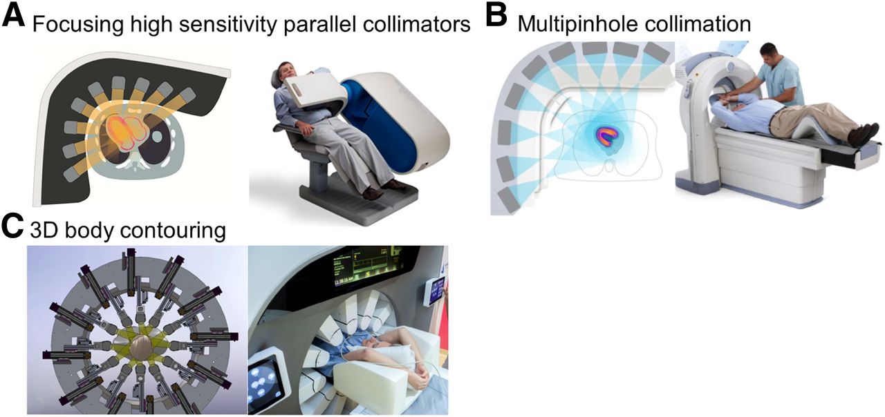
- FIGURE 1.
Three novel collimator designs used in solid-state camera systems. (A) High-sensitivity parallel collimators, as used in D-SPECT camera system. Collimators are square with parallel holes, each coupled to a single detector. (B) Multipinhole collimator design, as used in Discovery NM 530c camera system. (C) Collimator design of new general-purpose Veriton for 3-dimensional body contouring.
- FIGURE 2.
Count sensitivity expressed as percentage of incoming photons for several nuclear imaging systems. Solid-state SPECT cameras (fast MPI) have almost 8 times higher count sensitivity than conventional Anger cameras. Therefore, count sensitivity is more similar to 2-dimensional PET than to conventional SPECT camera.
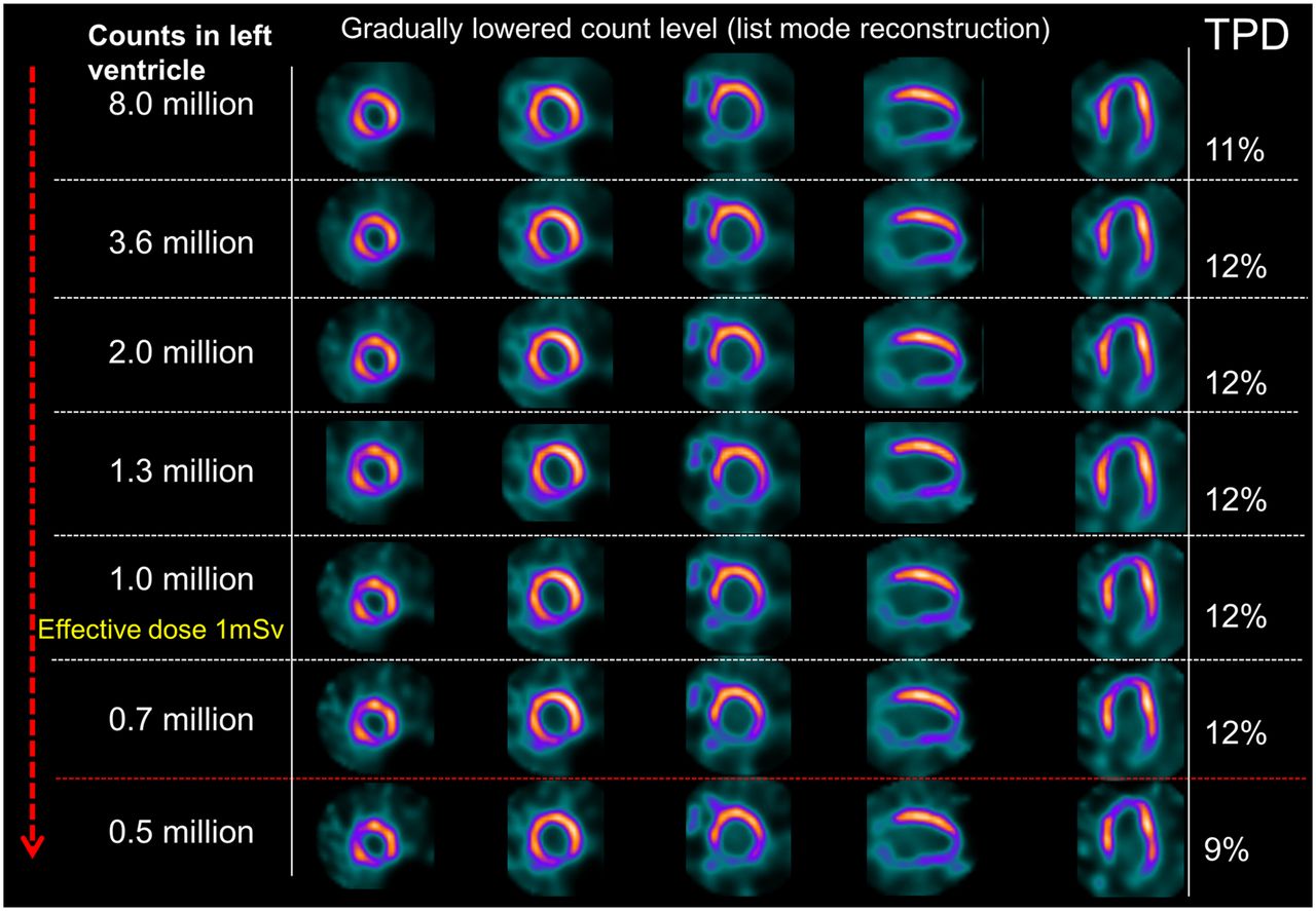
- FIGURE 3.
Patient with abnormal perfusion in low-dose simulation study performed by Nakazato et al. (18). TPD, which is quantitative measure of ischemia, was not significantly different except in simulations representing effective radiation dose < 0.5 mSv. (Reprinted from (18).)
- FIGURE 4.
Two-position imaging. In this case, combination of supine and prone imaging offers superior visual interpretation, but findings were still interpreted as abnormal. Quantitative perfusion analysis is also improved by integrating supine TPD (S-TPD), and prone TPD (P-TPD) into single, combined measure (C-TPD), which shows no perfusion deficit. Patient had no significant coronary disease on invasive coronary angiography. Case is from REFINE SPECT.
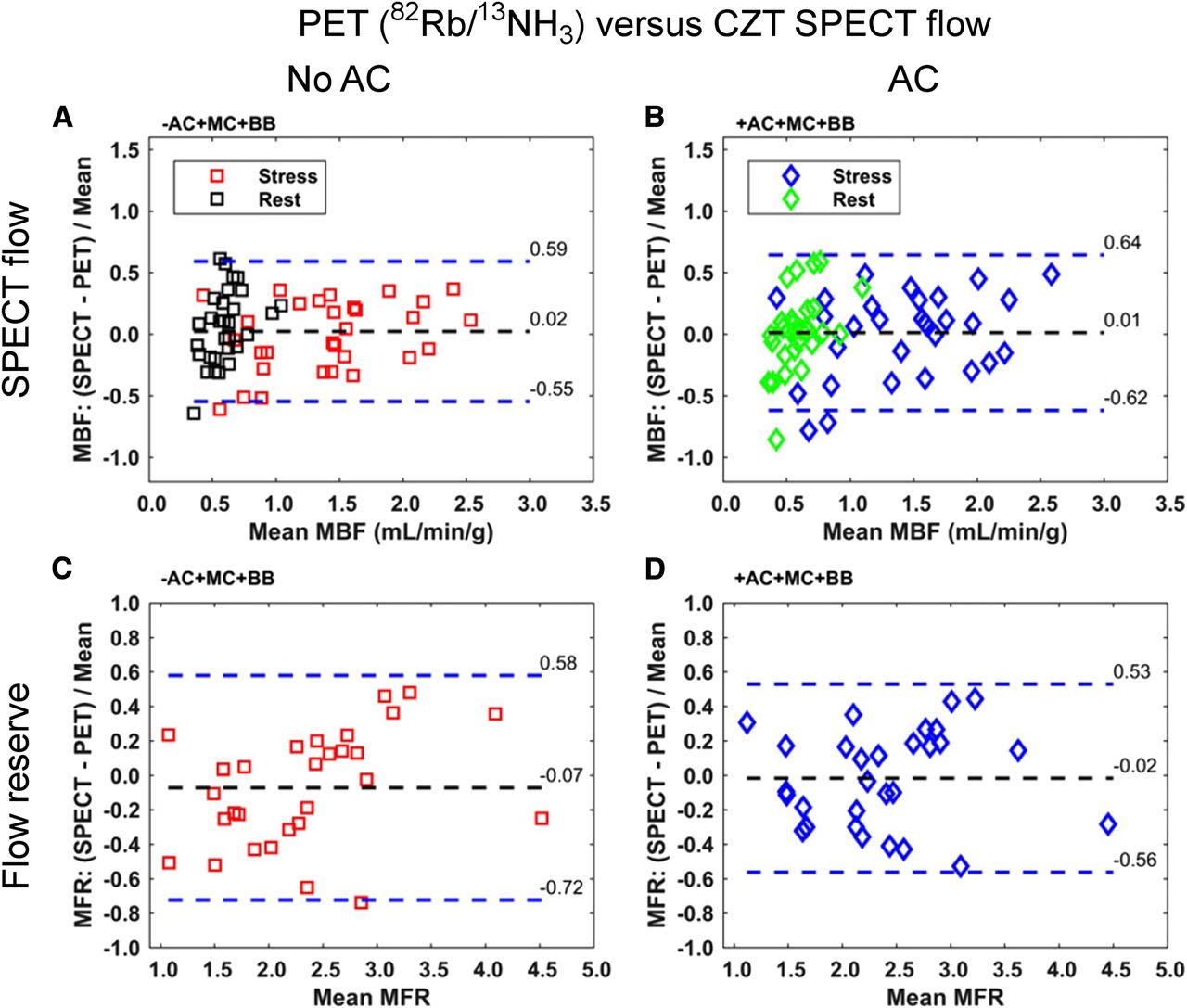
- FIGURE 5.
SPECT MBF assessments with and without AC compared with PET. SPECT MBF measurements at rest or with stress were not significantly different from PET values, with or without AC. Similar results were obtained with MFR measurements. BB = binding to red blood cells; MC = motion correction. (Reprinted from (59).)
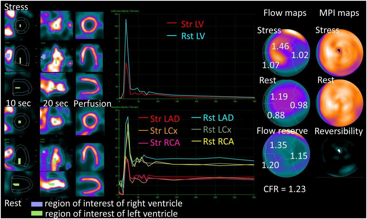
- FIGURE 6.
SPECT MBF information. One-day 99mTc sestamibi stress/rest protocol (259 MBq for stress and 777 MBq for rest) was used. Stress and rest flows are computed from early dynamic imaging for each vascular territory, by deriving regional time–activity curves for myocardium and for input blood in left atrium or left ventricle and applying compartmental models. These values can be used to calculate MFR as ratio between stress and rest MBF. Reduced MFR (<2.0) may be useful to identify presence of multivessel disease leading to balanced ischemia. This patient had normal regional myocardial perfusion but decreased MFR and was ultimately found to have obstructive triple-vessel disease on coronary angiography. LAD = left anterior descending; LCx = left circumflex; LV = left ventricle; RCA = right coronary artery; Rst = rest; Str = stress. (Courtesy of Dr. Alejandro Meretta, Instituto Cardiovascular de Buenos Aires.)
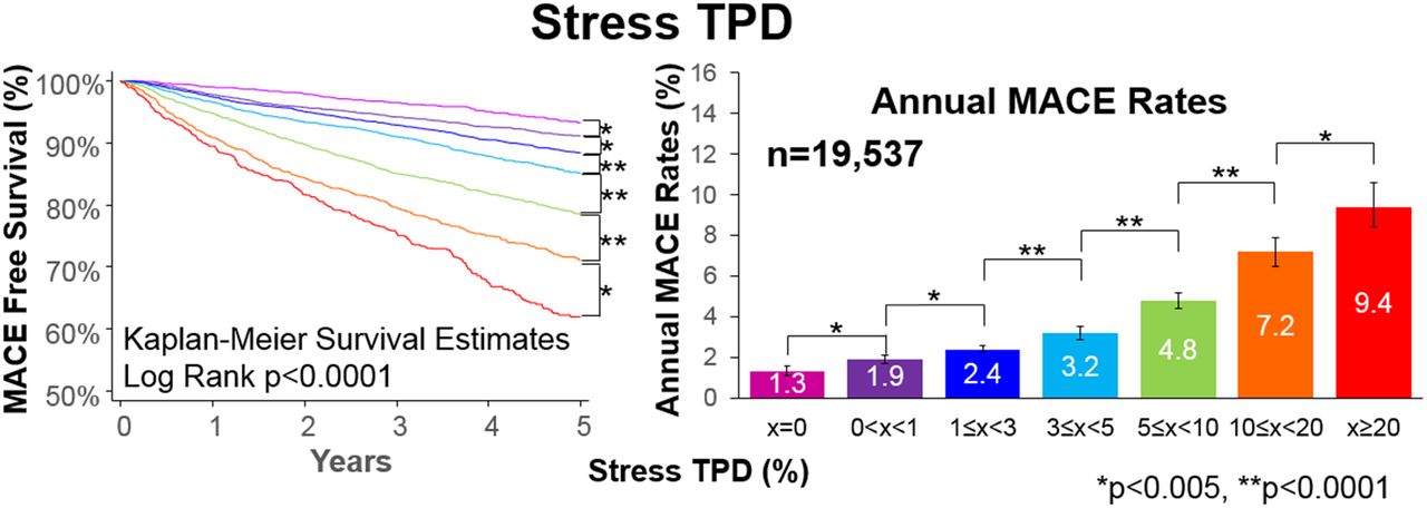
- FIGURE 7.
Prognostic value of quantitative perfusion analysis from REFINE SPECT with solid-state MPI. Quantitative perfusion assessments with TPD provide fine gradation of cardiovascular risk. Each increase in TPD extent, including <1% ischemia compared with no ischemia, was associated with significant increase in combined outcome of death, nonfatal myocardial infarction, unstable angina, or late revascularization. MACE = major adverse cardiovascular events. (Reprinted with permission of (67).)


Tables
Study Cases (n) System Tracer Validated with… Model used Ben-Haim (57) 95 D-SPECT Sestamibi Perfusion angiography Factor analysis Ben Bouallègue (63) 23 Discovery 530c Tetrofosmin iFFR 1TC Nkoulou (62) 28 Discovery 570c Tetrofosmin 13N-ammonia 1TC Miyagawa (73) 69 Discovery 530c Tetrofosmin/sestamibi iFFR 1TC Wells (59) 31 Discovery 530c Tetrofosmin 82Rb or 13N-ammonia 1TC Agostini (58) 30 D-SPECT Sestamibi 15O-water iFFR Net retention model Han (60) 34 Discovery 530c Thallium/tetrofosmin iFFR 1TC Ma (61) 40 Discovery 530c Sestamibi RRG vs. solid-state 1TC Zavadovsky (74) 23 Discovery 570c Sestamibi iFFR Net retention model iFFR = invasive fractional flow reserve; 1TC = 1-tissue-compartment model; RRG = rapid rotating gantry.
- TABLE 2
Major Diagnostic (n > 100) and Prognostic (n > 1,000) Validation Studies for Solid-State SPECT MPI
Study Cases (n) System Comparator Tracer Dose Stress/rest time (min) Diagnostic validation Nakazato (20) 56 ICA, 86 LLk D-SPECT ICA Sestamibi, dual-isotope SD 2/4 Gimelli (15) 137 Discovery 530c ICA Tetrofosmin LD 7/6 Duvall (22) 160 Discovery 530c ICA Sestamibi LD, SD 3–5/3–5 Mouden (75) 100 Discovery 570c iFFR Tetrofosmin SD 5/4 Nakazato (21) 67 ICA, 51 LLk D-SPECT ICA Sestamibi, dual-isotope SD 2–6/6–12 Chikamori (76) 102 Discovery 530c ICA/iFFR Tetrofosmin, sestamibi LD 10/6 Sharir (25) 208 ICA, 76 LLk Discovery 530c ICA Sestamibi LD vs. SD LD: 5–7/5–7; SD: 5/3 Betancur (45) 1,160 D-SPECT ICA Sestamibi LD, SD 4–6/6–10 Betancur (68) 1,638 D-SPECT, Discovery 530c or 570c ICA Sestamibi, tetrofosmin, dual-isotope LD, SD 4–6/6–10 Gimelli (77) 1,161 Discovery 530c ICA Tetrofosmin LD 7/6 Prognostic validation Nakazato (78) 1,613 D-SPECT All-cause mortality Sestamibi, dual-isotope SD Sestamibi: 2/4; dual: 6/4 Chowdhury (79) 1,109 (165 with ICA) Discovery 530c MACE, ICA Tetrofosmin SD 3–6/3–6 De Lorenzo (80) 1,396 Discovery 530c All-cause mortality Sestamibi SD 4/8 Yokota (81) 1,288 solid-state, 362 conventional Discovery 570c MACE Tetrofosmin LD 5/— Engbers (82) 4,057 Discovery 570c MACE Tetrofosmin SD 5/4 Lima (83) 3,554 Discovery 530c MACE Sestamibi LD 3/6 Lima (84) 2,930 Discovery 530c MACE Sestamibi SD, LD 3/6 Betancur (85) 2,619 D-SPECT MACE Sestamibi SD 4–6/6–10 van Dijk (86) 1,255 Discovery 570c MACE Tetrofosmin SD vs. LD SD: 5/4; LD: 8/6 Otaki (67) 19,495 D-SPECT, Discovery 530c MACE Tetrofosmin, sestamibi, dual-isotope LD, SD 4–6/6–10 ICA = invasive coronary angiography; LLk = low likelihood of coronary artery disease; SD = standard dose; LD = low-dose; iFFR = invasive fractional flow reserve; MACE = major adverse cardiac events.
Overall effective dose of test is LD if dose is ≤6 mSv; SD if above 6 mSv. Acquisition time is for default view if more than 1 position is taken.


Jump to section
- Article
- Abstract
- SOLID-STATE DETECTORS
- DEDICATED CARDIAC DETECTORS
- DEDICATED CARDIAC COLLIMATORS AND GEOMETRIES
- SOLID-STATE SPECT/CT SYSTEMS
- RECONSTRUCTION INCLUDING RESOLUTION RECOVERY AND ANATOMIC CONSTRAINTS
- PERFORMANCE
- CURRENT CLINICAL USE
- 2-POSITION IMAGING: UPRIGHT/SUPINE OR SUPINE/PRONE
- LOW-DOSE PROTOCOLS
- SIMULTANEOUS DUAL-ISOTOPE MPI
- NORMAL PERFUSION LIMITS FOR SOLID-STATE CAMERAS
- COMBINED QUANTIFICATION FROM 2 POSITIONS
- MOTION CORRECTION ON SOLID-STATE CAMERAS
- POTENTIAL PITFALLS
- SPECT MBF
- EARLY LEFT VENTRICULAR EJECTION FRACTION
- LARGE-SCALE CLINICAL VALIDATION
- FUTURE HARDWARE DESIGNS
- SUMMARY
- DISCLOSURE
- Footnotes
- REFERENCES
- Figures & Data
- Info & Metrics