


Article Figures & Data
Figures
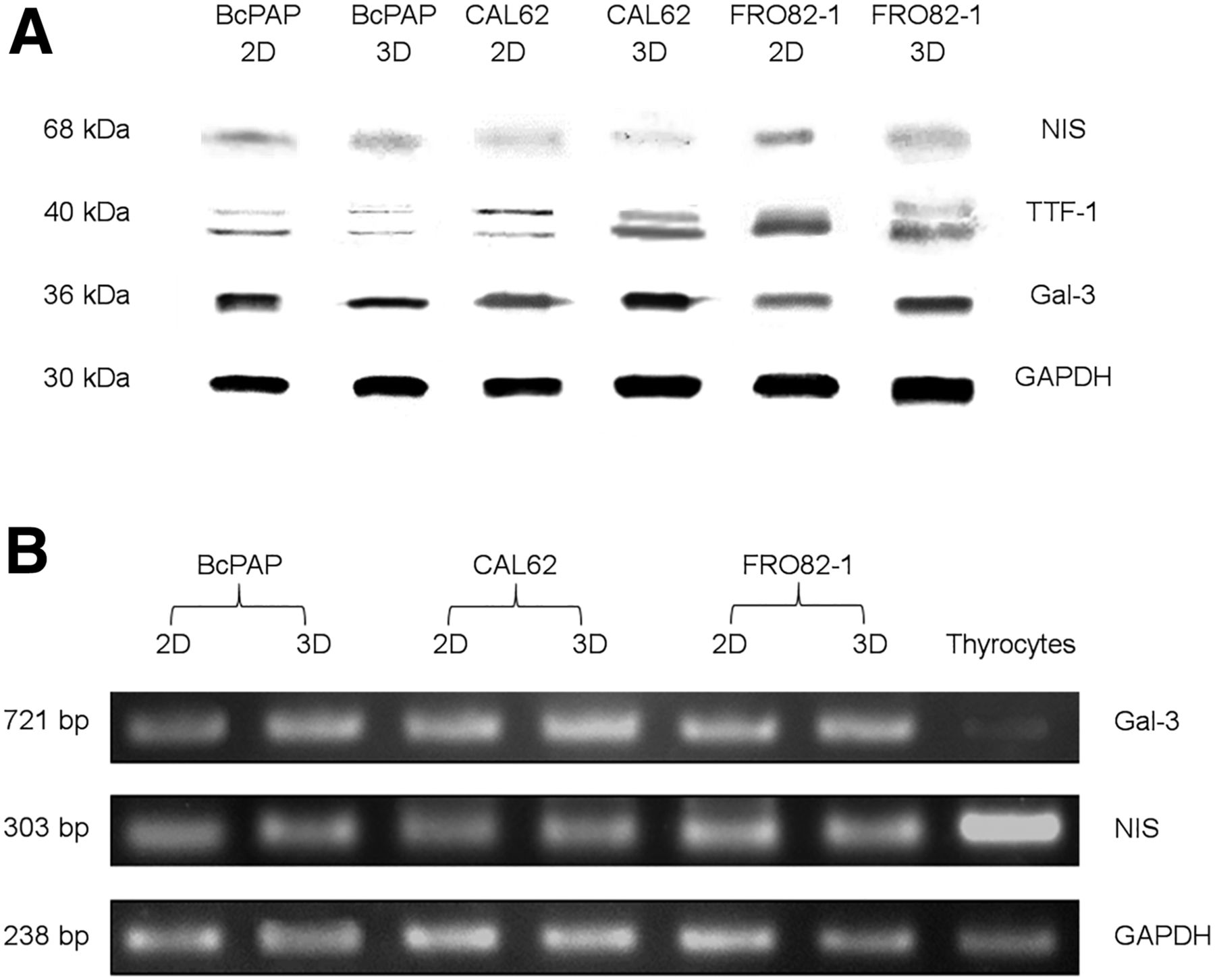
- FIGURE 1.
2D and 3D cell culture characterization. (A) Cell lysates from monolayer (2D) and spheroid (3D) cell cultures analyzed via Western blot. Bands were separated with 100 V for 90 min. (B) Electrophoretic separation of complementary DNA encoding for gal-3 and human NIS on 1% agarose gel, using 80 V for 60 min, and staining with ethidium bromide. TTF = thyroid transcription factor.
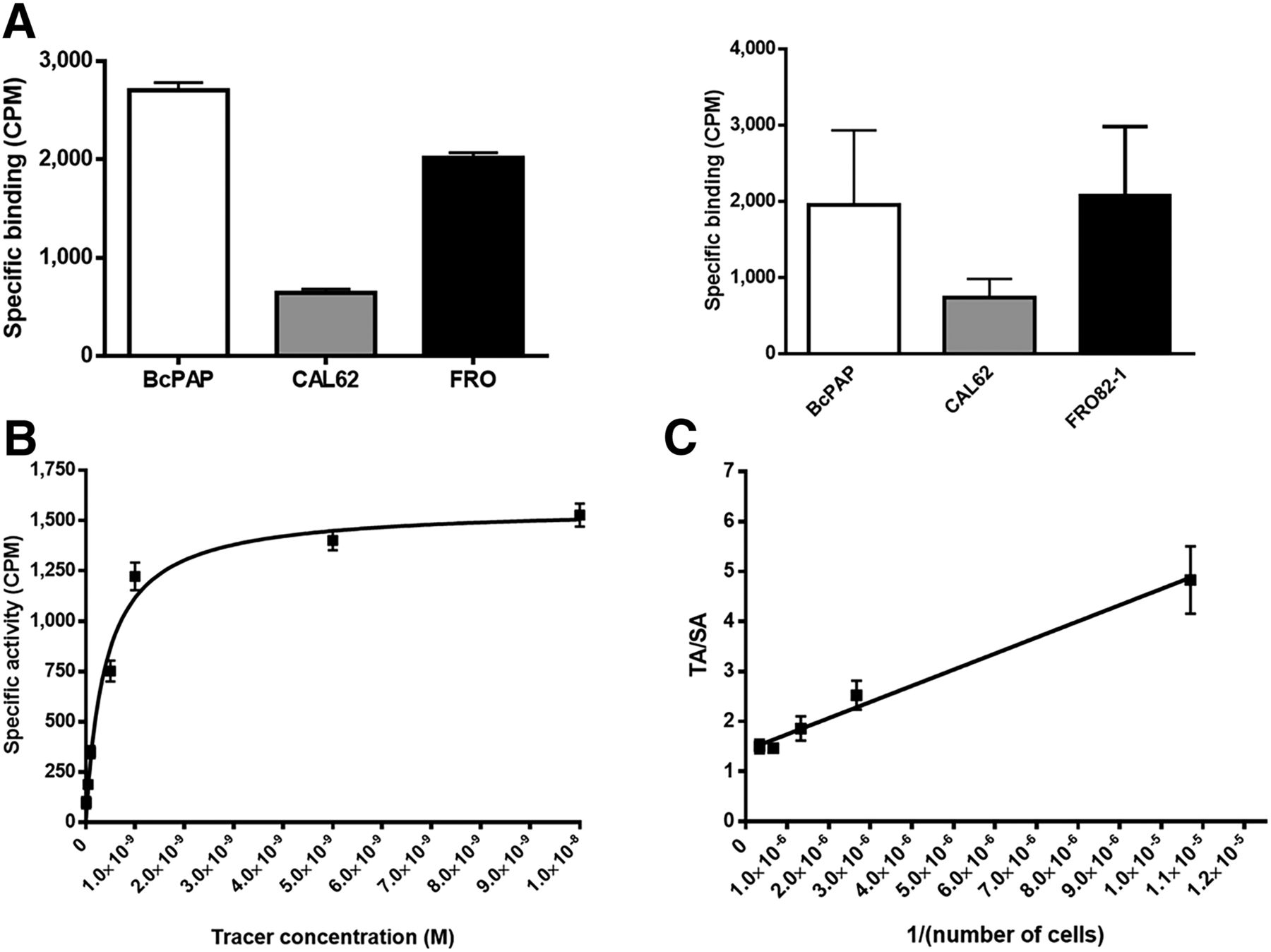
- FIGURE 2.
125I and 89Zr-Df-aGal3-F(ab′)2 cell binding analysis. (A) 125I uptake assay performed on 2D (left) and 3D (right) cell cultures. (B) Binding affinity test of 89Zr-Df-aGal3-F(ab′)2 on 2D cells incubated with increasing concentration of tracer. (C) Immunoreactivity assessed on cell dilutions from 6.0 × 10⁶ to 1 × 10⁵ incubated with constant concentration of tracer. Data represent 3 independent experiments performed each time in triplicate and are expressed as mean ± SD. TA/SA = total bounded activity/specific bounded activity.
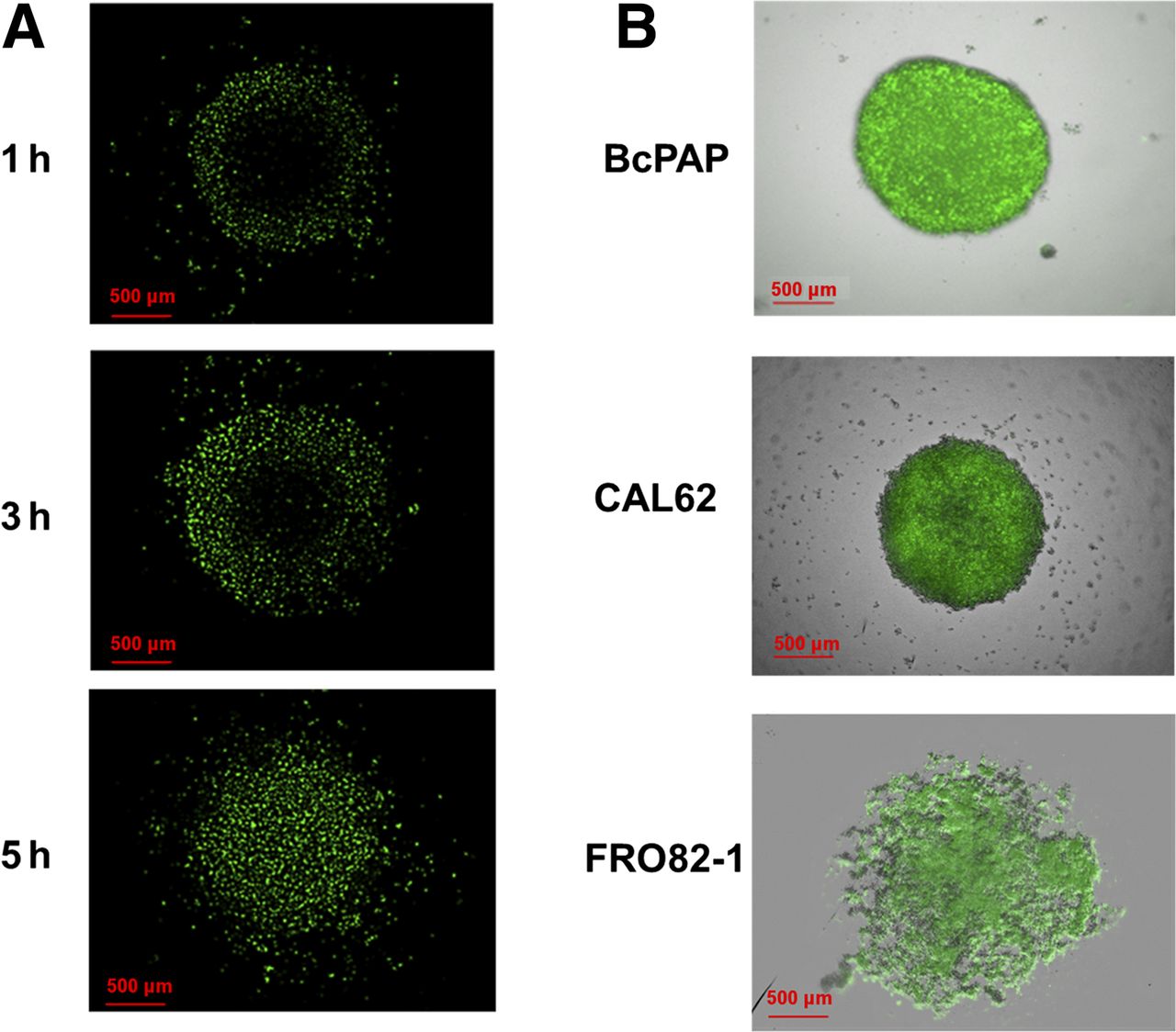
- FIGURE 3.
Characterization of gal-3–specific F(ab′)2 binding to spheroids. (A) Time-dependent penetration of AlexaFluor 488–aGal3-F(ab′)2 into BcPAP tumor spheroids during incubation with 10 μg/mL concentration of AlexaFluor 488 conjugate (representative image). (B) Full focus confocal and fluorescent overlay of tumor spheroids.
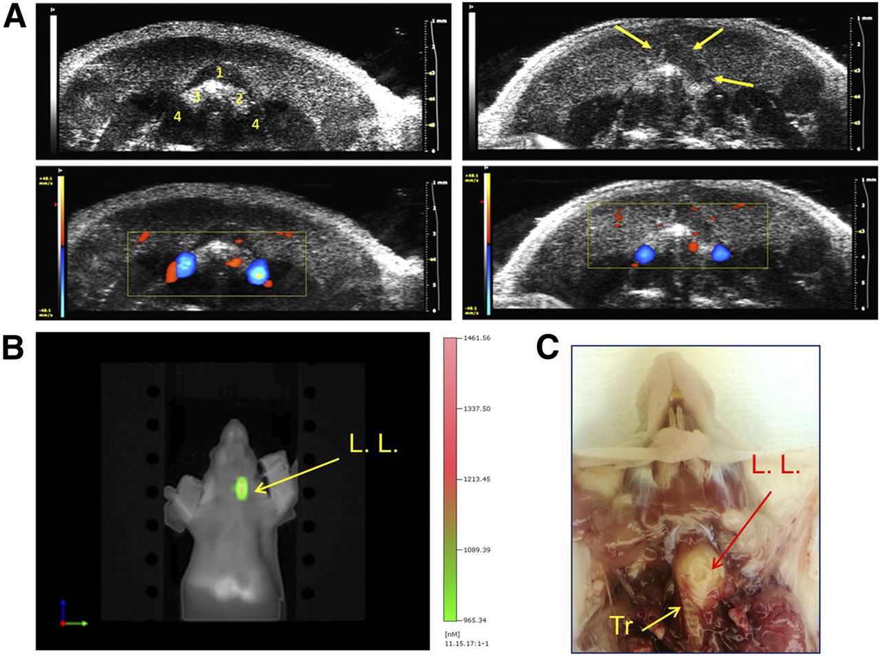
- FIGURE 4.
Orthotopic tumor growth monitoring. (A) Representative ultrasound transversal image of mouse neck before thyroid cancer cell transplantation (top left): strap muscle (1), left thyroid lobe (2), right thyroid lobe (3), carotid arteries (4); ultrasound image showing orthotopic tumor (arrows) expanding from left lobe (top right); and Doppler mode enhancement of activity of carotid arteries and correct localization of thyroid (bottom). (B) FMT imaging of same mouse performed 48 h after injection of Cy5.5-labeled aGal3-F(ab′)2 and visualizing gal-3–positive mass in neck. (C) Anatomic analysis of thyroid orthotopic tumor at necropsy. Tr = trachea; L. L. = left lobe.
- FIGURE 5.
Head-to-head comparison of 124I vs. 89Zr-DFO-aGal3-F(ab′)2 detection of thyroid orthotopic tumors. Representative small-animal PET images were acquired 1 h after injection of 124I and 48 h after injection of 89Zr-DFO-aGal3-F(ab′)2. Each row presents axial PET/CT fusion projection and 3D projection for both tracers. L. L. = left lobe; R. L. = right lobe.
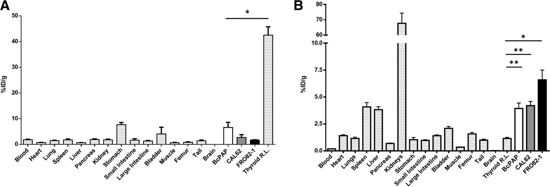
- FIGURE 6.
Biodistribution analysis of 124I vs. 89Zr-DFO-aGal3-F(ab′)2. Three groups of mice (3 per tumor type) were injected with 124I and 2 groups of mice (5 per tumor type) were injected with 89Zr-DFO-aGal3-F(ab′)2. (A) Low NIS expression in orthotopic tumors yielded low 124I accumulation in left lobe compared with right lobe. Differences were statistically significant (*P < 0.01). (B) Strong 89Zr-DFO-aGal3-F(ab′)2 retention was measured in orthotopic tumors, with background accumulation in right lobes. Differences in 89Zr-DFO-aGal3-F(ab′)2 accumulation were statistically significant (*P = <0.01 for FRO82-1; **P < 0.05 for BcPAP and CAL62). R. L. = right lobe.
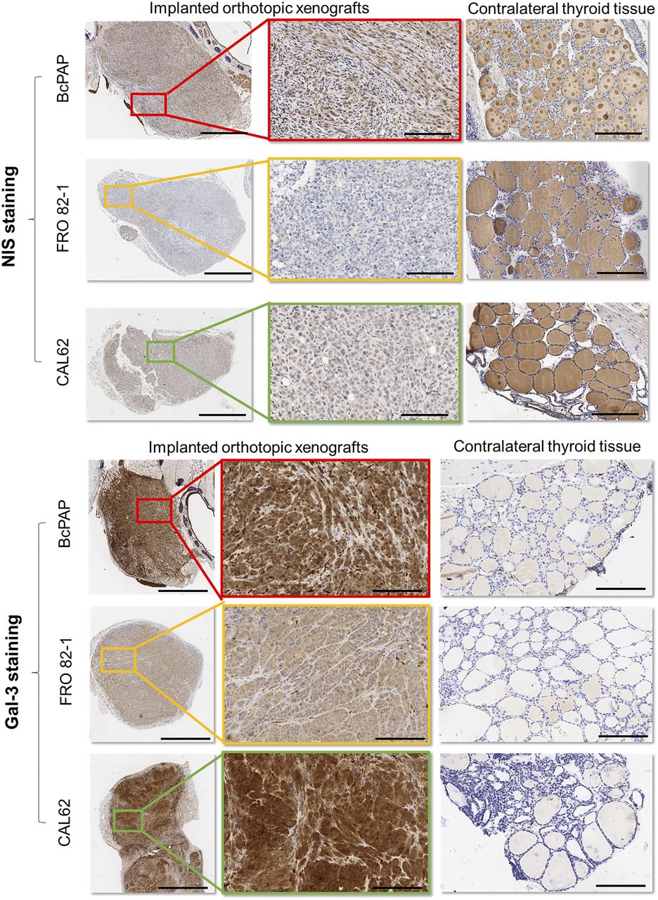
- FIGURE 7.
Immunohistochemical analysis for NIS and gal-3 expression. Tissue sections derived from normal and tumor-infiltrated thyroid lobes were stained for NIS and gal-3 expression using rabbit polyclonal antibody anti-NIS (GTX37599, 0.5 mg/mL; Genetex) and horseradish peroxidase–conjugated rat mAb to gal-3 (Mabtech) (10 μg/mL). (Top) Insets show weak membrane staining on tumor cells, and right column shows normal lobe with thyrocytes positive for NIS staining. (Bottom) On Gal-3 staining, insets show cytoplasmic staining, and right column shows normal lobe without any visible signal for gal-3. Left = ×4 with 2.0-mm bar; middle = ×20 with 300-μm bar; right = ×10 with 1-mm bar.


Additional Files
Supplemental Data
Files in this Data Supplement:


Jump to section
Related Articles
Cited By...
- No citing articles found.