


Article Figures & Data
Figures
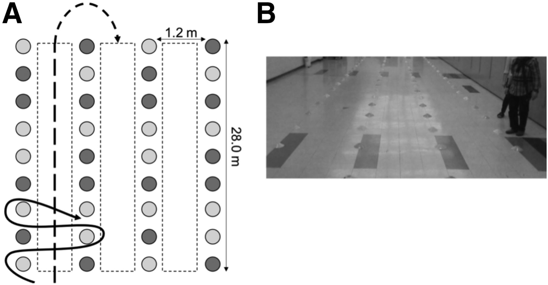
- FIGURE 1.
Experimental setup. (A) Solid line illustrates steering trajectory, and dashed line depicts straight walking. Light gray circles represent yellow cones, and dark gray circles represent orange cones. Complete experimental setup had 30 cones spanning entire length. (B) Participant performing steering task with experimenter following behind.
- FIGURE 2.
Statistical parametric maps showing steering-related rCGM group difference. Activations and deactivations are represented by warm and cool colors, respectively. P < 0.005 (uncorrected); cluster extent threshold = 30.
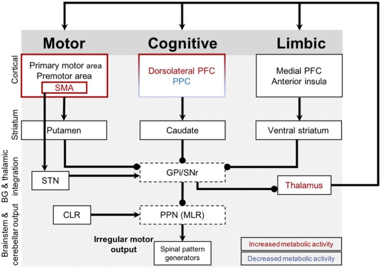
- FIGURE 3.
Complex locomotor control in FOG. Arrows indicate excitatory connections, and spheric ends denote inhibitory connections. Red and blue labels are regions with increased and decreased cerebral glucose metabolism, respectively, in FOG+ group compared with FOG− group. FOG+ group demonstrates changed metabolic activity in cognitive corticobasal ganglia-thalamocortical circuitry (less activation of parietal and less deactivation of prefrontal cortices). At the same time, there is less deactivation of thalamus during steering and increased activity of supplementary motor area, known to have hyperdirect connections with subthalamic nucleus, with overall inhibitory effect on already impaired basal ganglia outputs (i.e., globus pallidus internal segment and substantia nigra) and brain stem locomotor nuclei (i.e., pedunculopontine nucleus). BG = basal ganglia; CLR = cerebellar locomotor region; GPi = globus pallidus internal segment; PFC = prefrontal cortex; PPC = posterior parietal cortex; PPN (MLR) = mesencephalic locomotor region; SMA = supplementary motor area; SNr = substantia nigra; STN = subthalamic nucleus.


Tables
Variable FOG+ (n = 9) FOG− (n = 9) P Sex (n) 0.066 Male 5 8 Female 4 1 Age (y) 68 ± 6 65 ± 5 0.235 Time since disease onset (y)* 9 ± 6 8 ± 3 0.863 Laterality of predominant motor symptoms (n) 0.500 Right 3 2 Left 6 7 Hoehn and Yahr Scale* 3 ± 1 2 ± 0 0.258 MDS-UPDRSIII score (off drug) 48 ± 8 41 ± 7 0.064 DOPA equivalent dose (mg) 893 ± 617 751 ± 272 0.557 NFOG Questionnaire score* 13 ± 8 0 ± 0 <0.001 Montreal Cognitive Assessment 28 ± 2 29 ± 2 0.321 Hospital Anxiety and Depression Scale Anxiety 6 ± 3 4 ± 2 0.067 Depression 7 ± 4 4 ± 2 0.103 ↵* Nonparametric tests used.
MDS-UPDRSIII = Movement Disorders Society Unified Parkinson’s Disease Rating Scale, part III.
Data are mean ± SD for all variables except sex and laterality, represented as median ± interquartile range.
Supplemental Data
Files in this Data Supplement:


Jump to section
Related Articles
Cited By...
- No citing articles found.