


Article Figures & Data
Figures
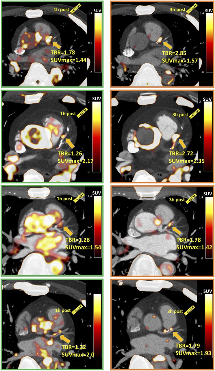
- FIGURE 1.
Four patients with significant coronary 18F-NaF uptake (TBR > 1.25) on 1-h PET in left anterior descending artery. Uptake can be difficult to differentiate in this region from blood pool in adjacent pulmonary artery. On 3-h PET, TBR increased significantly, and blood-pool activity reduced, improving image quality.
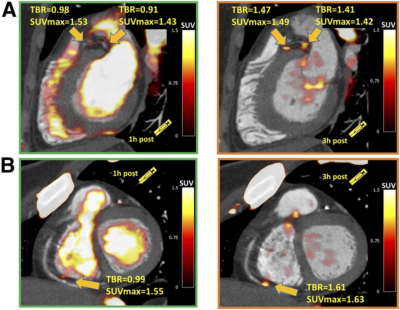
- FIGURE 2.
Examples of coronary plaques with significant uptake on 3-h PET and low tracer activity on 1-h PET. These short-axis images of proximal left anterior descending, proximal circumflex (A), and distal right (B) coronary artery plaques show TBR of less than 1.0 on 1-h PET and uptake exceeding 1.25 TBR threshold at 3 h.
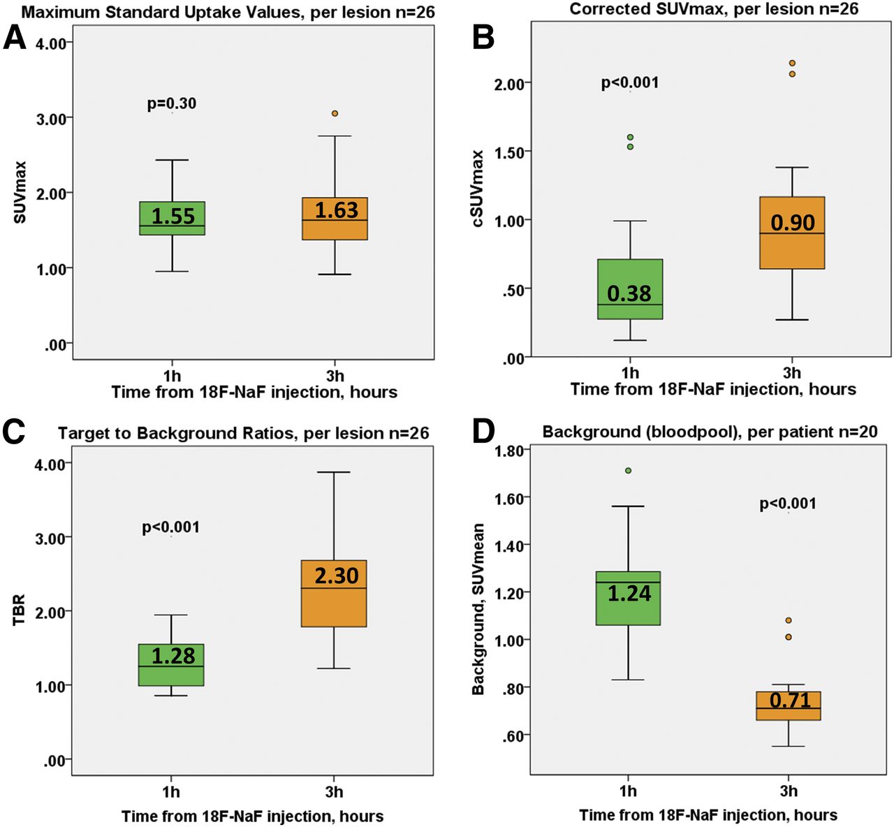
- FIGURE 3.
18F-NaF coronary uptake on 1-h and 3-h PET. (A) SUVmax was comparable on both scans. (B and C) cSUVmax and TBR were higher on 3-h PET. (D) Background (right atrium blood pool SUVmean) was lower on 3-h imaging.
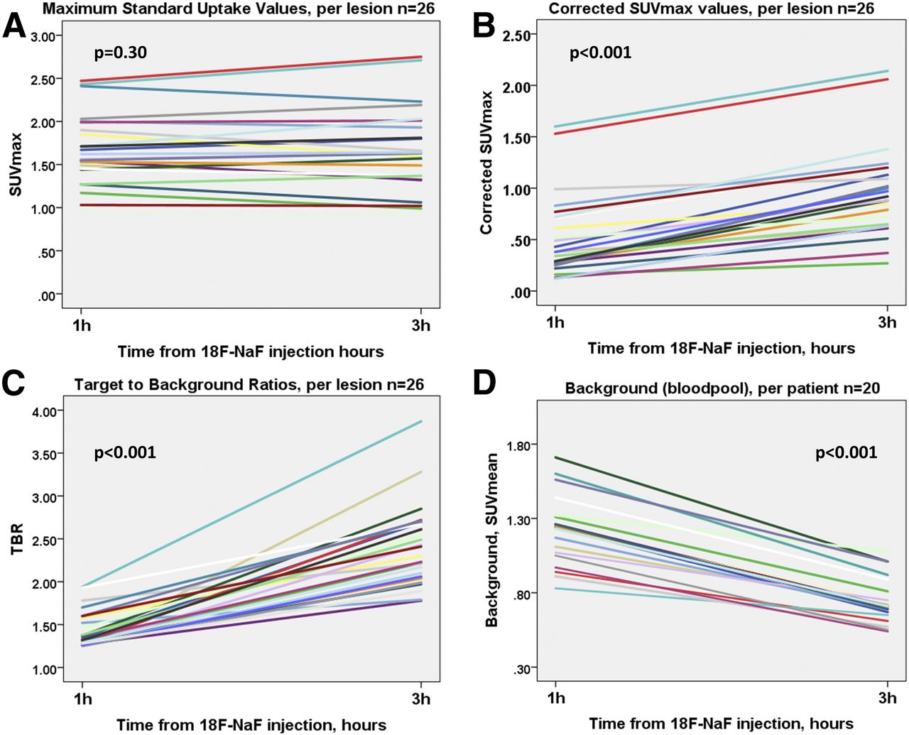
- FIGURE 4.
Line plots of 18F-NaF coronary uptake on 1-h and 3-h PET. (A) SUVmax was comparable on both scans. (B and C) cSUVmax and TBR were higher on 3-h PET. (D) Background (right atrium blood pool SUVmean) was lower on 3-h imaging.

Tables
Characteristic Data Mean age ± SD (y) 67 ± 7 Male (n) 11 (55%) Diabetes (n) 4 (20%) Hyperlipidemia (n) 5 (25%) Hypertension (n) 12 (60%) Tobacco use (n) 6 (30%) Family history of coronary artery disease (n) 3 (15%) Serum biomarkers Total cholesterol (mg/dL) 162 (135–189) High-density lipoprotein (mg/dL) 43 (38–46) Low-density lipoprotein (mg/dL) 93 (71–107) Triglyceride (mg/dL) 121 (86–140) Creatine (mg/dL) 0.8 (0.7–0.9) Medications Aspirin (n) 13 (65%) Statin (n) 6 (30%) ACEI/ARB (n) 6 (30%) β-blocker (n) 7 (35%) Leading clinical indication for CTA Chest pain (n) 15 (75%) Dyspnea (n) 3 (15%) Risk assessment (asymptomatic patient) (n) 2 (10%) Coronary CTA Segment involvement score 6 (3–8) Multivessel disease (n) 6 (30%) Coronary calcium score 312 (50–770) ACEI/ARB = angiotensin-converting enzyme inhibitor/angiotensin receptor blocker.
Qualitative data are expressed as number and percentage; continuous data, except for age, are expressed as median and interquartile range.
Measurement 1-h PET 3-h PET P SUVmax 1.55 (1.43–1.89) 1.63 (1.37–1.98) 0.30 TBR 1.28 (0.98–1.56) 2.30 (1.70–2.68) <0.001 cSUVmax 0.38 (0.27–0.70) 0.90 (0.64–1.17) <0.001 Background 1.24 (1.05–1.31) 0.71 (0.65–0.81) <0.001 Noise 0.07 (0.06–0.09) 0.10 (0.09–0.12) 0.02 Segments with TBR > 1.25 26 (8%) 33 (10%) 0.01 Patients with TBR > 1.25 12 (60%) 15 (75%) 0.004 Qualitative data are expressed as number and percentage; continuous data are expressed as median and interquartile range.
Supplemental Data
Files in this Data Supplement:


