
Main menu

  • Home
  • Content
    • Current
    • Ahead of print
    • Past Issues
    • JNM Supplement
    • SNMMI Annual Meeting Abstracts
    • Continuing Education
    • JNM Podcasts
  • Subscriptions
    • Subscribers
    • Institutional and Non-member
    • Rates
    • Journal Claims
    • Corporate & Special Sales
  • Authors
    • Submit to JNM
    • Information for Authors
    • Assignment of Copyright
    • AQARA requirements
  • Info
    • Reviewers
    • Permissions
    • Advertisers
  • About
    • About Us
    • Editorial Board
    • Contact Information
  • More
    • Alerts
    • Feedback
    • Help
    • SNMMI Journals
  • SNMMI
    • JNM
    • JNMT
    • SNMMI Journals
    • SNMMI

User menu

  • Subscribe
  • My alerts
  • Log in
  • My Cart

Search

  • Advanced search
Journal of Nuclear Medicine
  • SNMMI
    • JNM
    • JNMT
    • SNMMI Journals
    • SNMMI
  • Subscribe
  • My alerts
  • Log in
  • My Cart
Journal of Nuclear Medicine

Advanced Search

  • Home
  • Content
    • Current
    • Ahead of print
    • Past Issues
    • JNM Supplement
    • SNMMI Annual Meeting Abstracts
    • Continuing Education
    • JNM Podcasts
  • Subscriptions
    • Subscribers
    • Institutional and Non-member
    • Rates
    • Journal Claims
    • Corporate & Special Sales
  • Authors
    • Submit to JNM
    • Information for Authors
    • Assignment of Copyright
    • AQARA requirements
  • Info
    • Reviewers
    • Permissions
    • Advertisers
  • About
    • About Us
    • Editorial Board
    • Contact Information
  • More
    • Alerts
    • Feedback
    • Help
    • SNMMI Journals
  • View or Listen to JNM Podcast
  • Visit JNM on Facebook
  • Join JNM on LinkedIn
  • Follow JNM on Twitter
  • Subscribe to our RSS feeds
Research ArticleOncology

18F-FDG PET/CT for Systemic Staging of Newly Diagnosed Breast Cancer in Men

Gary A. Ulaner, Jessica Juarez, Christopher C. Riedl and Debra A. Goldman
Journal of Nuclear Medicine April 2019, 60 (4) 472-477; DOI: https://doi.org/10.2967/jnumed.118.217836
Gary A. Ulaner
1Department of Radiology, Memorial Sloan Kettering Cancer Center, New York, New York
2Department of Radiology, Weill Cornell Medical College, New York, New York; and
  • Find this author on Google Scholar
  • Find this author on PubMed
  • Search for this author on this site
Jessica Juarez
1Department of Radiology, Memorial Sloan Kettering Cancer Center, New York, New York
  • Find this author on Google Scholar
  • Find this author on PubMed
  • Search for this author on this site
Christopher C. Riedl
1Department of Radiology, Memorial Sloan Kettering Cancer Center, New York, New York
2Department of Radiology, Weill Cornell Medical College, New York, New York; and
  • Find this author on Google Scholar
  • Find this author on PubMed
  • Search for this author on this site
Debra A. Goldman
3Department of Epidemiology and Biostatistics, Memorial Sloan Kettering Cancer Center, New York, New York
  • Find this author on Google Scholar
  • Find this author on PubMed
  • Search for this author on this site
  • Article
  • Figures & Data
  • Info & Metrics
  • PDF
Loading

Article Figures & Data

Figures

  • Tables
  • FIGURE 1.
    • Download figure
    • Open in new tab
    • Download powerpoint
    FIGURE 1.

    Standards for Reporting of Diagnostic Accuracy Diagram (STARD) for patients in this study. MSKCC = Memorial Sloan Kettering Cancer Center.

  • FIGURE 2.
    • Download figure
    • Open in new tab
    • Download powerpoint
    FIGURE 2.

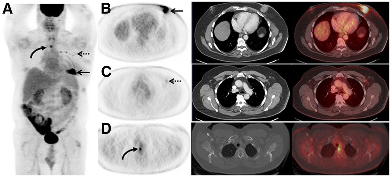
    A 53-y-old man with a pre-PET/CT stage IIB breast cancer, which was upstaged to stage IV by 18F-FDG PET/CT. (A) 18F-FDG maximum-intensity-projection image demonstrates 18F-FDG avidity in left breast (arrow), left axilla (dashed arrow), as well as overlying midline chest (curved arrow). (B) Axial 18F-FDG PET, CT on soft-tissue window, and fused 18F-FDG PET/CT demonstrate the 18F-FDG–avid known primary left breast malignancy (arrow). (C) Axial 18F-FDG PET, CT on soft-tissue window, and fused 18F-FDG PET/CT demonstrate 18F-FDG–avid known left axillary nodal metastases (dashed arrow). (D) Axial 18F-FDG PET, CT on bone window, and fused 18F-FDG PET/CT demonstrate 18F-FDG avidity in midline chest on the maximum-intensity-projection image corresponding to a thoracic vertebra without CT correlate. This lesion was biopsied and proven to be a previously unsuspected osseous metastasis, increasing patient’s stage to IV. This altered patient’s management from surgical management to systemic therapy without surgical management.

  • FIGURE 3.
    • Download figure
    • Open in new tab
    • Download powerpoint
    FIGURE 3.

    A 58-y-old man with a pre-PET/CT stage IIB breast cancer, which was upstaged to stage IV on 18F-FDG PET/CT. (A) 18F-FDG maximum-intensity-projection image demonstrates mild 18F-FDG avidity in left mastectomy bed (arrow) and left axillary node (dashed arrow), as well as overlying midline chest (curved arrow). (B) Axial 18F-FDG PET, CT on bone window, and fused 18F-FDG PET/CT demonstrate an 18F-FDG–avid thoracic vertebra (curved arrow) corresponding only with small, benign-appearing sclerotic focus on CT. This patient was subsequently treated with systemic therapy. (C) Axial 18F-FDG PET, CT on bone tissue window, and fused 18F-FDG PET/CT after systemic therapy demonstrate resolution of 18F-FDG avidity (curved arrow) and increased sclerosis of the vertebra (white arrow), consistent with therapy response in an osseous metastasis and osseous healing. (D) 18F-FDG maximum-intensity-projection image after systemic therapy demonstrates persistence of 18F-FDG–avid postmastectomy inflammation (arrow) but resolution of avidity in the node and osseous metastasis.

Tables

  • Figures
    • View popup
    TABLE 1

    Characteristics of Patients with Male Breast Cancer and Their Primary Tumors

    Characteristicn (%)
    No. of patients39
    Median age (y)62 (range, 31–90)
    Race
     African American4 (10)
     Asian3 (8)
     Caucasian31 (79)
     Other1 (3)
    Histology
     Ductal37 (95)
     Adenoid cystic1 (3)
     Papillary1 (3)
    Tumor grade
     High28 (72)
     Intermediate9 (23)
     Unknown2 (5)
    Receptor status
     ER+/HER2−33 (84)
     HER2+5 (13)
     Triple-negative1 (3)
    BRCA1 or 2 mutation
     Yes4 (10)
     No16 (41)
     Unknown19 (49)
    Initial staging
     Pre-PET/CT stage
      I2 (5)
      IIA6 (15)
      IIB19 (49)
      IIIA5 (13)
      IIIB4 (10)
      IIIC3 (8)
     T stage
      T15 (13)
      T229 (74)
      T31 (3)
      T44 (10)
     N stage
      N010 (26)
      N122 (56)
      N24 (10)
      N33 (8)
    M stage, M039 (100)
    • Data in parentheses are percentages, unless otherwise indicated.

    • View popup
    TABLE 2

    Summary of Patients with Male Breast Cancer Upstaged by 18F-FDG PET/CT Stratified by Pre-PET/CT Stage

    Post-PET/CT stage
    Pre-PET/CT stageTotalIIIAIIBIIIAIIIBIIICIV (%)
    I220 (0)
    IIA660 (0)
    IIB19163 (16)
    IIIA541 (20)
    IIIB4103 (75)
    IIIC330 (0)
    Total3926164137 (18)
PreviousNext
Back to top

In this issue

Journal of Nuclear Medicine: 60 (4)
Journal of Nuclear Medicine
Vol. 60, Issue 4
April 1, 2019
  • Table of Contents
  • Table of Contents (PDF)
  • About the Cover
  • Index by author
Print
Download PDF
Article Alerts
Sign In to Email Alerts with your Email Address
Email Article

Thank you for your interest in spreading the word on Journal of Nuclear Medicine.

NOTE: We only request your email address so that the person you are recommending the page to knows that you wanted them to see it, and that it is not junk mail. We do not capture any email address.

Enter multiple addresses on separate lines or separate them with commas.
18F-FDG PET/CT for Systemic Staging of Newly Diagnosed Breast Cancer in Men
(Your Name) has sent you a message from Journal of Nuclear Medicine
(Your Name) thought you would like to see the Journal of Nuclear Medicine web site.
Citation Tools
18F-FDG PET/CT for Systemic Staging of Newly Diagnosed Breast Cancer in Men
Gary A. Ulaner, Jessica Juarez, Christopher C. Riedl, Debra A. Goldman
Journal of Nuclear Medicine Apr 2019, 60 (4) 472-477; DOI: 10.2967/jnumed.118.217836

Citation Manager Formats

  • BibTeX
  • Bookends
  • EasyBib
  • EndNote (tagged)
  • EndNote 8 (xml)
  • Medlars
  • Mendeley
  • Papers
  • RefWorks Tagged
  • Ref Manager
  • RIS
  • Zotero
Share
18F-FDG PET/CT for Systemic Staging of Newly Diagnosed Breast Cancer in Men
Gary A. Ulaner, Jessica Juarez, Christopher C. Riedl, Debra A. Goldman
Journal of Nuclear Medicine Apr 2019, 60 (4) 472-477; DOI: 10.2967/jnumed.118.217836
Twitter logo Facebook logo LinkedIn logo Mendeley logo
  • Tweet Widget
  • Facebook Like
  • Google Plus One
Bookmark this article

Jump to section

  • Article
    • Abstract
    • MATERIALS AND METHODS
    • RESULTS
    • DISCUSSION
    • CONCLUSION
    • DISCLOSURE
    • Acknowledgments
    • Footnotes
    • REFERENCES
  • Figures & Data
  • Info & Metrics
  • PDF

Related Articles

  • This Month in JNM
  • PubMed
  • Google Scholar

Cited By...

  • No citing articles found.
  • Google Scholar

More in this TOC Section

Oncology

  • Comparative PSMA expression at early (1hour) vs late (2hour) in primary and secondary sites of involvement in prostate cancer.
  • Imaging spectrum of peritoneal carcinomatosis associated with various etiologies on 18F-FDG PET/CT
  • Hybrid Imaging Features of Musculoskeletal Tumors
Show more Oncology

Clinical

  • Comparative PSMA expression at early (1hour) vs late (2hour) in primary and secondary sites of involvement in prostate cancer.
  • Imaging spectrum of peritoneal carcinomatosis associated with various etiologies on 18F-FDG PET/CT
  • Hybrid Imaging Features of Musculoskeletal Tumors
Show more Clinical

Similar Articles

Keywords

  • Male breast cancer
  • 18F-FDG PET/CT
  • staging
SNMMI

© 2025 SNMMI

Powered by HighWire