


Article Figures & Data
Figures
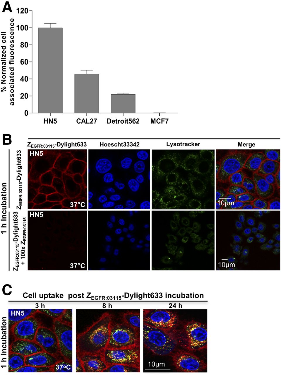
- FIGURE 1.
(A) EGFR expression as determined by flow cytometry in selected cancer cell lines. (B) In vitro binding specificity of ZEGFR:03115-Dylight633 in HN5 cells as shown by confocal microscopy. (C) Internalization studies of ZEGFR:03115-Dylight633 3, 8, and 24 h after 1 h of incubation in HN5 cells.
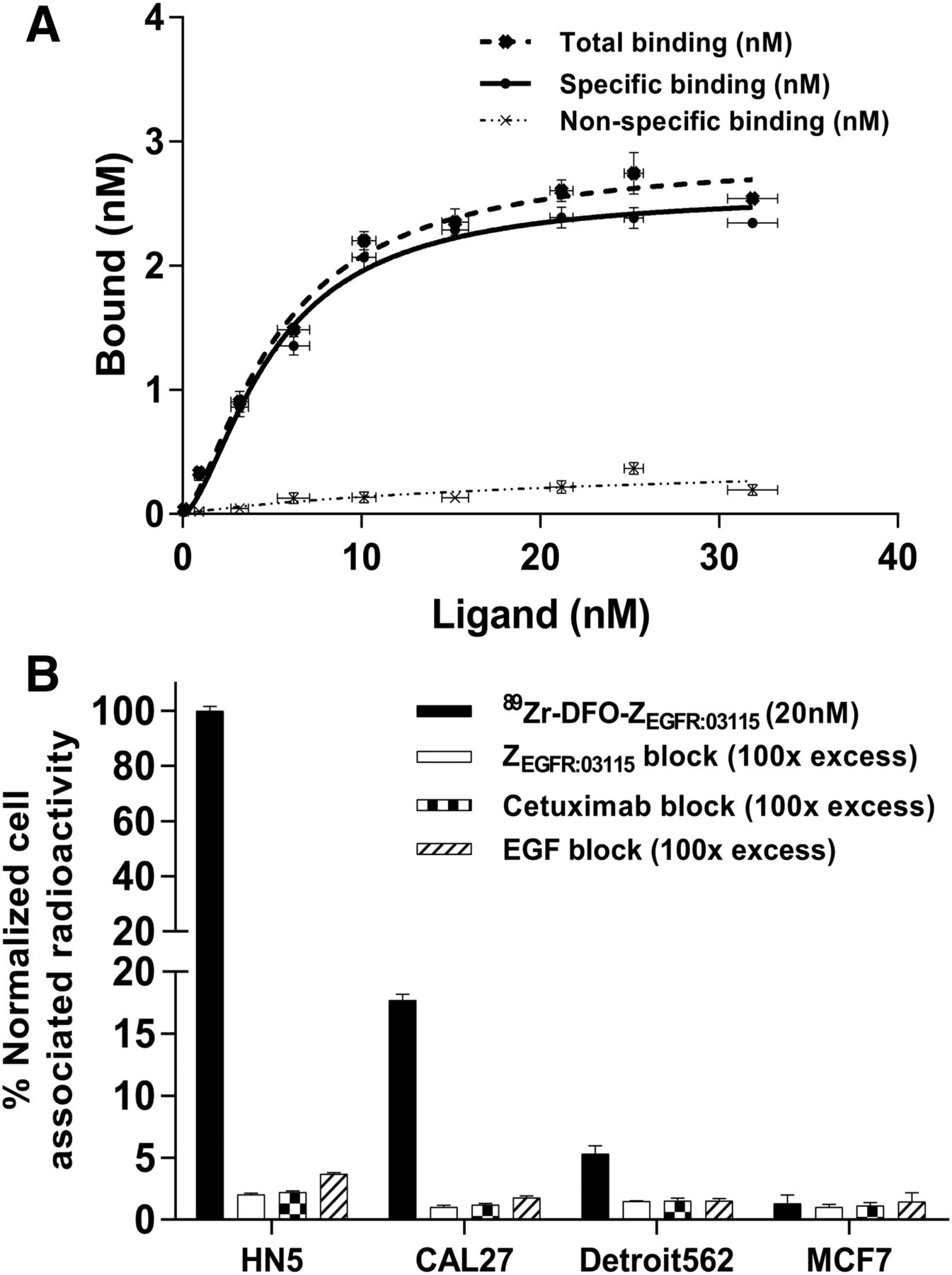
- FIGURE 2.
(A) Saturation curve obtained for CAL27 cells incubated with increasing concentrations of 89Zr-DFO-ZEGFR:03115 bound vs. 89Zr-DFO-ZEGFR:03115 incubation concentration. (B) In vitro binding specificity of 89Zr-DFO-ZEGFR:03115 in selected cell lines with and without blocking using unlabeled Affibody, cetuximab, or EGF. Data are normalized to maximum cell-associated radioactivity per experiment.
- FIGURE 3.
Whole-body coronal PET/CT images acquired 3 h after 89Zr-DFO-ZEGFR:03115 administration spiked with different amounts of nonlabeled ZEGFR:03115 in mice bearing CAL27 tumors: 2 μg of 89Zr-DFO-ZEGFR:03115 injected (A); 1 μg of ZEGFR:03115 injected 30 min before injection of 2 μg of 89Zr-DFO-ZEGFR:03115 (B); 5 μg of ZEGFR:03115 injected 30 min before injection of 2 μg of 89Zr-DFO-ZEGFR:03115 (C); 10 μg of ZEGFR:03115 injected 30 min before injection of 2 μg of 89Zr-DFO-ZEGFR:03115 (D); 10 μg of ZEGFR:03115 coinjected with 2 μg of 89Zr-DFO-ZEGFR:03115 (E).
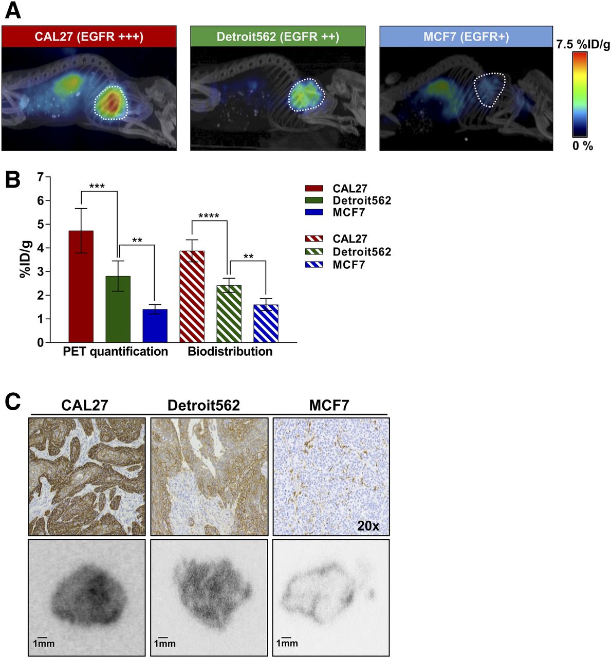
- FIGURE 4.
Radioconjugate uptake in xenografts with varying EGFR expression. (A) Representative whole-body sagittal PET/CT images acquired 3 h after injection. (B) PET quantification of radiotracer uptake in tumors (outlined on image) 3 h after injection in comparison with data obtained from biodistribution studies. Data are mean ± SD. (C) Histopathologic analysis of EGFR expression in xenograft models (top), and representative autoradiography tumor sections 3 h after radioconjugate administration (bottom). **P < 0.01. ***P < 0.001. ****P < 0.0001.
- FIGURE 5.
18F-AlF-NOTA-ZEGFR:03115 uptake assessed 1 h after injection. (A) Representative sagittal whole-body PET/CT images of mice bearing HN5 tumors (outlined on image) with or without treatment with cetuximab. (B) PET quantification in control and cetuximab-treated HN5 tumors and corresponding biodistribution %ID/g values. Data are mean ± SD (n ≥ 6). ****P < 0.0001.
- FIGURE 6.
(A) Western blot of tumor tissue lysates from control and cetuximab-treated mice demonstrating EGFR protein expression, activation, and downstream signaling. (B) Spearman rank correlation analysis for EGFR expression as determined by Western blot against 18F-AlF-NOTA-ZEGFR:03115 tumor uptake as quantified by PET image analysis. Dashed lines represent 95% confidence levels. (C) Histopathologic analysis of EGFR expression and hematoxylin and eosin (H&E) staining in HN5 xenografts in both control and cetuximab-treated mouse. A.U. = arbitrary units.


Tables
- TABLE 1
Ex Vivo Biodistribution 3 Hours After Intravenous Administration of Increasing Amounts of Nonlabeled ZEGFR:03115 30 Minutes Before 2 μg of 89Zr-DFO-ZEGFR:03115 or 2 μg of 89Zr-DFO-ZTaq in CAL27 Xenografts
2 μg 89Zr-DFO-ZEGFR:03115 Organ 0 μg ZEGFR:03115 1 μg ZEGFR:03115 5 μg ZEGFR:03115 10 μg ZEGFR:03115 20 μg ZEGFR:03115 2 μg 89Zr-DFO-ZTaq (10 μg ZEGFR:03115) Blood 3.52 ± 1.42 6.01 ± 0.18 3.87 ± 0.30 4.46 ± 1.65 3.06 ± 0.20 0.64 ± 0.04 Heart 0.89 ± 0.22 1.53 ± 0.05 0.95 ± 0.09 1.03 ± 0.29 0.77 ± 0.06 0.25 ± 0.03 Lungs 1.45 ± 0.45 2.55 ± 0.23 1.84 ± 0.50 2.05 ± 0.1.06 1.45 ± 0.08 0.47 ± 0.05 Kidney 37.14 ± 1.17 54.52 ± 17.13 73.26 ± 12.91 140.84 ± 47.70 109.14 ± 22.30 172.04 ± 20.07 Spleen 0.91 ± 0.10 1.58 ± 0.23 0.94 ± 0.20 1.28 ± 0.13 0.94 ± 0.13 0.37 ± 0.08 Liver 20.74 ± 8.31 10.36 ± 1.49 3.82 ± 0.39 4.25 ± 1.55 3.19 ± 0.14 0.82 ± 0.22 Pancreas 0.73 ± 0.19 1.62 ± 0.24 0.71 ± 0.12 0.69 ± 0.13 0.46 ± 0.04 0.14 ± 0.03 Tumor 1.75 ± 0.21 1.70 ± 0.68 1.87 ± 0.58 3.69 ± 1.19 2.59 ± 0.48 0.26 ± 0.05 Bone 1.35 ± 0.24 1.24 ± 0.21 0.81 ± 0.15 0.87 ± 0.14 0.66 ± 0.09 0.21 ± 0.05 Intestine 0.59 ± 0.28 1.62 ± 0.57 0.90 ± 0.11 0.89 ± 0.11 0.81 ± 0.05 0.19 ± 0.03 Muscle 0.24 ± 0.01 0.35 ± 0.07 0.37 ± 0.09 0.36 ± 0.05 0.33 ± 0.04 0.08 ± 0.03 Ratio Tumor-to-blood 0.50 0.28 0.48 0.83 0.85 0.41 Tumor-to-muscle 7.29 4.86 5.05 10.25 7.85 3.15 Tumor-to-liver 0.08 0.16 0.49 0.87 0.81 0.32 Data are mean ± SD (n ≥ 3) %ID/g.
- TABLE 2
Ex Vivo Biodistribution 3, 24, and 48 Hours After Intravenous Injection of 2 μg of 89Zr-DFO-ZEGFR:03115 Coinjected with 10 μg of Nonlabeled ZEGFR:03115 in Mice Bearing CAL27 Xenografts
Organ 3 h 24 h 48 h Blood 3.70 ± 0.55 0.87 ± 0.32 0.23 ± 0.06 Heart 0.99 ± 0.19 0.37 ± 0.12 0.31 ± 0.04 Lungs 2.74 ± 0.93 0.88 ± 0.13 0.70 ± 0.08 Kidney 130.42 ± 22.25 108.80 ± 19.60 104.85 ± 11.10 Spleen 1.14 ± 0.18 0.72 ± 0.20 0.79 ± 0.37 Liver 4.99 ± 0.85 4.31 ± 1.00 4.11 ± 1.41 Pancreas 0.67 ± 0.14 0.36 ± 0.10 0.35 ± 0.25 Tumor 3.88 ± 0.46 2.43 ± 0.27 2.13 ± 0.12 Bone 0.99 ± 0.28 2.28 ± 0.27 3.44 ± 0.59 Intestine 1.09 ± 0.16 0.42 ± 0.03 0.40 ± 0.02 Muscle 0.38 ± 0.09 0.14 ± 0.04 0.16 ± 0.07 Ratio Tumor-to-blood 1.04 2.79 9.34 Tumor-to-muscle 10.31 17.35 13.56 Tumor-to-liver 0.78 0.56 0.52 Data are mean ± SD (n ≥ 3) %ID/g.
Supplemental Data
Files in this Data Supplement:


Jump to section
Related Articles
Cited By...
- No citing articles found.