


Article Figures & Data
Figures
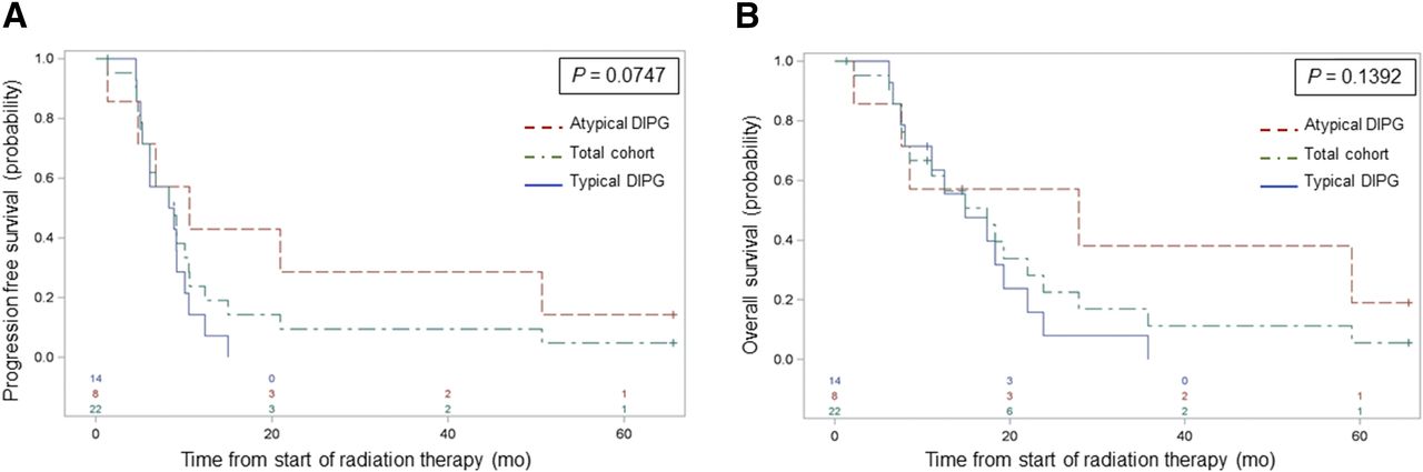
- FIGURE 1.
PFS (A) and OS (B) estimates for total cohort, patients with typical DIPG, and patients with atypical DIPG, showing trend toward significant difference in PFS by DIPG type. CI = confidence interval.
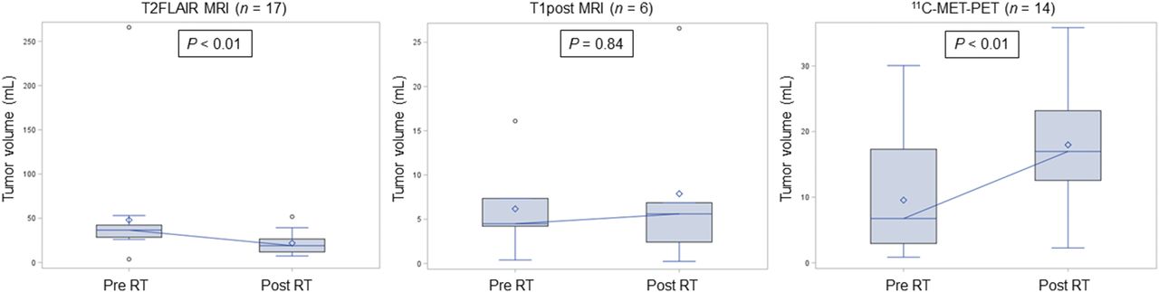
- FIGURE 2.
Box-and-whisker plots of imaging modality–defined tumor-volume change over time in patients with imaging abnormalities noted at both time points. Significant volume reductions were observed after RT for T2FLAIR MRI–defined tumor and for 11C-methionine PET–defined tumor. Included are medians (connecting horizontal lines), means (diamonds), interquartile ranges (boxes), minimum and maximum values (whiskers), and outliers, that is, values beyond 1.5 interquartile ranges (circles).
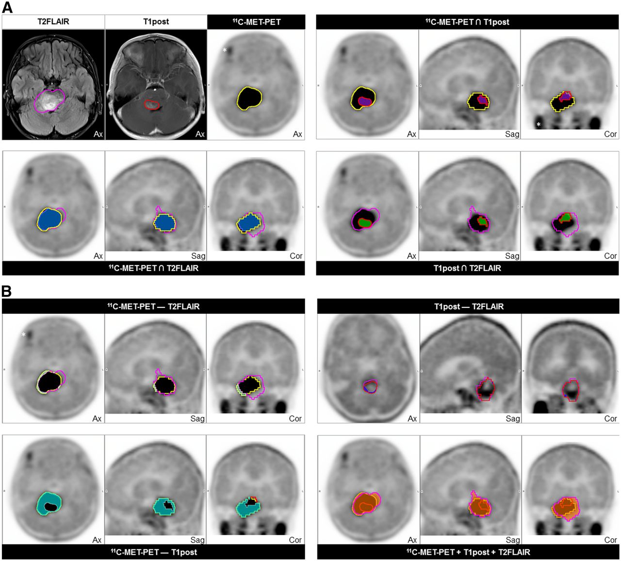
- FIGURE 3.
Example of concordant and discordant segmented tumor volumes based on T2FLAIR (magenta) and T1post (red) abnormalities on MR images and 11C-methionine abnormality (yellow) on 11C-methionine PET. (A) Concordance volumes with coregistered MRI- and 11C-methionine PET–defined tumor (upper left) and indicated concordance volumes (blue = 11C-methionine PET ∩ T2FLAIR; purple = 11C-methionine PET ∩ T1post; green = T1post ∩ T2FLAIR). (B) Indicated discordance volumes (light green = 11C-methionine PET − T2FLAIR; aqua = 11C-methionine PET − T1post; dark blue = T1post − T2FLAIR) and concordant total tumor volume delineated on MRI and 11C-methionine PET (red-orange, bottom right). *Physiologic uptake in exocrine glands.
- FIGURE 4.
Waterfall plot of concordance and discordance of imaging modality–defined tumor volumes at diagnosis (A) and first surveillance (B) and contribution of 11C-methionine PET volume to T2FLAIR-defined tumor volume at defined time points (C). Blue = typical DIPG; red = atypical DIPG; MET = methionine.
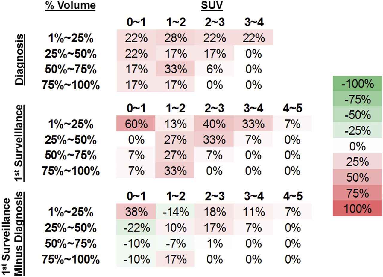
- FIGURE 5.
Descriptive analysis of tumor volume–SUV proportions over indicated time points. Percentage of tumor volume in quartiles with SUV greater than or equal to specified SUV range is displayed with percentage of patients within each SUV range who had indicated volume–SUV relationships. Matrix subtraction of volume–SUV proportions between first surveillance and diagnosis are displayed on bottom row. Colorimetric scale (far right) uses progressively darker red values to indicate relative increase in percentage of patients’ specified volume–SUV metrics, whereas progressively darker green values indicate decrease.


Tables
- TABLE 1
Cox Proportional Hazards Models of Associations of Baseline Covariates with PFS and OS
Variable Survival type Hazard ratio P Contrast enhancement (yes vs. no) PFS 1.11 0.83 OS 1.86 0.23 DIPG status (typical vs. atypical) PFS 2.75 0.06 OS 2.36 0.13 11C-methionine PET intensity grade (1 and 2 vs. 3) PFS 1.62 0.43 OS 1.64 0.42 11C-methionine PET uniformity grade Continuous PFS 1.02 0.23 OS 1.02 0.20 ≤10% vs. >10% PFS 0.59 0.32 OS 0.52 0.26
Supplemental Data
Files in this Data Supplement:


