


Article Figures & Data
Figures
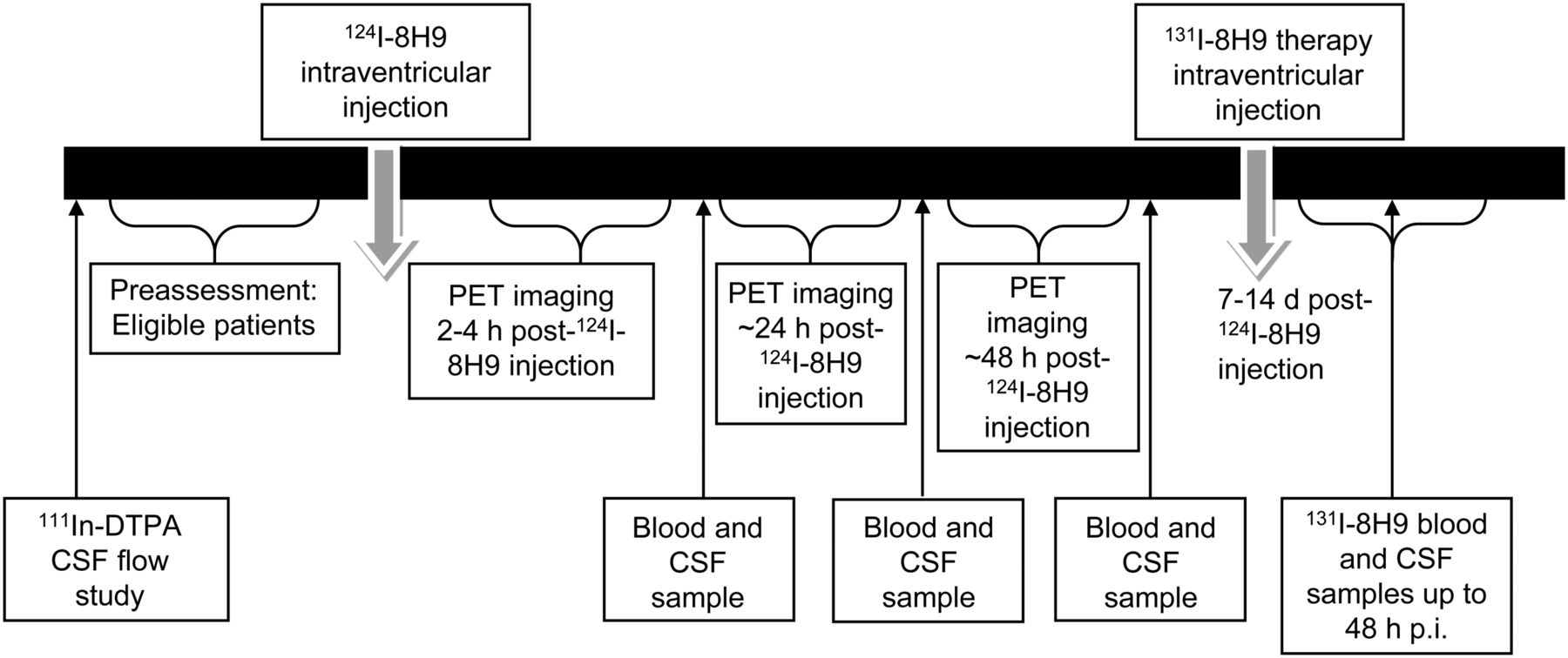
- FIGURE 1.
Study schema.
- FIGURE 2.
Serial 124I-omburtamab images in patient with metastatic neuroblastoma with leptomeningeal disease: anterior maximum-intensity projections (A), sagittal PET projections (B), and fused images (C) from D0, D1, and D2 show activity within ventricles and CSF canal that decreases over time. Systemic activity is seen in liver and bladder in D1 and D2. Clearance from ventricles is slower (long arrows). Pooling is also seen along cauda equina (short arrow).
- FIGURE 3.
CSF dose estimates for 131I omburtamab: 124I-omburtamab PET estimates vs. actual 124I-omburtamab CSF samples. (A) Scatterplot showing estimated radiation doses for 131I-omburtamab in units of cGy/MBq. x-axis shows dose values estimated from 124I-8H9 PET region-of-interest data vs. doses estimated from 124I-8H9 CSF sample counts on y-axis. Pearson correlation coefficient was calculated to show correlation between PET region of interest vs. sample count data. This coefficient is 0.00655 with P value of 0.96, indicating no correlation. Solid and dashed lines represent lines of correlation and identity, respectively. (B) Bland–Altman plot showing mean difference between PET region-of-interest dose and CSF sample–based dose estimates on y-axis vs. average of 2 dose estimates on x-axis; 95% confidence intervals for mean difference was determined using R package “Bland–Altman Leh.” (C) Scatterplot comparing radiation doses (cGy/MBq) from posttherapy 131I-omburtamab samples (y-axis) vs. pretherapy 124I-omburtamab samples (x-axis). Pearson coefficient is 0.084 with P value of 0.52, indicating no correlation. Solid and dashed lines represent lines of correlation and identity, respectively.


Tables
Demographic Data Total number of patients 42 1 dose 20 2 doses 22 Primary malignancy Neuroblastoma 32 Medulloblastoma 2 Sarcoma 3 Ependymoma 1 Rhabdoid tumor 1 Melanoma 1 Chordoma 1 Choroid plexus tumor 1 Age range 3 mo–42 y Sex Male 26 Female 16 Data are n, except for age.
- TABLE 2
Normal Organ-Absorbed Dose and Effective Dose Estimates for 124I-Omburtamab Administered Intraventricularly
Site Mean SD Median Minimum Maximum Salivary gland 0.52 0.33 0.44 0.05 1.45 Adrenals 0.41 0.23 0.40 0.06 1.10 Brain 1.13 0.55 1.01 0.20 3.03 Gallbladder wall 0.46 0.24 0.46 0.07 1.13 Lower large intestine wall 0.36 0.22 0.34 0.06 1.02 Small intestine 0.40 0.24 0.38 0.06 1.02 Stomach wall 0.86 0.57 0.71 0.17 2.65 Upper large intestine wall 0.40 0.23 0.39 0.06 1.08 Heart wall 0.39 0.23 0.37 0.06 1.06 Kidneys 0.39 0.22 0.38 0.06 1.05 Liver 1.58 1.04 1.62 0.06 4.22 Lungs 0.36 0.21 0.34 0.05 1.00 Muscle 0.34 0.21 0.31 0.05 0.98 Pancreas 0.44 0.25 0.44 0.07 1.16 Red marrow 0.37 0.27 0.30 0.05 1.21 Osteogenic cells 0.67 0.43 0.59 0.12 1.99 Skin 0.30 0.18 0.27 0.05 0.85 Spleen 0.57 0.32 0.55 0.04 1.16 Thymus 0.35 0.21 0.32 0.05 0.99 Thyroid 0.58 0.74 0.40 0.07 5.29 Urinary bladder wall 0.35 0.21 0.34 0.06 0.98 Total body 0.45 0.27 0.45 0.07 1.22 Effective dose (mSv/MBq) 0.49 0.27 0.47 0.10 1.23 Effective dose equivalent (mSv/MBq) 0.55 0.28 0.53 0.10 1.32 Units are mGy/MBq unless otherwise noted.
- TABLE 3
PET Imaging and CSF-Derived Biologic Clearance Half-Lives of 124I-Omburtamab from CSF
Mean Median Parameter Total median 25% Q 50% Q 75% Q Range <5 y 5–10 y >10 y <5 y 5–10 y >10 y 124I-omburtamab PET Ventricle 8.19 6.61 8.89 9.57 3.6–18.2 9.22 8.62 8.81 8.14 8.3 9.01 Cervical 6.86 6.36 9.08 8.89 3.9–16.9 8.13 9.72 9.54 6.78 6.65 9.7 Thoracic 7.83 6.71 11.01 10.39 5.0–25.0 9.14 12.84 10.82 7.51 7.94 9.87 Lumbar 7.02 6.32 9.84 11.05 4.1–39.6 9.46 9.46 14.91 6.62 6.94 11.47 124I-omburtamab samples 6.3 5.49 10.31 11.63 3.7–46.2 10.15 9.33 9.31 8.41 6.62 5.74 Q = quartile.
Data are in hours and exclude outliers 26 h and 44.4 h in single patient each.
Parameter Mean 25% quartile Median 75% quartile Range 124I-omburtamab PET-derived doses 0.620 0.436 0.523 0.755 0.21–2.948 Ventricle Cervical 0.445 0.326 0.387 0.494 0.170–0.711 Thoracic 0.538 0.373 0.442 0.555 0.189–1.978 Lumbar 0.567 0.454 0.552 0.652 0.235–1.051 Whole CSF From 124I-omburtamab CSF samples 2.253 0.961 1.443 2.512 0.100–10.243 From 131I-omburtamab posttherapy CSF samples 1.534 0.695 1.1 1.841 0.041−8.386 Data are cGy/MBq.
- TABLE 5
Blood Dosimetry Estimates for 131I-mAb-Omburtamab Derived from Pretherapy 124I-Omburtamab Administration and Actual 131I-Omburtamab Therapy
Omburtamab blood sample Mean dose (cGy/MBq) Median dose (cGy/MBq) Dose range (cGy/MBq) 124I 0.051 0.039 0.0035–0.244 131I 0.068 0.069 0.0032–0.158


Jump to section
Related Articles
Cited By...
- Determination of the Intralesional Distribution of Theranostic 124I-Omburtamab Convection-Enhanced Delivery in Treatment of Diffuse Intrinsic Pontine Glioma
- Theranostic Intratumoral Convection-Enhanced Delivery of 124I-Omburtamab in Patients with Diffuse Intrinsic Pontine Glioma: Pharmacokinetics and Lesion Dosimetry
- Radioimmunoscintigraphy and Pretreatment Dosimetry of 131I-Omburtamab for Planning Treatment of Leptomeningeal Disease
- Dosimetry in Radiopharmaceutical Therapy
- Biodistribution and Radiation Dosimetry of Intraperitoneally Administered 124I-Omburtamab in Patients with Desmoplastic Small Round Cell Tumors
- {gamma}-Tocotrienol-Loaded Liposomes for Radioprotection from Hematopoietic Side Effects Caused by Radiotherapeutic Drugs