
Main menu

  • Home
  • Content
    • Current
    • Ahead of print
    • Past Issues
    • JNM Supplement
    • SNMMI Annual Meeting Abstracts
    • Continuing Education
    • JNM Podcasts
  • Subscriptions
    • Subscribers
    • Institutional and Non-member
    • Rates
    • Journal Claims
    • Corporate & Special Sales
  • Authors
    • Submit to JNM
    • Information for Authors
    • Assignment of Copyright
    • AQARA requirements
  • Info
    • Reviewers
    • Permissions
    • Advertisers
  • About
    • About Us
    • Editorial Board
    • Contact Information
  • More
    • Alerts
    • Feedback
    • Help
    • SNMMI Journals
  • SNMMI
    • JNM
    • JNMT
    • SNMMI Journals
    • SNMMI

User menu

  • Subscribe
  • My alerts
  • Log in
  • My Cart

Search

  • Advanced search
Journal of Nuclear Medicine
  • SNMMI
    • JNM
    • JNMT
    • SNMMI Journals
    • SNMMI
  • Subscribe
  • My alerts
  • Log in
  • My Cart
Journal of Nuclear Medicine

Advanced Search

  • Home
  • Content
    • Current
    • Ahead of print
    • Past Issues
    • JNM Supplement
    • SNMMI Annual Meeting Abstracts
    • Continuing Education
    • JNM Podcasts
  • Subscriptions
    • Subscribers
    • Institutional and Non-member
    • Rates
    • Journal Claims
    • Corporate & Special Sales
  • Authors
    • Submit to JNM
    • Information for Authors
    • Assignment of Copyright
    • AQARA requirements
  • Info
    • Reviewers
    • Permissions
    • Advertisers
  • About
    • About Us
    • Editorial Board
    • Contact Information
  • More
    • Alerts
    • Feedback
    • Help
    • SNMMI Journals
  • View or Listen to JNM Podcast
  • Visit JNM on Facebook
  • Join JNM on LinkedIn
  • Follow JNM on Twitter
  • Subscribe to our RSS feeds
Research ArticleOncology

68Ga-DOTA-E[c(RGDfK)]2 PET Imaging of SHARPIN-Regulated Integrin Activity in Mice

Riikka Siitonen, Emilia Peuhu, Anu Autio, Heidi Liljenbäck, Elina Mattila, Olli Metsälä, Meeri Käkelä, Tiina Saanijoki, Ingrid Dijkgraaf, Sirpa Jalkanen, Johanna Ivaska and Anne Roivainen
Journal of Nuclear Medicine October 2019, 60 (10) 1380-1387; DOI: https://doi.org/10.2967/jnumed.118.222026
Riikka Siitonen
1Turku PET Centre, University of Turku, Turku, Finland
  • Find this author on Google Scholar
  • Find this author on PubMed
  • Search for this author on this site
Emilia Peuhu
2Turku Centre for Biotechnology, University of Turku and Åbo Akademi University, Turku, Finland
3FICAN West Cancer Research Laboratory, University of Turku and Turku University Hospital, Turku, Finland
  • Find this author on Google Scholar
  • Find this author on PubMed
  • Search for this author on this site
Anu Autio
1Turku PET Centre, University of Turku, Turku, Finland
  • Find this author on Google Scholar
  • Find this author on PubMed
  • Search for this author on this site
Heidi Liljenbäck
1Turku PET Centre, University of Turku, Turku, Finland
4Turku Center for Disease Modeling, University of Turku, Turku, Finland
  • Find this author on Google Scholar
  • Find this author on PubMed
  • Search for this author on this site
Elina Mattila
2Turku Centre for Biotechnology, University of Turku and Åbo Akademi University, Turku, Finland
  • Find this author on Google Scholar
  • Find this author on PubMed
  • Search for this author on this site
Olli Metsälä
1Turku PET Centre, University of Turku, Turku, Finland
  • Find this author on Google Scholar
  • Find this author on PubMed
  • Search for this author on this site
Meeri Käkelä
1Turku PET Centre, University of Turku, Turku, Finland
  • Find this author on Google Scholar
  • Find this author on PubMed
  • Search for this author on this site
Tiina Saanijoki
1Turku PET Centre, University of Turku, Turku, Finland
  • Find this author on Google Scholar
  • Find this author on PubMed
  • Search for this author on this site
Ingrid Dijkgraaf
5Department of Biochemistry, University of Maastricht, Maastricht, the Netherlands
  • Find this author on Google Scholar
  • Find this author on PubMed
  • Search for this author on this site
Sirpa Jalkanen
6MediCity Research Laboratory, University of Turku, Turku, Finland
  • Find this author on Google Scholar
  • Find this author on PubMed
  • Search for this author on this site
Johanna Ivaska
2Turku Centre for Biotechnology, University of Turku and Åbo Akademi University, Turku, Finland
7Department of Biochemistry, University of Turku, Turku, Finland; and
  • Find this author on Google Scholar
  • Find this author on PubMed
  • Search for this author on this site
Anne Roivainen
1Turku PET Centre, University of Turku, Turku, Finland
4Turku Center for Disease Modeling, University of Turku, Turku, Finland
8Turku PET Centre, Turku University Hospital, Turku, Finland
  • Find this author on Google Scholar
  • Find this author on PubMed
  • Search for this author on this site
  • Article
  • Figures & Data
  • Supplemental
  • Info & Metrics
  • PDF
Loading

Article Figures & Data

Figures

  • Tables
  • Additional Files
  • FIGURE 1.
    • Download figure
    • Open in new tab
    • Download powerpoint
    FIGURE 1.

    Increased tissue uptake of 68Ga-DOTA-E[c(RGDfK)]2 in Sharpincpdm mice. (A) Alopecia on dorsal skin of Sharpincpdm mouse, with wt littermate for comparison. (B) Ex vivo uptake of 68Ga-DOTA-E[c(RGDfK)]2 in Sharpincpdm and wt mice without tumors. (C) Competition with nonlabeled DOTA-E[c(RGDfK)]2 peptide and imaging with control peptide 68Ga-DOTA-E[c(RGEfK)]2 revealing specific binding of tracer. Ex vivo results are expressed as percentage of injected radioactivity dose per gram of tissue (%ID/g). n = 4–9/group. *P < 0.05. **P < 0.01. ***P < 0.001.

  • FIGURE 2.
    • Download figure
    • Open in new tab
    • Download powerpoint
    FIGURE 2.

    SHARPIN deficiency increases metastasis but not growth in tumor microenvironment. (A) Growth curves of B16 melanoma tumors during follow-up period (n = 8–9/group). (B) Tumor volume at end of experiment in wt and Sharpincpdm mice. (C) Pie-chart presenting lymph node metastasis (red) vs. no metastasis (black) in B16 melanoma tumor–bearing wt and Sharpincpdm mice. (D) Quantification of blood flow in B16 melanoma tumors.
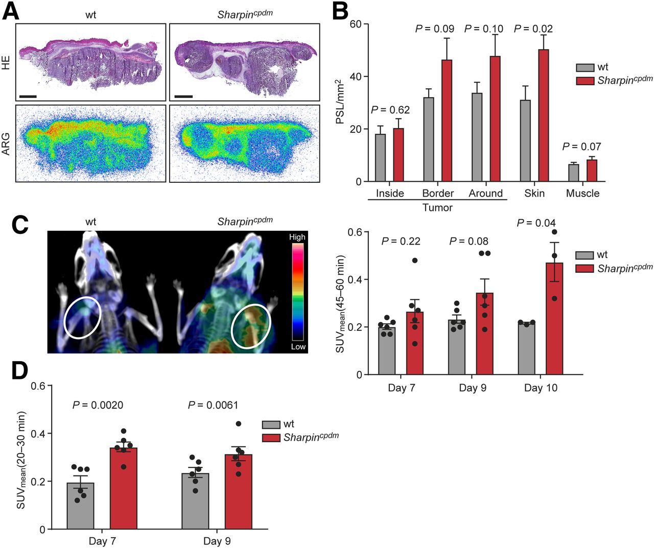
  • FIGURE 3.
    • Download figure
    • Open in new tab
    • Download powerpoint
    FIGURE 3.

    68Ga-DOTA-E[c(RGDfK)]2 binding is enhanced in SHARPIN-deficient tumor microenvironment. (A) Representative autoradiographs (ARG) and corresponding hematoxylin–eosin (HE) staining of B16 melanoma tumors (scale bar, 2 mm). (B) Quantification of autoradiographs showing distribution of 68Ga-DOTA-E[c(RGDfK)]2 radioactivity concentration in tumor, skin, and muscle (n = 12/group). (C) Representative coronal PET/CT images of wt and Sharpincpdm tumor–bearing mice and in vivo tumor uptake of 68Ga-DOTA-E[c(RGDfK)]2 in wt and Sharpincpdm mice. Bars show SUVmean 45–60 min after injection. (D) In vivo tumor uptake of 68Ga-DOTA-Siglec-9 in wt and Sharpincpdm mice. Bars show SUVmean 20–30 min after injection. PSL = photostimulated luminescence.

  • FIGURE 4.
    • Download figure
    • Open in new tab
    • Download powerpoint
    FIGURE 4.

    Stromal SHARPIN regulates tumor vascularization. (A–C) Representative cryosections of B16 tumors from wt and Sharpincpdm mouse immunolabeled with VAP-1 (A), CD31 (B), and β3 integrin antibody (C). Scale bar, 200 μm. Bars show VAP-1–positive, CD31-positive, and β3 integrin–positive tumor areas from B16 tumors implanted into wt and Sharpincpdm mice.

Tables

  • Figures
  • Additional Files
    • View popup
    TABLE 1

    Ex Vivo Biodistribution of 68Ga-DOTA-E[c(RGDfK)]2 in Tumor-Bearing Mice at Days 9–10 After Inoculation

    SiteSharpincpdmwtP
    Aorta4.3 ± 0.812.1 ± 0.18<0.05
    Brown adipose tissue0.92 ± 0.150.51 ± 0.037<0.05
    Blood1.5 ± 0.430.60 ± 0.058NS
    Bone1.4 ± 0.150.87 ± 0.037<0.05
    Heart0.82 ± 0.140.51 ± 0.032<0.05
    Lungs3.0 ± 0.372.0 ± 0.079<0.05
    Lymph nodes2.0 ± 0.280.81 ± 0.056<0.01
    Muscle0.58 ± 0.0790.36 ± 0.016<0.05
    Skin2.9 ± 0.411.3 ± 0.070<0.01
    Small intestine5.5 ± 0.623.2 ± 0.39<0.05
    Thymus1.4 ± 0.210.79 ± 0.041<0.05
    Tumor1.9 ± 0.451.0 ± 0.15<0.05
    White adipose tissue0.68 ± 0.160.43 ± 0.11NS
    • Results are expressed as percentage of injected radioactivity dose per gram of tissue (mean ± SEM).

    • NS = not statistically significant.

Additional Files

  • Figures
  • Tables
  • Supplemental Data

    Files in this Data Supplement:

    • Supplemental Data
PreviousNext
Back to top

In this issue

Journal of Nuclear Medicine: 60 (10)
Journal of Nuclear Medicine
Vol. 60, Issue 10
October 1, 2019
  • Table of Contents
  • Table of Contents (PDF)
  • About the Cover
  • Index by author
Print
Download PDF
Article Alerts
Sign In to Email Alerts with your Email Address
Email Article

Thank you for your interest in spreading the word on Journal of Nuclear Medicine.

NOTE: We only request your email address so that the person you are recommending the page to knows that you wanted them to see it, and that it is not junk mail. We do not capture any email address.

Enter multiple addresses on separate lines or separate them with commas.
68Ga-DOTA-E[c(RGDfK)]2 PET Imaging of SHARPIN-Regulated Integrin Activity in Mice
(Your Name) has sent you a message from Journal of Nuclear Medicine
(Your Name) thought you would like to see the Journal of Nuclear Medicine web site.
Citation Tools
68Ga-DOTA-E[c(RGDfK)]2 PET Imaging of SHARPIN-Regulated Integrin Activity in Mice
Riikka Siitonen, Emilia Peuhu, Anu Autio, Heidi Liljenbäck, Elina Mattila, Olli Metsälä, Meeri Käkelä, Tiina Saanijoki, Ingrid Dijkgraaf, Sirpa Jalkanen, Johanna Ivaska, Anne Roivainen
Journal of Nuclear Medicine Oct 2019, 60 (10) 1380-1387; DOI: 10.2967/jnumed.118.222026

Citation Manager Formats

  • BibTeX
  • Bookends
  • EasyBib
  • EndNote (tagged)
  • EndNote 8 (xml)
  • Medlars
  • Mendeley
  • Papers
  • RefWorks Tagged
  • Ref Manager
  • RIS
  • Zotero
Share
68Ga-DOTA-E[c(RGDfK)]2 PET Imaging of SHARPIN-Regulated Integrin Activity in Mice
Riikka Siitonen, Emilia Peuhu, Anu Autio, Heidi Liljenbäck, Elina Mattila, Olli Metsälä, Meeri Käkelä, Tiina Saanijoki, Ingrid Dijkgraaf, Sirpa Jalkanen, Johanna Ivaska, Anne Roivainen
Journal of Nuclear Medicine Oct 2019, 60 (10) 1380-1387; DOI: 10.2967/jnumed.118.222026
Twitter logo Facebook logo LinkedIn logo Mendeley logo
  • Tweet Widget
  • Facebook Like
  • Google Plus One
Bookmark this article

Jump to section

  • Article
    • Abstract
    • MATERIALS AND METHODS
    • RESULTS
    • DISCUSSION
    • CONCLUSION
    • DISCLOSURE
    • KEY POINTS
    • Acknowledgments
    • Footnotes
    • REFERENCES
  • Figures & Data
  • Supplemental
  • Info & Metrics
  • PDF

Related Articles

  • This Month in JNM
  • PubMed
  • Google Scholar

Cited By...

  • No citing articles found.
  • Google Scholar

More in this TOC Section

Oncology

  • Comparative PSMA expression at early (1hour) vs late (2hour) in primary and secondary sites of involvement in prostate cancer.
  • Imaging spectrum of peritoneal carcinomatosis associated with various etiologies on 18F-FDG PET/CT
  • Diagnostic Role of 18F-FDG PET/CT in Urothelial carcinoma and its impact on management, and prognosis : A case based approach
Show more Oncology

Basic

  • Dopamine D1 Receptor Agonist PET Tracer Development: Assessment in Nonhuman Primates
  • Optical Navigation of the Drop-In γ-Probe as a Means to Strengthen the Connection Between Robot-Assisted and Radioguided Surgery
  • Synthesis and Preclinical Evaluation of a 68Ga-Labeled Adnectin, 68Ga-BMS-986192, as a PET Agent for Imaging PD-L1 Expression
Show more Basic

Similar Articles

Keywords

  • SHARPIN
  • αvβ3 integrin
  • RGD
  • melanoma
  • PET
SNMMI

© 2025 SNMMI

Powered by HighWire