
Main menu

  • Home
  • Content
    • Current
    • Ahead of print
    • Past Issues
    • JNM Supplement
    • SNMMI Annual Meeting Abstracts
    • Continuing Education
    • JNM Podcasts
  • Subscriptions
    • Subscribers
    • Institutional and Non-member
    • Rates
    • Journal Claims
    • Corporate & Special Sales
  • Authors
    • Submit to JNM
    • Information for Authors
    • Assignment of Copyright
    • AQARA requirements
  • Info
    • Reviewers
    • Permissions
    • Advertisers
  • About
    • About Us
    • Editorial Board
    • Contact Information
  • More
    • Alerts
    • Feedback
    • Help
    • SNMMI Journals
  • SNMMI
    • JNM
    • JNMT
    • SNMMI Journals
    • SNMMI

User menu

  • Subscribe
  • My alerts
  • Log in
  • Log out
  • My Cart

Search

  • Advanced search
Journal of Nuclear Medicine
  • SNMMI
    • JNM
    • JNMT
    • SNMMI Journals
    • SNMMI
  • Subscribe
  • My alerts
  • Log in
  • Log out
  • My Cart
Journal of Nuclear Medicine

Advanced Search

  • Home
  • Content
    • Current
    • Ahead of print
    • Past Issues
    • JNM Supplement
    • SNMMI Annual Meeting Abstracts
    • Continuing Education
    • JNM Podcasts
  • Subscriptions
    • Subscribers
    • Institutional and Non-member
    • Rates
    • Journal Claims
    • Corporate & Special Sales
  • Authors
    • Submit to JNM
    • Information for Authors
    • Assignment of Copyright
    • AQARA requirements
  • Info
    • Reviewers
    • Permissions
    • Advertisers
  • About
    • About Us
    • Editorial Board
    • Contact Information
  • More
    • Alerts
    • Feedback
    • Help
    • SNMMI Journals
  • View or Listen to JNM Podcast
  • Visit JNM on Facebook
  • Join JNM on LinkedIn
  • Follow JNM on Twitter
  • Subscribe to our RSS feeds
Research ArticleNeurology

Evaluation of a PET Radioligand to Image O-GlcNAcase in Brain and Periphery of Rhesus Monkey and Knock-Out Mouse

Soumen Paul, Mohammad B. Haskali, Jeih-San Liow, Sami S. Zoghbi, Vanessa N. Barth, Marcy Comly Kolodrubetz, Michelle R. Bond, Cheryl L. Morse, Robert L. Gladding, Michael P. Frankland, Nancy Kant, Lawrence Slieker, Sergey Shcherbinin, Hugh N. Nuthall, Paolo Zanotti-Fregonara, John A. Hanover, Cynthia Jesudason, Victor W. Pike and Robert B. Innis
Journal of Nuclear Medicine January 2019, 60 (1) 129-134; DOI: https://doi.org/10.2967/jnumed.118.213231
Soumen Paul
1Molecular Imaging Branch, NIMH, National Institutes of Health, Bethesda, Maryland
  • Find this author on Google Scholar
  • Find this author on PubMed
  • Search for this author on this site
Mohammad B. Haskali
1Molecular Imaging Branch, NIMH, National Institutes of Health, Bethesda, Maryland
2Peter MacCallum Cancer Centre, Melbourne, Australia
  • Find this author on Google Scholar
  • Find this author on PubMed
  • Search for this author on this site
Jeih-San Liow
1Molecular Imaging Branch, NIMH, National Institutes of Health, Bethesda, Maryland
  • Find this author on Google Scholar
  • Find this author on PubMed
  • Search for this author on this site
Sami S. Zoghbi
1Molecular Imaging Branch, NIMH, National Institutes of Health, Bethesda, Maryland
  • Find this author on Google Scholar
  • Find this author on PubMed
  • Search for this author on this site
Vanessa N. Barth
3Eli Lilly and Company, Indianapolis, Indiana
  • Find this author on Google Scholar
  • Find this author on PubMed
  • Search for this author on this site
Marcy Comly Kolodrubetz
4LCMB, NIDDK, National Institutes of Health, Bethesda, Maryland
  • Find this author on Google Scholar
  • Find this author on PubMed
  • Search for this author on this site
Michelle R. Bond
4LCMB, NIDDK, National Institutes of Health, Bethesda, Maryland
  • Find this author on Google Scholar
  • Find this author on PubMed
  • Search for this author on this site
Cheryl L. Morse
1Molecular Imaging Branch, NIMH, National Institutes of Health, Bethesda, Maryland
  • Find this author on Google Scholar
  • Find this author on PubMed
  • Search for this author on this site
Robert L. Gladding
1Molecular Imaging Branch, NIMH, National Institutes of Health, Bethesda, Maryland
  • Find this author on Google Scholar
  • Find this author on PubMed
  • Search for this author on this site
Michael P. Frankland
1Molecular Imaging Branch, NIMH, National Institutes of Health, Bethesda, Maryland
  • Find this author on Google Scholar
  • Find this author on PubMed
  • Search for this author on this site
Nancy Kant
3Eli Lilly and Company, Indianapolis, Indiana
  • Find this author on Google Scholar
  • Find this author on PubMed
  • Search for this author on this site
Lawrence Slieker
3Eli Lilly and Company, Indianapolis, Indiana
  • Find this author on Google Scholar
  • Find this author on PubMed
  • Search for this author on this site
Sergey Shcherbinin
3Eli Lilly and Company, Indianapolis, Indiana
  • Find this author on Google Scholar
  • Find this author on PubMed
  • Search for this author on this site
Hugh N. Nuthall
5Eli Lilly and Company, Windlesham, United Kingdom; and
  • Find this author on Google Scholar
  • Find this author on PubMed
  • Search for this author on this site
Paolo Zanotti-Fregonara
6Houston Methodist Research Institute, Houston, Texas
  • Find this author on Google Scholar
  • Find this author on PubMed
  • Search for this author on this site
John A. Hanover
4LCMB, NIDDK, National Institutes of Health, Bethesda, Maryland
  • Find this author on Google Scholar
  • Find this author on PubMed
  • Search for this author on this site
Cynthia Jesudason
3Eli Lilly and Company, Indianapolis, Indiana
  • Find this author on Google Scholar
  • Find this author on PubMed
  • Search for this author on this site
Victor W. Pike
1Molecular Imaging Branch, NIMH, National Institutes of Health, Bethesda, Maryland
  • Find this author on Google Scholar
  • Find this author on PubMed
  • Search for this author on this site
Robert B. Innis
1Molecular Imaging Branch, NIMH, National Institutes of Health, Bethesda, Maryland
  • Find this author on Google Scholar
  • Find this author on PubMed
  • Search for this author on this site
  • Article
  • Figures & Data
  • Supplemental
  • Info & Metrics
  • PDF
Loading

Article Figures & Data

Figures

  • Additional Files
  • FIGURE 1.
    • Download figure
    • Open in new tab
    • Download powerpoint
    FIGURE 1.

    Structure of 18F-LSN3316612.

  • FIGURE 2.
    • Download figure
    • Open in new tab
    • Download powerpoint
    FIGURE 2.

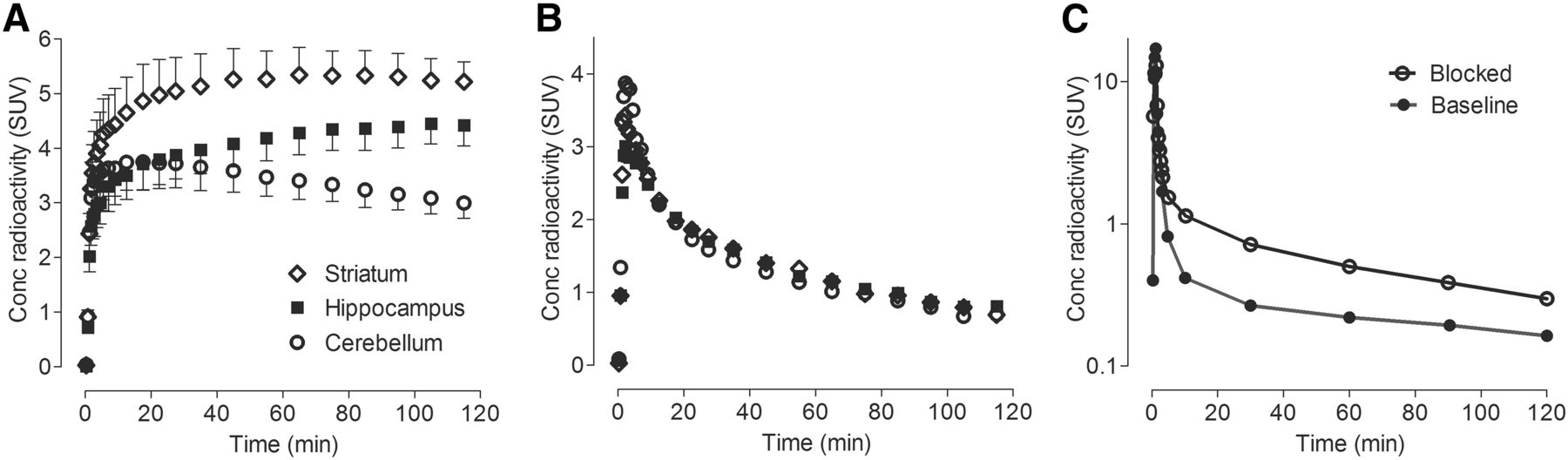
    Concentration of radioactivity in brain at baseline (A) and after blockade (B) and concentration of parent radioligand in plasma (C). In baseline graph, symbols represent mean ± SD from 4 monkeys. The other 2 graphs show results from 1 animal. For blockade, thiamet-G (10 mg/kg intravenously) was injected 45 min before radioligand.

  • FIGURE 3.
    • Download figure
    • Open in new tab
    • Download powerpoint
    FIGURE 3.

    Parametric images of VT in rhesus monkey brain at baseline and after thiamet-G blockade. Uptake at baseline was especially high in striatum, hippocampus, frontal cortex, amygdala, and pituitary (arrows). Thiamet-G (10 mg/kg intravenously) decreased uptake by >90% in all regions. Skull shows negligible uptake of radioactivity. These parametric images quantified VT at voxel level using Logan plot.

  • FIGURE 4.
    • Download figure
    • Open in new tab
    • Download powerpoint
    FIGURE 4.

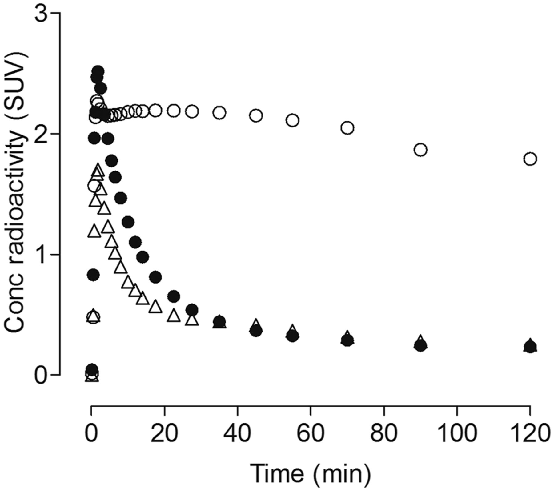
    Uptake of radioactivity in corpus callosum (white matter region) of rhesus macaque brain. Uptake at baseline (○) was significantly blocked by nonradioactive LSN3316612 (●), 1 mg/kg intravenously, administered 45 min before radioligand.

  • FIGURE 5.
    • Download figure
    • Open in new tab
    • Download powerpoint
    FIGURE 5.

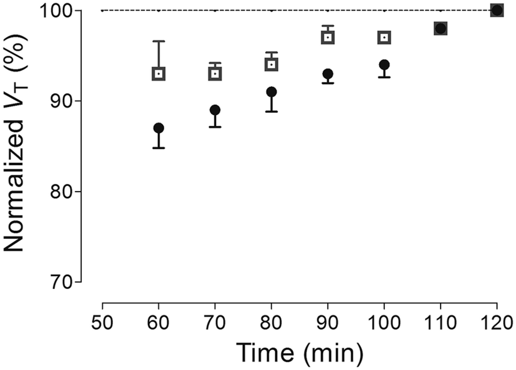
    Time stability curves of total VT for 2 regions: striatum (□), which had high uptake, and cerebellum (●), which had lowest uptake. VT was calculated using unconstrained 2-tissue-compartment model with increasingly truncated acquisition times. All values of VT were normalized as percentage of terminal value attained from 120 min of imaging. Data represent mean ± SD of 4 monkeys.

  • FIGURE 6.
    • Download figure
    • Open in new tab
    • Download powerpoint
    FIGURE 6.

    Uptake of radioactivity in whole brain from 2 controls (○) and 2 Oga∆Br (●) mice. Uptake in control mice preblocked with thiamet-G (△) (10 mg/kg intravenously) was similar to that in Oga∆Br mice.
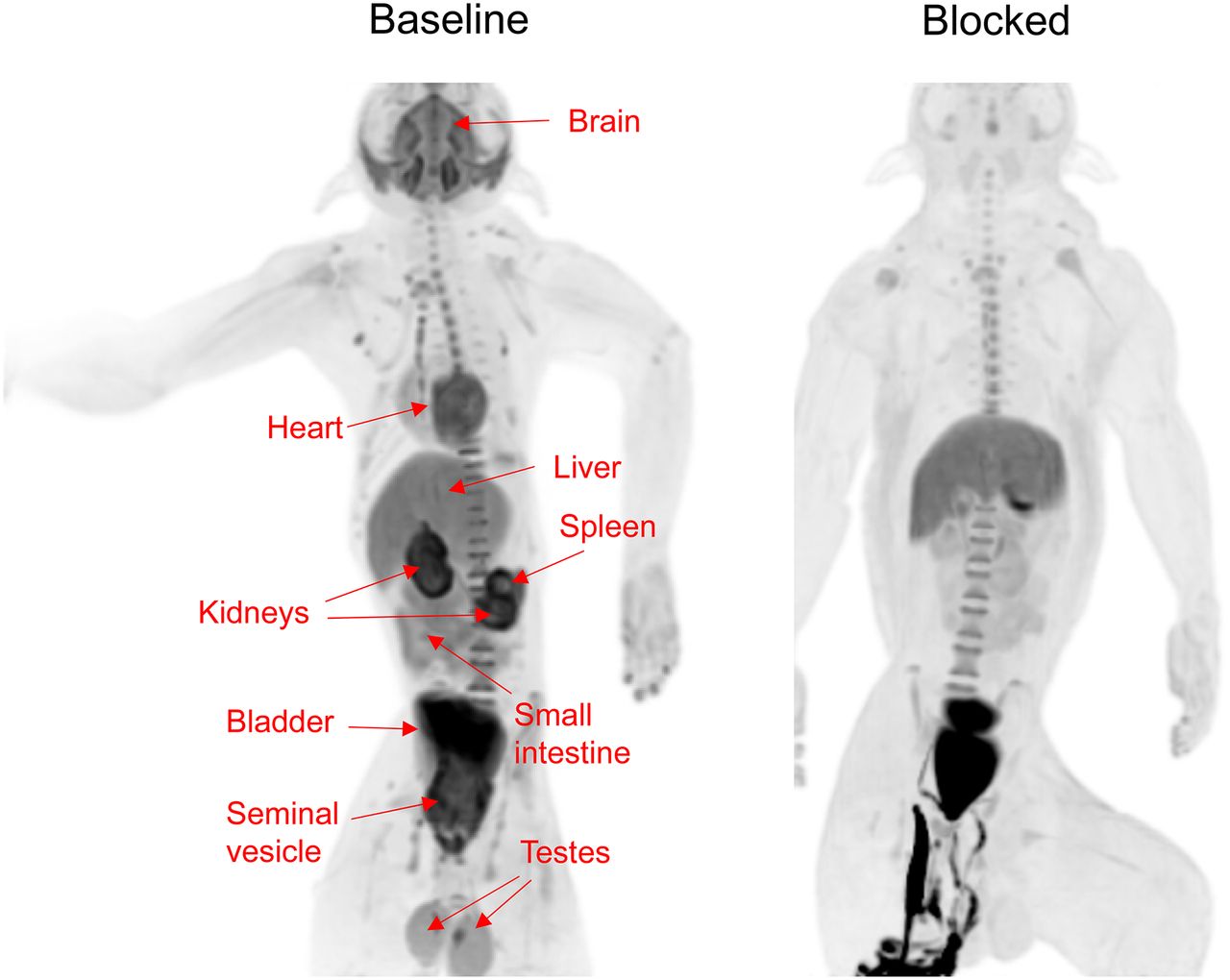
  • FIGURE 7.
    • Download figure
    • Open in new tab
    • Download powerpoint
    FIGURE 7.

    Whole-body distribution of radioactivity in male monkey after injection of 18F- LSN3316612 at baseline and with thiamet-G blockade (10 mg/kg, intravenously). Images were averaged for entire 120-min scan and presented as maximal-intensity projection, which enhances contrast and allows easier visualization of organs. Clear blocking effect was seen in brain, heart, kidneys, small intestine, spleen, testes, and seminal vesicle. In blocked scan, radioactivity below pelvis was due to urine excreted into animal’s diaper.

Additional Files

  • Figures
  • Supplemental Data

    Files in this Data Supplement:

    • Supplemental Data
PreviousNext
Back to top

In this issue

Journal of Nuclear Medicine: 60 (1)
Journal of Nuclear Medicine
Vol. 60, Issue 1
January 1, 2019
  • Table of Contents
  • Table of Contents (PDF)
  • About the Cover
  • Index by author
Print
Download PDF
Article Alerts
Sign In to Email Alerts with your Email Address
Email Article

Thank you for your interest in spreading the word on Journal of Nuclear Medicine.

NOTE: We only request your email address so that the person you are recommending the page to knows that you wanted them to see it, and that it is not junk mail. We do not capture any email address.

Enter multiple addresses on separate lines or separate them with commas.
Evaluation of a PET Radioligand to Image O-GlcNAcase in Brain and Periphery of Rhesus Monkey and Knock-Out Mouse
(Your Name) has sent you a message from Journal of Nuclear Medicine
(Your Name) thought you would like to see the Journal of Nuclear Medicine web site.
Citation Tools
Evaluation of a PET Radioligand to Image O-GlcNAcase in Brain and Periphery of Rhesus Monkey and Knock-Out Mouse
Soumen Paul, Mohammad B. Haskali, Jeih-San Liow, Sami S. Zoghbi, Vanessa N. Barth, Marcy Comly Kolodrubetz, Michelle R. Bond, Cheryl L. Morse, Robert L. Gladding, Michael P. Frankland, Nancy Kant, Lawrence Slieker, Sergey Shcherbinin, Hugh N. Nuthall, Paolo Zanotti-Fregonara, John A. Hanover, Cynthia Jesudason, Victor W. Pike, Robert B. Innis
Journal of Nuclear Medicine Jan 2019, 60 (1) 129-134; DOI: 10.2967/jnumed.118.213231

Citation Manager Formats

  • BibTeX
  • Bookends
  • EasyBib
  • EndNote (tagged)
  • EndNote 8 (xml)
  • Medlars
  • Mendeley
  • Papers
  • RefWorks Tagged
  • Ref Manager
  • RIS
  • Zotero
Share
Evaluation of a PET Radioligand to Image O-GlcNAcase in Brain and Periphery of Rhesus Monkey and Knock-Out Mouse
Soumen Paul, Mohammad B. Haskali, Jeih-San Liow, Sami S. Zoghbi, Vanessa N. Barth, Marcy Comly Kolodrubetz, Michelle R. Bond, Cheryl L. Morse, Robert L. Gladding, Michael P. Frankland, Nancy Kant, Lawrence Slieker, Sergey Shcherbinin, Hugh N. Nuthall, Paolo Zanotti-Fregonara, John A. Hanover, Cynthia Jesudason, Victor W. Pike, Robert B. Innis
Journal of Nuclear Medicine Jan 2019, 60 (1) 129-134; DOI: 10.2967/jnumed.118.213231
Twitter logo Facebook logo LinkedIn logo Mendeley logo
  • Tweet Widget
  • Facebook Like
  • Google Plus One
Bookmark this article

Jump to section

  • Article
    • Abstract
    • MATERIALS AND METHODS
    • RESULTS
    • DISCUSSION
    • CONCLUSION
    • DISCLOSURE
    • Acknowledgments
    • Footnotes
    • REFERENCES
  • Figures & Data
  • Supplemental
  • Info & Metrics
  • PDF

Related Articles

  • This Month in JNM
  • PubMed
  • Google Scholar

Cited By...

  • Development of a PET Tracer for OGA with Improved Kinetics in the Living Brain
  • Neuroinflammation PET Imaging: Current Opinion and Future Directions
  • PET ligands [18F]LSN3316612 and [11C]LSN3316612 quantify O-linked-{beta}-N-acetyl-glucosamine hydrolase in the brain
  • Google Scholar

More in this TOC Section

Neurology

  • Dopamine D1 Receptor Agonist PET Tracer Development: Assessment in Nonhuman Primates
  • Hypermetabolism on Pediatric PET Scans of Brain Glucose Metabolism: What Does It Signify?
  • TauIQ: A Canonical Image Based Algorithm to Quantify Tau PET Scans
Show more Neurology

Basic

  • Dopamine D1 Receptor Agonist PET Tracer Development: Assessment in Nonhuman Primates
  • Optical Navigation of the Drop-In γ-Probe as a Means to Strengthen the Connection Between Robot-Assisted and Radioguided Surgery
  • Synthesis and Preclinical Evaluation of a 68Ga-Labeled Adnectin, 68Ga-BMS-986192, as a PET Agent for Imaging PD-L1 Expression
Show more Basic

Similar Articles

Keywords

  • PET
  • O-GlcNAcase
  • tau
  • Alzheimer disease
SNMMI

© 2025 SNMMI

Powered by HighWire