


Article Figures & Data
Figures
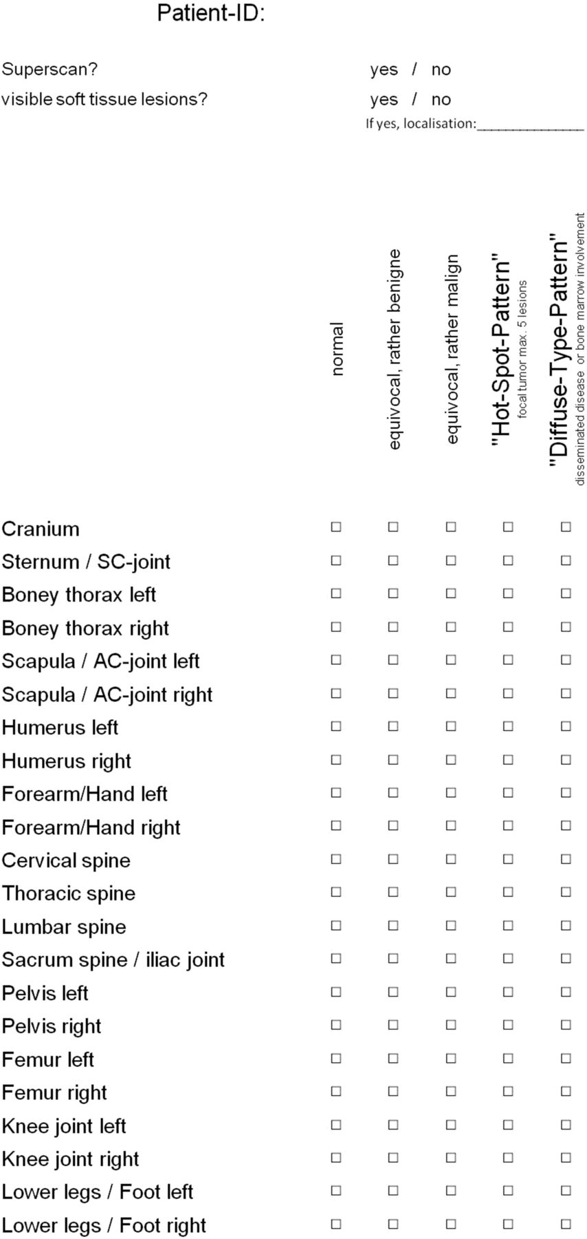
- FIGURE 1.
Report form for standardized evaluation of masked patients.
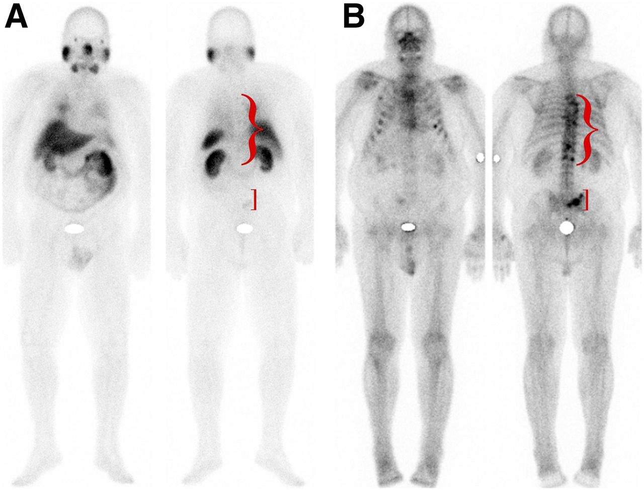
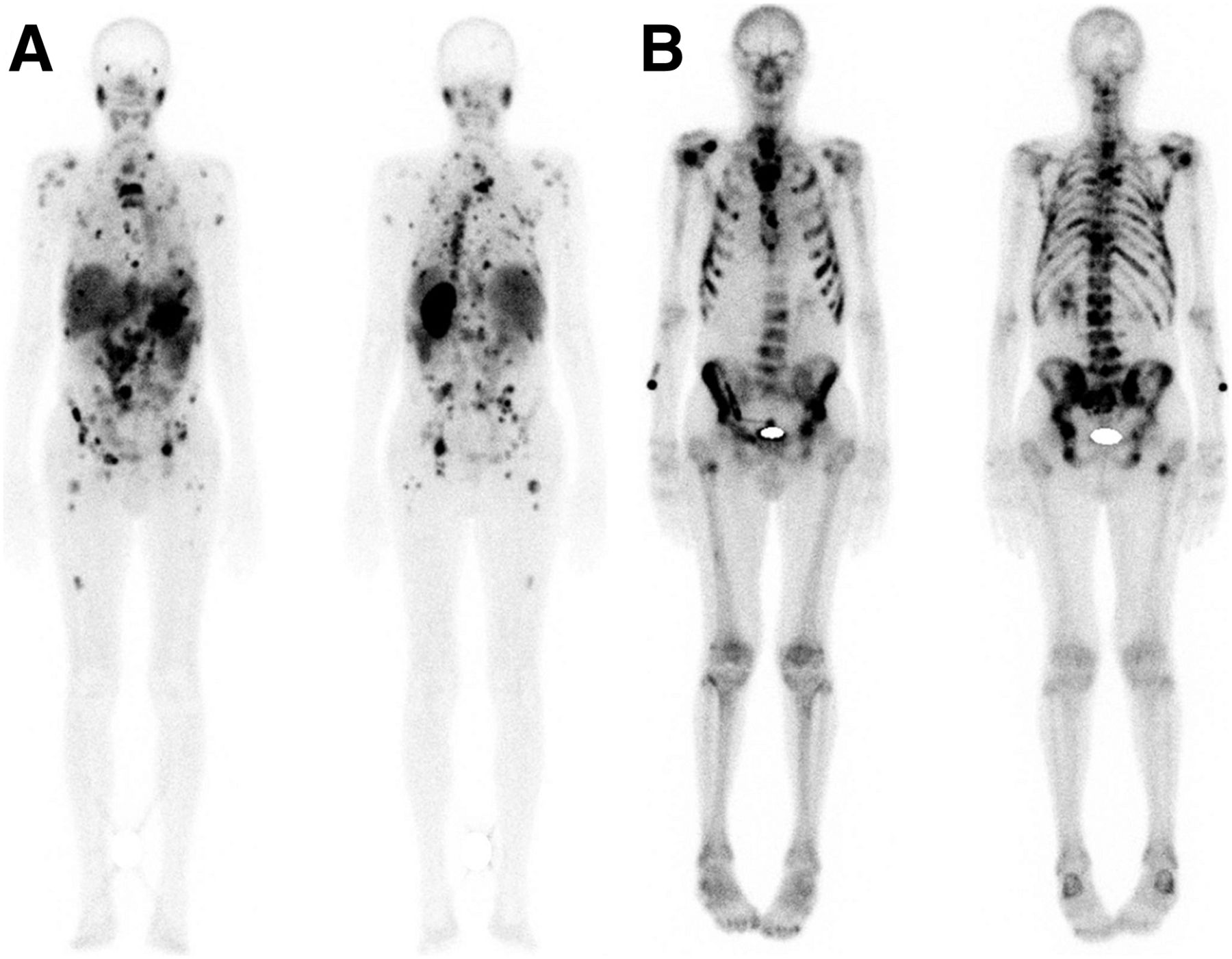
- FIGURE 2.
Patient with PSMA-negative tumor phenotype. No pathologic findings in spine or pelvis were depicted with PSMA (A) but were successfully diagnosed by BS (B).
- FIGURE 3.
Intrahepatic lesion and pleural carcinosis. Three-dimensional imaging was pivotal for correct allocation of respective lesions. In particular, liver and lung lesions were often misinterpreted and falsely assigned to overlapping bone structures.
- FIGURE 4.
Patient with intracerebral metastasis (A, green arrow) that was misinterpreted as skull lesion on planar scan (B, green arrow). Similarly, pulmonary lesion (C, red arrow) was also misinterpreted as rib lesion (B, red arrow). (A) MRI. (B) 99mTc-PSMA whole-body scan. (C) 99mTc-PSMA SPECT/CT.
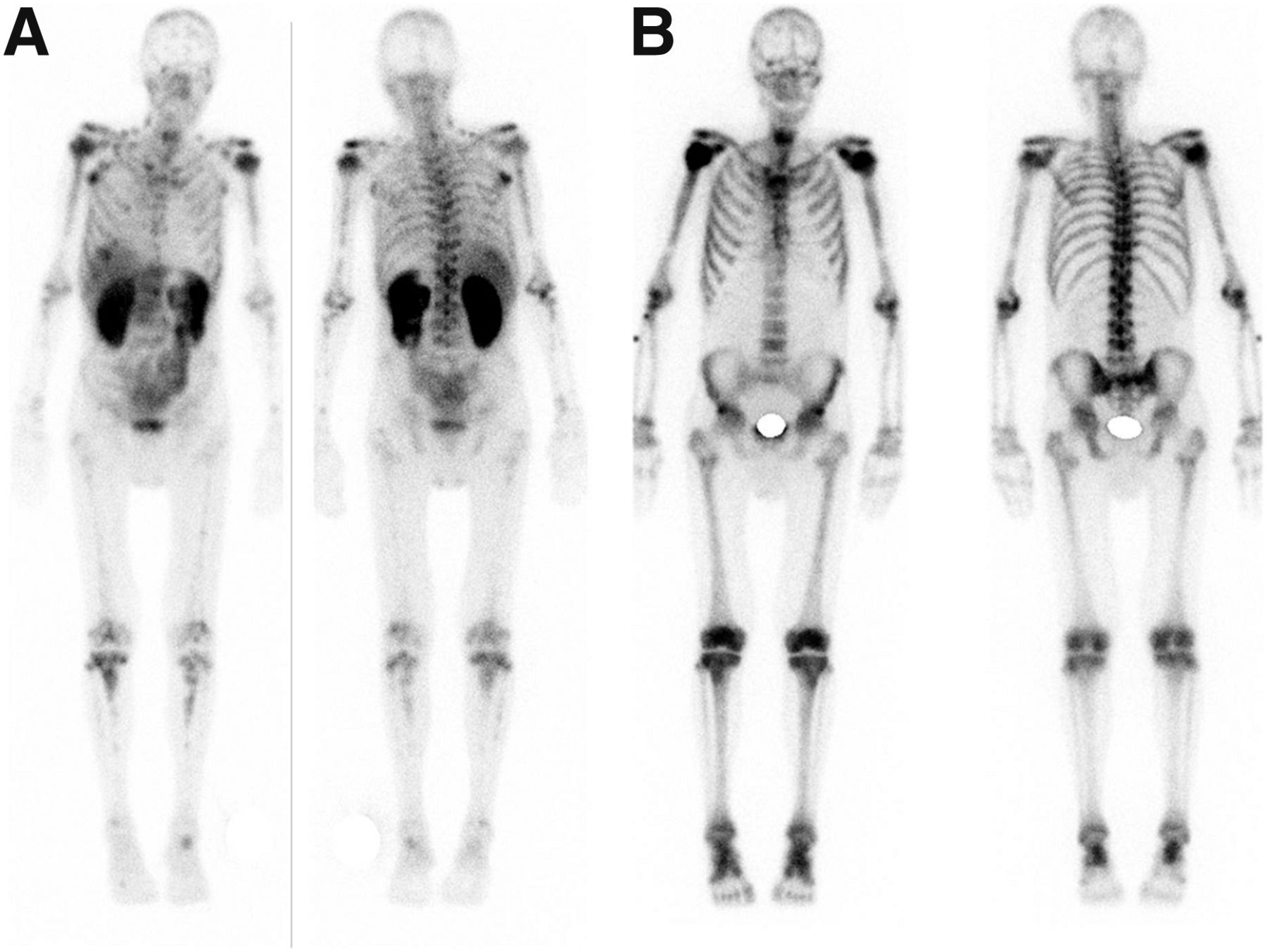
- FIGURE 5.
In patient with heterogeneously PSMA-expressing lesions, PSMA scanning (A) presented highly discordant lesion distribution pattern in comparison to 99mTc-MDP BS (B).
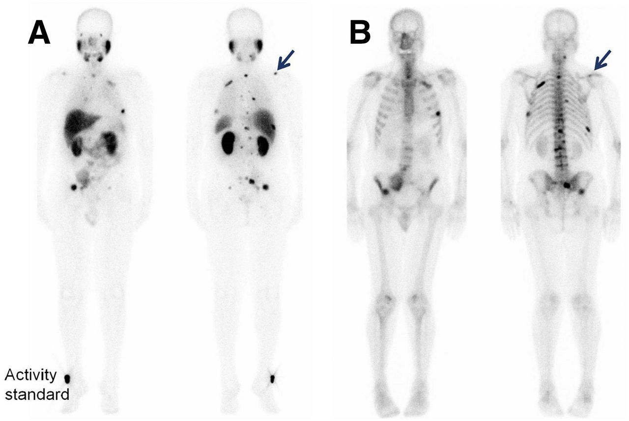
- FIGURE 6.
PSMA imaging (A, arrow) of typical malignant lesion that was scored as equivocal by BS (B, arrow).
- FIGURE 7.
Example of superscan pattern in patient imaged by PSMA scanning (A) and BS (B). Excessive tumor uptake in highly perfused red marrow resulted in at least relatively or even absolutely reduced uptake in salivary glands (PSMA scanning) and kidneys (BS).


Tables
Patient characteristic Value* Age (y) Median 75.5 Range 57–85 Gleason score Median 8 No. of patients with Gleason score of: <7 2 7 6 8 2 9 6 Unknown 5 PSA (ng/mL) Median 502 Range 6–1,855 Alkaline phosphatase (U/L) Mean 166.8 SD 121.8 Localization of metastases Lymph node(s) 9 Bone 21 Liver 1 Lung 5 Brain 1 Other 2 Local recurrence 1 Previous therapy RPx 10 LRTx 13 CRPC 21 Abiraterone (Zytiga; Janssen Biotech, Inc.) 17 Enzalutamide (Xtandi; Astellas Pharma Inc.) 12 Zoledronic acid (Zometa; Novartis)/denosumab (Xgeva; Amgen Inc.) 8 223RaCl2 (Xofigo; Bayer) 9 CTx 13 ↵* Values are reported as numbers of patients unless otherwise indicated.
RPx = radical prostatectomy; LRTx = local radiation therapy; CRPC = castration-resistant prostate cancer; CTx = chemotherapy.
Type of lesion Tracer Description of lesion No. of findings Ratio Benign 99mTc-MDP Normal 687 1.9:1 Equivocal 366 PSMA Normal 858 27.7:1 Equivocal 31 Malignant 99mTc-MDP Malignant 1,049 4.9:1 Equivocal 214 PSMA Malignant 1,271 8.1:1 Equivocal 156 2-Step evaluation 5-Step evaluation Parameter PSMA 99mTc-MDP PSMA 99mTc-MDP Median 0.36 0.55 1.55 2.23 Mean 0.43 0.76 1.45 2.41 Misclassification rate 9% 15% 29% 48% SD 0.39 0.66 0.36 0.64 P 0.039 <0.001 In 2-step evaluation, only correspondence according to benign vs. malignant was evaluated. In 5-step evaluation, correct alignment of normal, equivocal benign, equivocal malignant, focal tumor, and diffuse tumor in comparison to consensus reading was evaluated. Mean indicates average number of misclassifications out of 5 raters. Misclassification rate indicates portion of raters with misclassification vs. consensus reading of mean.


