


Article Figures & Data
Figures
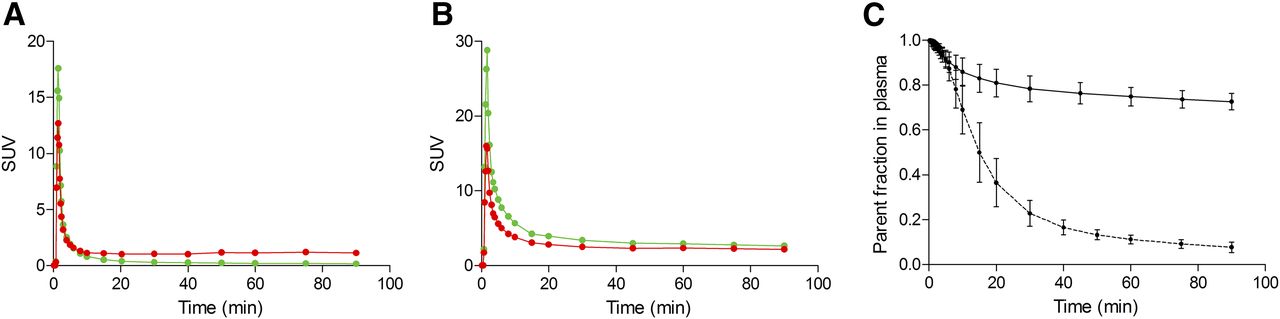
- FIGURE 1.
SUV concentrations of parent compound (green lines) and whole blood (red lines) for 11C-PBR28 (A) and 18F-GE180 (B) in representative subject. Although parent concentration of 11C-PBR28 is generally lower than whole-blood concentration, opposite is true for 18F-GE180. (C) Average and SD from all subjects of parent fraction in plasma for 11C-PBR28 (dashed line) and 18F-GE180 (solid line). Metabolism rate is much slower for 18F-GE180, as parent constitutes about 70%–80% of total plasma activity at end of 90-min scan.
- FIGURE 2.
Time–activity curves, expressed in SUVs, of 3 representative regions from right hemisphere of brain of healthy volunteer with high-affinity binding. (A) Temporal cortex. (B) Putamen. (C) Cerebellum. SUVs of 18F-GE180 curves (○) were substantially lower (peak value of <1) than those of 11C-PBR28 curves (●) (peak value of >2), and curves were almost flat. Lines represented fitting by 2-tissue-compartment model.
- FIGURE 3.
Parametric images of 11C-PBR28 and 18F-GE180, obtained with Logan plot, for healthy volunteer with high-affinity binding and corresponding MR images for anatomic reference. 18F-GE180 had very low uptake in brain; therefore, vascular structures are prominently visible.


Tables
- TABLE 1
Comparison of Kinetic Modeling Parameters for 11C-PBR28 and 18F-GE180 Using 2-Tissue-Compartment Model
K1 (mL ⋅ cm−3 ⋅ min−1) VT (mL ⋅ cm−3) Region* 11C-PBR28 18F-GE180 11C-PBR28 18F-GE180 Superior frontal cortex 0.090 ± 0.012 (2.0) 0.0066 ± 0.0006 (5.4) 3.22 ± 0.69 (2.1) 0.15 ± 0.03 (6.3) Temporal cortex 0.096 ± 0.013 (2.3) 0.0070 ± 0.0006 (5.6) 3.21 ± 0.75 (2.1) 0.15 ± 0.02 (3.4) Parietal cortex 0.085 ± 0.012 (2.8) 0.0082 ± 0.0023 (9.6) 3.04 ± 0.70 (3.0) 0.14 ± 0.04 (3.8) Cerebellum 0.115 ± 0.014 (1.8) 0.0077 ± 0.0011 (11.7) 3.30 ± 0.70 (2.2) 0.16 ± 0.05 (10.7) Average 0.094 ± 0.017 (3.5) 0.0070 ± 0.0016 (14.7) 3.27 ± 0.66 (4.0) 0.15 ± 0.03 (7.0) ↵* Representative brain regions from right hemisphere.
Values are reported as mean ± SD. Average SEs are shown in parentheses and are expressed as percentages of variables.
Parameter 11C-PBR28 18F-GE180 11C-LY2428703 (28) 18F-FMPEP-d2 (30) 18F-SP203 (50) 11C-(R)-rolipram (31) 11C-NOP-1A (32) Target TSPO TSPO mGluR1 CB1 mGluR5 PDE4 NOP Brain SUVpeak ∼2 ∼0.7 ∼0.5 3–4 ∼6 2–2.5 5–7 Exposure SUV (0–20 min) 41.1 169.8 202.2 47.8 37.0 124.9 36.7 fp (%) 4.1 3.5 0.094 0.63 5.2 6.4 10.1 Effective exposure* 1.7 5.9 0.19 0.30 1.9 8.0 3.7 ↵* Area under curve of input function during first 20 min multiplied by free fraction.
mGluR1 = metabotropic glutamate receptor 1; CB1 = cannabinoid receptors type 1; mGluR5 = metabotropic glutamate receptor 5; PDE4 = phosphodiesterase 4; NOP = nociceptin/orphanin FQ peptide.
Brain exposure for 11C-PBR28 and 18F-GE180, compared with that of the 5 other radioligands analyzed in Zanotti-Fregonara (28).


Jump to section
Related Articles
Cited By...
- Imaging Neuroinflammation in Neurodegenerative Disorders
- Safety, Biodistribution, and Radiation Dosimetry of 18F-rhPSMA-7.3 in Healthy Adult Volunteers
- Quantification of Macrophage-Driven Inflammation During Myocardial Infarction with 18F-LW223, a Novel TSPO Radiotracer with Binding Independent of the rs6971 Human Polymorphism