
Main menu

  • Home
  • Content
    • Current
    • Ahead of print
    • Past Issues
    • JNM Supplement
    • SNMMI Annual Meeting Abstracts
    • Continuing Education
    • JNM Podcasts
  • Subscriptions
    • Subscribers
    • Institutional and Non-member
    • Rates
    • Journal Claims
    • Corporate & Special Sales
  • Authors
    • Submit to JNM
    • Information for Authors
    • Assignment of Copyright
    • AQARA requirements
  • Info
    • Reviewers
    • Permissions
    • Advertisers
  • About
    • About Us
    • Editorial Board
    • Contact Information
  • More
    • Alerts
    • Feedback
    • Help
    • SNMMI Journals
  • SNMMI
    • JNM
    • JNMT
    • SNMMI Journals
    • SNMMI

User menu

  • Subscribe
  • My alerts
  • Log in
  • My Cart

Search

  • Advanced search
Journal of Nuclear Medicine
  • SNMMI
    • JNM
    • JNMT
    • SNMMI Journals
    • SNMMI
  • Subscribe
  • My alerts
  • Log in
  • My Cart
Journal of Nuclear Medicine

Advanced Search

  • Home
  • Content
    • Current
    • Ahead of print
    • Past Issues
    • JNM Supplement
    • SNMMI Annual Meeting Abstracts
    • Continuing Education
    • JNM Podcasts
  • Subscriptions
    • Subscribers
    • Institutional and Non-member
    • Rates
    • Journal Claims
    • Corporate & Special Sales
  • Authors
    • Submit to JNM
    • Information for Authors
    • Assignment of Copyright
    • AQARA requirements
  • Info
    • Reviewers
    • Permissions
    • Advertisers
  • About
    • About Us
    • Editorial Board
    • Contact Information
  • More
    • Alerts
    • Feedback
    • Help
    • SNMMI Journals
  • View or Listen to JNM Podcast
  • Visit JNM on Facebook
  • Join JNM on LinkedIn
  • Follow JNM on Twitter
  • Subscribe to our RSS feeds
Research ArticlePhysics and Instrumentation

Evaluation of Penalized-Likelihood Estimation Reconstruction on a Digital Time-of-Flight PET/CT Scanner for 18F-FDG Whole-Body Examinations

Elin Lindström, Anders Sundin, Carlos Trampal, Lars Lindsjö, Ezgi Ilan, Torsten Danfors, Gunnar Antoni, Jens Sörensen and Mark Lubberink
Journal of Nuclear Medicine July 2018, 59 (7) 1152-1158; DOI: https://doi.org/10.2967/jnumed.117.200790
Elin Lindström
1Radiology and Nuclear Medicine Division, Department of Surgical Sciences, Uppsala University, Uppsala, Sweden
2Department of Medical Physics, Uppsala University Hospital, Uppsala, Sweden
  • Find this author on Google Scholar
  • Find this author on PubMed
  • Search for this author on this site
Anders Sundin
1Radiology and Nuclear Medicine Division, Department of Surgical Sciences, Uppsala University, Uppsala, Sweden
  • Find this author on Google Scholar
  • Find this author on PubMed
  • Search for this author on this site
Carlos Trampal
3PET Centre, Uppsala University Hospital, Uppsala, Sweden; and
  • Find this author on Google Scholar
  • Find this author on PubMed
  • Search for this author on this site
Lars Lindsjö
3PET Centre, Uppsala University Hospital, Uppsala, Sweden; and
  • Find this author on Google Scholar
  • Find this author on PubMed
  • Search for this author on this site
Ezgi Ilan
1Radiology and Nuclear Medicine Division, Department of Surgical Sciences, Uppsala University, Uppsala, Sweden
2Department of Medical Physics, Uppsala University Hospital, Uppsala, Sweden
  • Find this author on Google Scholar
  • Find this author on PubMed
  • Search for this author on this site
Torsten Danfors
1Radiology and Nuclear Medicine Division, Department of Surgical Sciences, Uppsala University, Uppsala, Sweden
  • Find this author on Google Scholar
  • Find this author on PubMed
  • Search for this author on this site
Gunnar Antoni
4Molecular Imaging Division, Department of Medicinal Chemistry, Uppsala University, Uppsala, Sweden
  • Find this author on Google Scholar
  • Find this author on PubMed
  • Search for this author on this site
Jens Sörensen
1Radiology and Nuclear Medicine Division, Department of Surgical Sciences, Uppsala University, Uppsala, Sweden
3PET Centre, Uppsala University Hospital, Uppsala, Sweden; and
  • Find this author on Google Scholar
  • Find this author on PubMed
  • Search for this author on this site
Mark Lubberink
1Radiology and Nuclear Medicine Division, Department of Surgical Sciences, Uppsala University, Uppsala, Sweden
2Department of Medical Physics, Uppsala University Hospital, Uppsala, Sweden
  • Find this author on Google Scholar
  • Find this author on PubMed
  • Search for this author on this site
  • Article
  • Figures & Data
  • Supplemental
  • Info & Metrics
  • PDF
Loading

Article Figures & Data

Figures

  • Tables
  • Additional Files
  • FIGURE 1.
    • Download figure
    • Open in new tab
    • Download powerpoint
    FIGURE 1.

    NEMA image quality phantom reconstruction data showing background variability (A) and CNR as function of sphere diameter (B). Reconstruction methods were TOF OSEM (3 iterations, 16 subsets, 5-mm gaussian postprocessing filter, and PSF) and BSREM (including TOF and PSF) with β-factors of 133, 267, 400, and 533 and 50-cm FOV.

  • FIGURE 2.
    • Download figure
    • Open in new tab
    • Download powerpoint
    FIGURE 2.

    Coronal whole-body 18F-FDG PET images of patient with inflammatory disease, demonstrating ovarian uptake (arrows) by SUVmax obtained with respective reconstruction method. Images were reconstructed with 70-cm FOV and 3 min/bp acquisition, using TOF OSEM (3 iterations, 16 subsets, 5-mm gaussian postprocessing filter, and PSF) and BSREM (including TOF and PSF) with β-factors of 133, 267, 400, and 533.
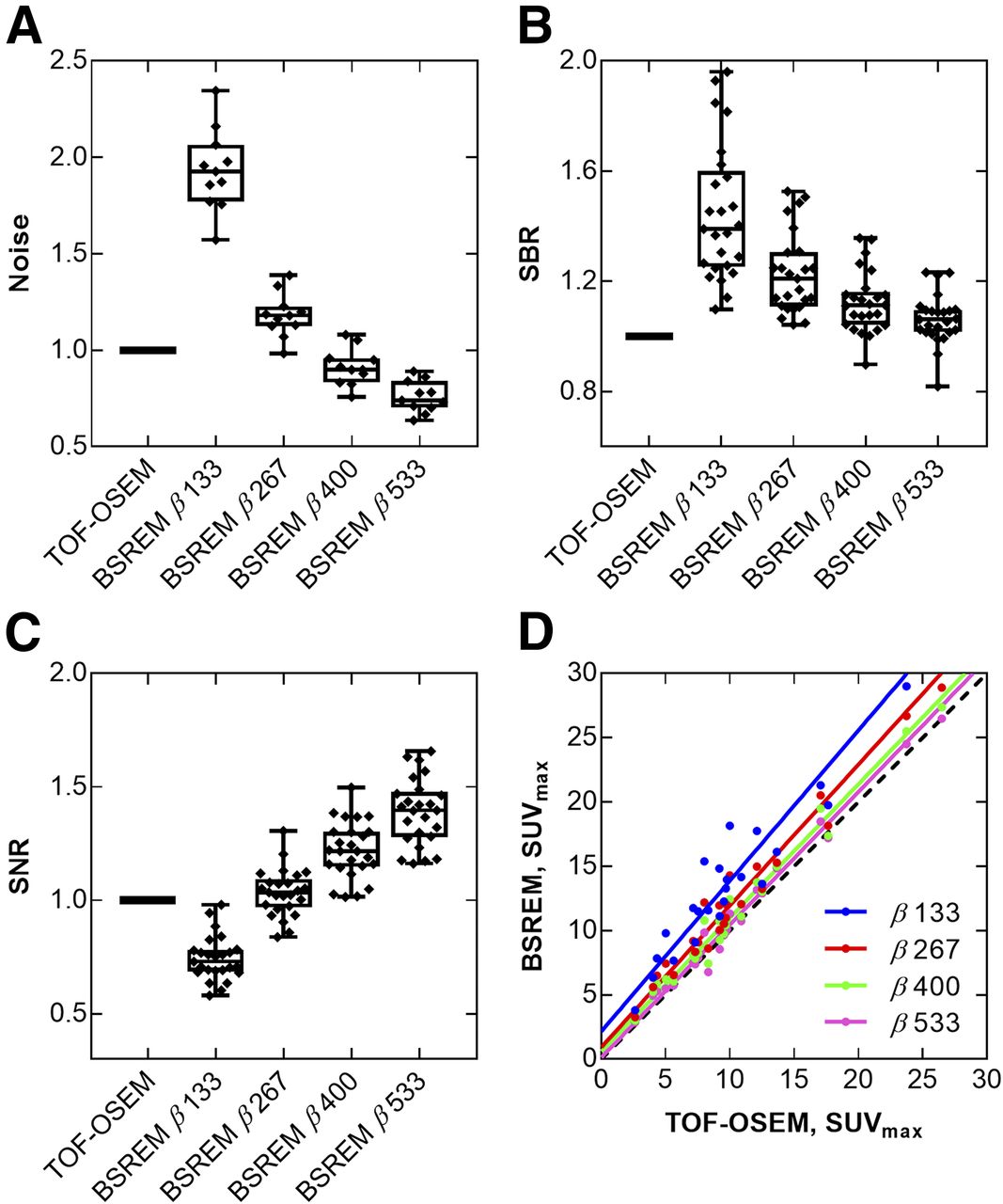
  • FIGURE 3.
    • Download figure
    • Open in new tab
    • Download powerpoint
    FIGURE 3.

    Quantitative data from 18F-FDG PET/CT examinations of 11 patients with total of 25 lesions. Box plots show noise level (A), SBR (B), and SNR (C) for different reconstruction methods, and correlation plot shows SUVmax (D) for BSREM compared with TOF OSEM. Reconstruction methods were TOF OSEM (3 iterations, 16 subsets, 5-mm gaussian postprocessing filter, and PSF) and BSREM (including TOF and PSF) with β-factors of 133, 267, 400, and 533; 70-cm FOV; and 3 min/bp acquisition. All data were normalized to data obtained by TOF OSEM. Dashed line represents line of identity.
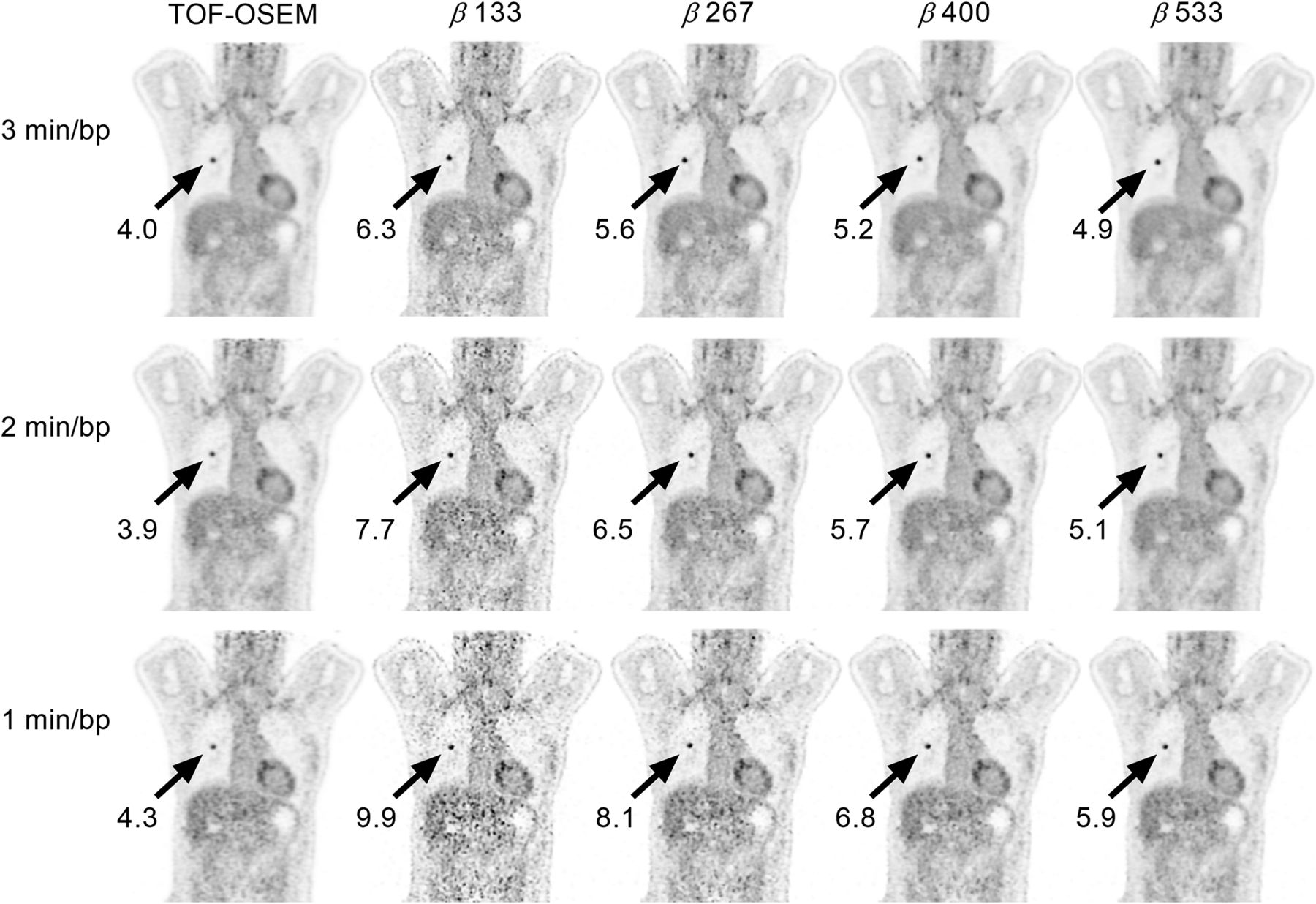
  • FIGURE 4.
    • Download figure
    • Open in new tab
    • Download powerpoint
    FIGURE 4.

    Coronal whole-body 18F-FDG PET images of patient with adenocarcinoma, demonstrating SUVmax of lesion (arrows) obtained with respective reconstruction method (gray scale 0–6). Images were reconstructed with 70-cm FOV using TOF OSEM (3 iterations, 16 subsets, 5-mm gaussian postprocessing filter, and PSF) and BSREM (including TOF and PSF) with β-factors of 133, 267, 400, and 533 for 3, 2, and 1 min/bp acquisitions.
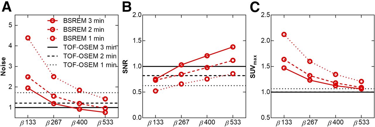
  • FIGURE 5.
    • Download figure
    • Open in new tab
    • Download powerpoint
    FIGURE 5.

    Mean noise level (A), SNR (B), and SUVmax (C) for different reconstruction methods and PET acquisition durations (3, 2, and 1 min/bp). Reconstruction methods were TOF OSEM (3 iterations, 16 subsets, 5-mm gaussian postprocessing filter, and PSF) and BSREM (including TOF and PSF) with β-factors of 133, 267, 400, and 533 and 70-cm FOV. All data were normalized to data obtained by TOF OSEM.
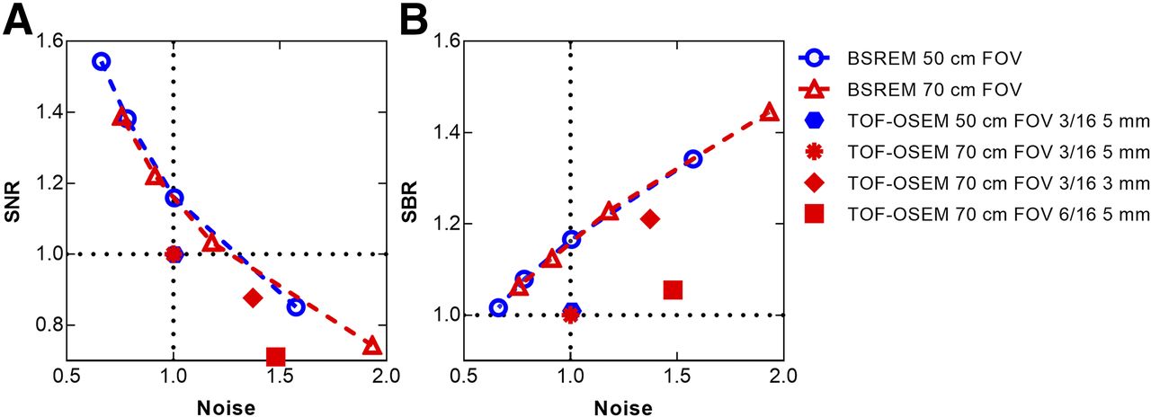
  • FIGURE 6.
    • Download figure
    • Open in new tab
    • Download powerpoint
    FIGURE 6.

    Trade-off curves showing SNR vs. noise (A) and SBR vs. noise (B) for reconstructions using TOF OSEM (3 iterations, 16 subsets, 5-mm gaussian postprocessing filter, and PSF) and BSREM (including TOF and PSF) with β-factors of 133, 267, 400, and 533 and transaxial FOV of 50 cm (blue) and 70 cm (red). Decreasing β-factor is seen with increasing noise for BSREM data points in both plots. All data were normalized to TOF OSEM with 70-cm FOV, and data points represent mean values. For reference, additional reconstructions with 70-cm FOV using TOF OSEM with PSF and 3 iterations, 16 subsets, and 3-mm gaussian postprocessing filter, and TOF OSEM with PSF and 6 iterations, 16 subsets, and 5-mm gaussian postprocessing filter, are also shown in both graphs.
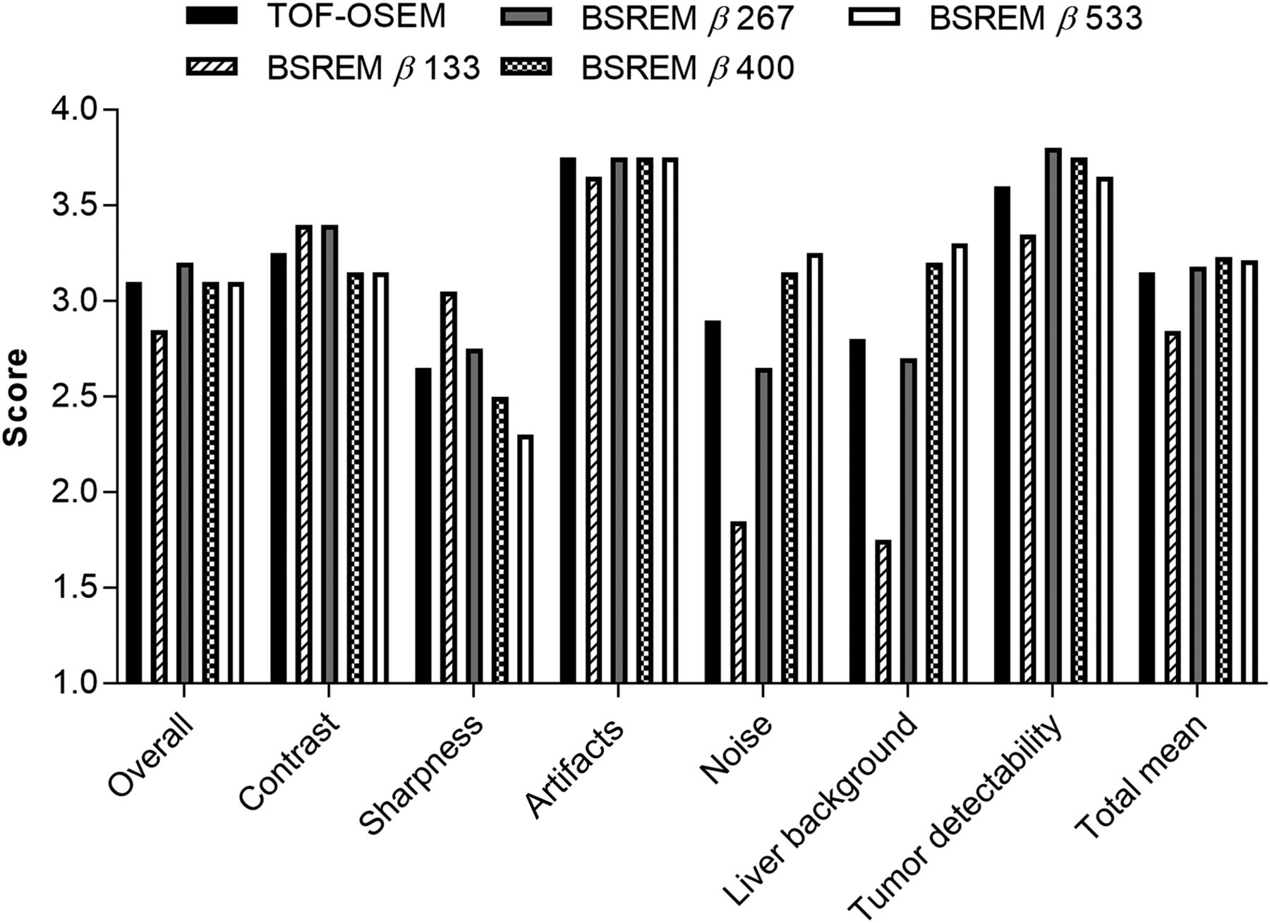
  • FIGURE 7.
    • Download figure
    • Open in new tab
    • Download powerpoint
    FIGURE 7.

    Visual scoring of image quality by 2 observers per arbitrary scale (1, poor; 2, moderate; 3, good; 4, very good). Seven different aspects of image quality were considered, and last category in plot represents summarized total mean score. There were 5 sets of images for each of 10 patients. Reconstruction used TOF OSEM (3 iterations, 16 subsets, 5-mm gaussian postprocessing filter, and PSF) and BSREM (including TOF and PSF) with β-factors of 133, 267, 400, and 533; 50-cm FOV, and 3 min/bp acquisition.

Tables

  • Figures
  • Additional Files
    • View popup
    TABLE 1

    Quantitative Measures of Reference Sphere in Healthy Liver Tissue Using TOF OSEM and BSREM

    BSREM
    MeasureTOF OSEM*β-factor, 133β-factor, 267β-factor, 400β-factor, 533
    Volume (cm3)19.2 (18.0–19.6)
    SUVmax2.9 (2.1–4.0)4.2 (2.7–5.4)3.1 (2.2–4.0)2.8 (2.0–3.7)2.6 (2.0–3.5)
    SUVmean2.1 (1.6–2.6)2.0 (1.6–2.5)2.0 (1.6–2.5)2.1 (1.6–2.6)2.1 (1.6–2.6)
    SUVSD0.2 (0.2–0.4)0.4 (0.3–0.6)0.3 (0.2–0.4)0.2 (0.2–0.3)0.2 (0.1–0.3)
    Noise level*0.11 (0.09–0.16)0.22 (0.18–0.28)0.13 (0.11–0.17)0.10 (0.08–0.13)0.09 (0.07–0.11)
    • ↵* Measured in liver and defined as SUVSD divided by SUVmean.

    • Data are mean followed by range. TOF OSEM used 3 iterations, 16 subsets, 5-mm gaussian postprocessing filter, and PSF. BSREM included TOF and PSF with β-factors of 133, 267, 400, and 533; 70-cm FOV; and 3 min/bp.

Additional Files

  • Figures
  • Tables
  • Supplemental Data

    Files in this Data Supplement:

    • Supplemental Data
PreviousNext
Back to top

In this issue

Journal of Nuclear Medicine: 59 (7)
Journal of Nuclear Medicine
Vol. 59, Issue 7
July 1, 2018
  • Table of Contents
  • Table of Contents (PDF)
  • About the Cover
  • Index by author
Print
Download PDF
Article Alerts
Sign In to Email Alerts with your Email Address
Email Article

Thank you for your interest in spreading the word on Journal of Nuclear Medicine.

NOTE: We only request your email address so that the person you are recommending the page to knows that you wanted them to see it, and that it is not junk mail. We do not capture any email address.

Enter multiple addresses on separate lines or separate them with commas.
Evaluation of Penalized-Likelihood Estimation Reconstruction on a Digital Time-of-Flight PET/CT Scanner for 18F-FDG Whole-Body Examinations
(Your Name) has sent you a message from Journal of Nuclear Medicine
(Your Name) thought you would like to see the Journal of Nuclear Medicine web site.
Citation Tools
Evaluation of Penalized-Likelihood Estimation Reconstruction on a Digital Time-of-Flight PET/CT Scanner for 18F-FDG Whole-Body Examinations
Elin Lindström, Anders Sundin, Carlos Trampal, Lars Lindsjö, Ezgi Ilan, Torsten Danfors, Gunnar Antoni, Jens Sörensen, Mark Lubberink
Journal of Nuclear Medicine Jul 2018, 59 (7) 1152-1158; DOI: 10.2967/jnumed.117.200790

Citation Manager Formats

  • BibTeX
  • Bookends
  • EasyBib
  • EndNote (tagged)
  • EndNote 8 (xml)
  • Medlars
  • Mendeley
  • Papers
  • RefWorks Tagged
  • Ref Manager
  • RIS
  • Zotero
Share
Evaluation of Penalized-Likelihood Estimation Reconstruction on a Digital Time-of-Flight PET/CT Scanner for 18F-FDG Whole-Body Examinations
Elin Lindström, Anders Sundin, Carlos Trampal, Lars Lindsjö, Ezgi Ilan, Torsten Danfors, Gunnar Antoni, Jens Sörensen, Mark Lubberink
Journal of Nuclear Medicine Jul 2018, 59 (7) 1152-1158; DOI: 10.2967/jnumed.117.200790
Twitter logo Facebook logo LinkedIn logo Mendeley logo
  • Tweet Widget
  • Facebook Like
  • Google Plus One
Bookmark this article

Jump to section

  • Article
    • Abstract
    • MATERIALS AND METHODS
    • RESULTS
    • DISCUSSION
    • CONCLUSION
    • DISCLOSURE
    • Acknowledgments
    • Footnotes
    • REFERENCES
  • Figures & Data
  • Supplemental
  • Info & Metrics
  • PDF

Related Articles

  • This Month in JNM
  • PubMed
  • Google Scholar

Cited By...

  • Performance Characteristics of a New-Generation Digital Bismuth Germanium Oxide PET/CT System, Omni Legend 32, According to NEMA NU 2-2018 Standards
  • Google Scholar

More in this TOC Section

  • Performance Evaluation of the uMI Panorama PET/CT System in Accordance with the National Electrical Manufacturers Association NU 2-2018 Standard
  • A Multicenter Study on Observed Discrepancies Between Vendor-Stated and PET-Measured 90Y Activities for Both Glass and Resin Microsphere Devices
  • Ultra-Fast List-Mode Reconstruction of Short PET Frames and Example Applications
Show more Physics and Instrumentation

Similar Articles

Keywords

  • PET/CT
  • FDG
  • Image Reconstruction
  • penalization factor
  • block-sequential regularized expectation maximization
SNMMI

© 2025 SNMMI

Powered by HighWire