
Main menu

  • Home
  • Content
    • Current
    • Ahead of print
    • Past Issues
    • JNM Supplement
    • SNMMI Annual Meeting Abstracts
    • Continuing Education
    • JNM Podcasts
  • Subscriptions
    • Subscribers
    • Institutional and Non-member
    • Rates
    • Journal Claims
    • Corporate & Special Sales
  • Authors
    • Submit to JNM
    • Information for Authors
    • Assignment of Copyright
    • AQARA requirements
  • Info
    • Reviewers
    • Permissions
    • Advertisers
  • About
    • About Us
    • Editorial Board
    • Contact Information
  • More
    • Alerts
    • Feedback
    • Help
    • SNMMI Journals
  • SNMMI
    • JNM
    • JNMT
    • SNMMI Journals
    • SNMMI

User menu

  • Subscribe
  • My alerts
  • Log in
  • My Cart

Search

  • Advanced search
Journal of Nuclear Medicine
  • SNMMI
    • JNM
    • JNMT
    • SNMMI Journals
    • SNMMI
  • Subscribe
  • My alerts
  • Log in
  • My Cart
Journal of Nuclear Medicine

Advanced Search

  • Home
  • Content
    • Current
    • Ahead of print
    • Past Issues
    • JNM Supplement
    • SNMMI Annual Meeting Abstracts
    • Continuing Education
    • JNM Podcasts
  • Subscriptions
    • Subscribers
    • Institutional and Non-member
    • Rates
    • Journal Claims
    • Corporate & Special Sales
  • Authors
    • Submit to JNM
    • Information for Authors
    • Assignment of Copyright
    • AQARA requirements
  • Info
    • Reviewers
    • Permissions
    • Advertisers
  • About
    • About Us
    • Editorial Board
    • Contact Information
  • More
    • Alerts
    • Feedback
    • Help
    • SNMMI Journals
  • View or Listen to JNM Podcast
  • Visit JNM on Facebook
  • Join JNM on LinkedIn
  • Follow JNM on Twitter
  • Subscribe to our RSS feeds
Research ArticleEndocrinology

PET/MRI in the Diagnosis of Hormone-Producing Pituitary Microadenoma: A Prospective Pilot Study

Hao Wang, Bo Hou, Lin Lu, Ming Feng, Jie Zang, Shaobo Yao, Feng Feng, Renzhi Wang, Fang Li and Zhaohui Zhu
Journal of Nuclear Medicine March 2018, 59 (3) 523-528; DOI: https://doi.org/10.2967/jnumed.117.191916
Hao Wang
1Department of Nuclear Medicine and Beijing Key Laboratory of Molecular Targeted Diagnosis and Therapy in Nuclear Medicine, Peking Union Medical College (PUMC) Hospital, Chinese Academy of Medical Science and PUMC, Beijing, China
  • Find this author on Google Scholar
  • Find this author on PubMed
  • Search for this author on this site
Bo Hou
2Department of Radiology, PUMC Hospital, Chinese Academy of Medical Science and PUMC, Beijing, China
  • Find this author on Google Scholar
  • Find this author on PubMed
  • Search for this author on this site
Lin Lu
3Department of Endocrinology and Key Laboratory of Endocrinology of National Health and Family Planning Commission, PUMC Hospital, Chinese Academy of Medical Science and PUMC, Beijing, China; and
  • Find this author on Google Scholar
  • Find this author on PubMed
  • Search for this author on this site
Ming Feng
4Department of Neurosurgery, PUMC Hospital, Chinese Academy of Medical Science and PUMC, Beijing, China
  • Find this author on Google Scholar
  • Find this author on PubMed
  • Search for this author on this site
Jie Zang
1Department of Nuclear Medicine and Beijing Key Laboratory of Molecular Targeted Diagnosis and Therapy in Nuclear Medicine, Peking Union Medical College (PUMC) Hospital, Chinese Academy of Medical Science and PUMC, Beijing, China
  • Find this author on Google Scholar
  • Find this author on PubMed
  • Search for this author on this site
Shaobo Yao
1Department of Nuclear Medicine and Beijing Key Laboratory of Molecular Targeted Diagnosis and Therapy in Nuclear Medicine, Peking Union Medical College (PUMC) Hospital, Chinese Academy of Medical Science and PUMC, Beijing, China
  • Find this author on Google Scholar
  • Find this author on PubMed
  • Search for this author on this site
Feng Feng
2Department of Radiology, PUMC Hospital, Chinese Academy of Medical Science and PUMC, Beijing, China
  • Find this author on Google Scholar
  • Find this author on PubMed
  • Search for this author on this site
Renzhi Wang
4Department of Neurosurgery, PUMC Hospital, Chinese Academy of Medical Science and PUMC, Beijing, China
  • Find this author on Google Scholar
  • Find this author on PubMed
  • Search for this author on this site
Fang Li
1Department of Nuclear Medicine and Beijing Key Laboratory of Molecular Targeted Diagnosis and Therapy in Nuclear Medicine, Peking Union Medical College (PUMC) Hospital, Chinese Academy of Medical Science and PUMC, Beijing, China
  • Find this author on Google Scholar
  • Find this author on PubMed
  • Search for this author on this site
Zhaohui Zhu
1Department of Nuclear Medicine and Beijing Key Laboratory of Molecular Targeted Diagnosis and Therapy in Nuclear Medicine, Peking Union Medical College (PUMC) Hospital, Chinese Academy of Medical Science and PUMC, Beijing, China
  • Find this author on Google Scholar
  • Find this author on PubMed
  • Search for this author on this site
  • Article
  • Figures & Data
  • Supplemental
  • Info & Metrics
  • PDF
Loading

Article Figures & Data

Figures

  • Tables
  • Additional Files
  • FIGURE 1.
    • Download figure
    • Open in new tab
    • Download powerpoint
    FIGURE 1.

    A 31-y-old woman with high adrenocorticotropic hormone level (patient 1 in Table 1). (A–D) MR images showing equivocal lesion at right end of sella region (from left to right: T1-weighted coronal, T2-weighted coronal, and contrast-enhanced T1-weighted coronal and sagittal views). (E–H) 18F-FDG PET/MR images showing definitely avid lesion (arrows) at same region as on MR images (from left to right: coronal PET, coronal PET/MRI, sagittal PET, and sagittal PET/MRI). (I–L) 68Ga-DOTATATE PET/MR images showing lower uptake in lesion than in normal pituitary tissue (arrowhead) (from left to right: coronal PET, coronal PET/MRI, sagittal PET, and sagittal PET/MRI). Follow-up surgery confirmed presence of right-sided 6 × 5 × 5 mm functional pituitary microadenoma, and pathologic stains were adrenocorticotropic hormone–positive, luteinizing hormone–positive, and growth hormone–positive.
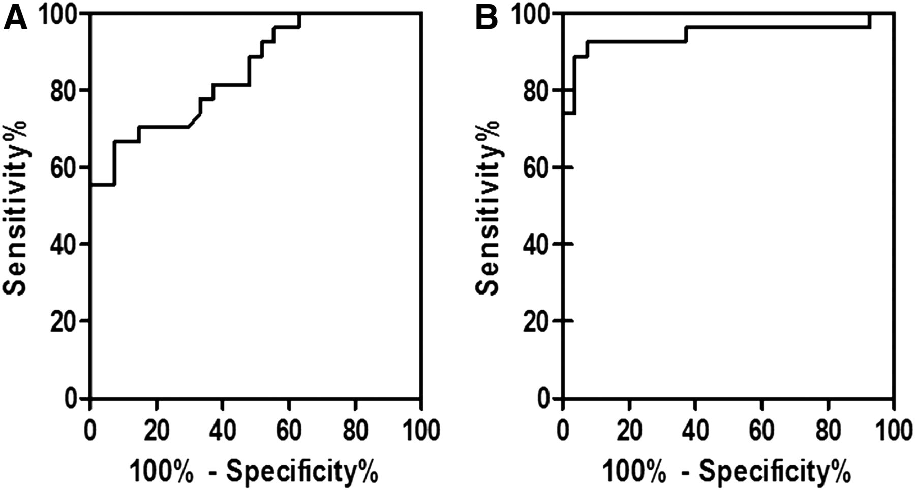
  • FIGURE 2.
    • Download figure
    • Open in new tab
    • Download powerpoint
    FIGURE 2.

    Use of 18F-FDG SUVmax (A) and 18F-FDG/68Ga-DOTATATE SUVmax ratio (B) ROC curves for differentiation of pituitary adenoma from normal pituitary tissue. Area under curve and standard error were 0.85 ± 0.05 for 18F-FDG and 0.94 ± 0.03 for 18F-FDG/68Ga-DOTATATE. Optimal diagnostic cutoff by ROC analysis was 3.88 and 1.04, respectively. Diagnostic performance was better with 18F-FDG/68Ga-DOTATATE than with 18F-FDG alone.

Tables

  • Figures
  • Additional Files
    • View popup
    TABLE 1

    Patient Characteristics

    MRI resultPET/MRI SUVmax
    Patient no.Age (y)SexP/RHormone levelHalf doseFull dose18F-FDG68Ga-DOTATATEAdenoma size (mm)Immunohistochemical staining result
    131FPACTH↑, F↑−+7.562.836 × 5 × 5ACTH+, GH+, LH+
    216MPACTH↑, F↑−+4.82.85 × 5 × 5ACTH+, GH+
    326FPACTH↑, F↑−−3.12.098 × 3 × 3ACTH+, GH+
    463FPGH↑, IGF1↑−−10.215.237 × 5 × 5ACTH+, GH+, LH+, PRL+
    538FPACTH↑, F↑−−4.811.245 × 4 × 3ACTH+
    638FPACTH↑, F↑−−9.074.615 × 4 × 3ACTH+
    716MPACTH↑, F↑±+6.815.36 × 5 × 5ACTH+, GH+, LH+
    828FPACTH↑, F↑±+2.9512.053 × 3 × 2ACTH+, GH+, LH+
    963MPACTH↑, F↑±−7.232.555 × 5 × 5ACTH+, GH+, LH+
    1026MPACTH↑, F↑±−3.686.153 × 2 × 2ACTH+, GH+, LH+
    1117MPACTH↑, F↑±−17.483.3510 × 8 × 8ACTH+, GH+
    1250FPACTH↑, F↑±+3.12.095 × 5 × 5ACTH+, GH+, LH+
    1338MPACTH↑, F↑−−9.692.285 × 3 × 3ACTH+, GH+
    1445FPACTH↑, F↑−+5.613.025 × 5 × 3ACTH+, GH+, LH+
    1520FPACTH↑, F↑±−8.562.25 × 5 × 5ACTH+, GH+, LH+
    1616MPACTH↑, F↑±−4.273.024 × 3 × 3ACTH+, GH+
    1720FRACTH↑, F↑−+5.585.385 × 5 × 5ACTH+, GH+
    1830FRACTH↑, F↑−+8.842.626 × 5 × 5ACTH+, GH+
    1925FRACTH↑, F↑±+6.64.366 × 5 × 5ACTH+
    2031FRACTH↑, F↑±+2.691.945 × 3 × 3ACTH−, FSH+, GH+, LH+
    2129FRACTH↑, F↑±+5.43.835 × 5 × 5ACTH+, GH+, LH+
    2210FRACTH↑, F↑−−2.492.393 × 3 × 3ACTH+, GH+, LH+
    2345FRACTH↑, F↑±+3.962.085 × 5 × 5ACTH+, GH+
    2435FRACTH↑, F↑−−13.32.355 × 5 × 5ACTH+, GH+
    2550FRACTH↑, F↑−−3.73.354 × 3 × 3ACTH+
    2641FRACTH↑, F↑±+10.492.025 × 6 × 6ACTH+, GH+
    2732MRACTH↑, F↑−+3.522.815 × 5 × 4ACTH+, GH+, LH+
    2811FPACTH↑, F↑−−7.453.92NANA
    2951MPACTH↑, F↑−+6.332.17NANA
    3059FPACTH↑, F↑±−4.232.15NANA
    3155FRGH↑, IGF1↑−−5.243.31NANA
    3257MRT4↑−+7.342.35NANA
    3330FRGH↑, IGF1↑−−9.563.22NANA
    3434FRACTH↑, F↑−−8.282.13NANA
    3517MRACTH↑, F↑−−5.213.28NANA
    3639FRACTH↑, F↑±−8.355.28NANA
    3735FRACTH↑, F↑±−4.383.17NANA
    • P = primary; R = recurrent; half dose = 0.05 mmol/kg dose of gadopentetate dimeglumine; full dose = 0.1 mmol/kg dose of gadopentetate dimeglumine; ACTH = adrenocorticotropic hormone; F = cortisol (compound F); GH = growth hormone; LH = luteinizing hormone; IGF1 = insulinlike growth factor 1; PRL = prolactin; FSH = follicle-stimulating hormone; NA = not available because patient accepted clinical follow-up or treatments other than surgery; T4 = thyroxine; + = positive; − = negative.

Additional Files

  • Figures
  • Tables
  • Supplemental Data

    Files in this Data Supplement:

    • Supplemental Data
PreviousNext
Back to top

In this issue

Journal of Nuclear Medicine: 59 (3)
Journal of Nuclear Medicine
Vol. 59, Issue 3
March 1, 2018
  • Table of Contents
  • Table of Contents (PDF)
  • About the Cover
  • Index by author
Print
Download PDF
Article Alerts
Sign In to Email Alerts with your Email Address
Email Article

Thank you for your interest in spreading the word on Journal of Nuclear Medicine.

NOTE: We only request your email address so that the person you are recommending the page to knows that you wanted them to see it, and that it is not junk mail. We do not capture any email address.

Enter multiple addresses on separate lines or separate them with commas.
PET/MRI in the Diagnosis of Hormone-Producing Pituitary Microadenoma: A Prospective Pilot Study
(Your Name) has sent you a message from Journal of Nuclear Medicine
(Your Name) thought you would like to see the Journal of Nuclear Medicine web site.
Citation Tools
PET/MRI in the Diagnosis of Hormone-Producing Pituitary Microadenoma: A Prospective Pilot Study
Hao Wang, Bo Hou, Lin Lu, Ming Feng, Jie Zang, Shaobo Yao, Feng Feng, Renzhi Wang, Fang Li, Zhaohui Zhu
Journal of Nuclear Medicine Mar 2018, 59 (3) 523-528; DOI: 10.2967/jnumed.117.191916

Citation Manager Formats

  • BibTeX
  • Bookends
  • EasyBib
  • EndNote (tagged)
  • EndNote 8 (xml)
  • Medlars
  • Mendeley
  • Papers
  • RefWorks Tagged
  • Ref Manager
  • RIS
  • Zotero
Share
PET/MRI in the Diagnosis of Hormone-Producing Pituitary Microadenoma: A Prospective Pilot Study
Hao Wang, Bo Hou, Lin Lu, Ming Feng, Jie Zang, Shaobo Yao, Feng Feng, Renzhi Wang, Fang Li, Zhaohui Zhu
Journal of Nuclear Medicine Mar 2018, 59 (3) 523-528; DOI: 10.2967/jnumed.117.191916
Twitter logo Facebook logo LinkedIn logo Mendeley logo
  • Tweet Widget
  • Facebook Like
  • Google Plus One
Bookmark this article

Jump to section

  • Article
    • Abstract
    • MATERIALS AND METHODS
    • RESULTS
    • DISCUSSION
    • CONCLUSION
    • DISCLOSURE
    • Acknowledgments
    • Footnotes
    • REFERENCES
  • Figures & Data
  • Supplemental
  • Info & Metrics
  • PDF

Related Articles

  • This Month in JNM
  • PubMed
  • Google Scholar

Cited By...

  • Using Molecular Imaging to Enhance Decision Making in the Management of Pituitary Adenomas
  • Automatic detection of pituitary microadenoma from magnetic resonance imaging using deep learning algorithms
  • Google Scholar

More in this TOC Section

Endocrinology

  • Assessment and Comparison of 18F-Fluorocholine PET and 99mTc-Sestamibi Scans in Identifying Parathyroid Adenomas: A Metaanalysis
  • Targeted Optical Imaging of the Glucagonlike Peptide 1 Receptor Using Exendin-4-IRDye 800CW
  • 18F-Fluorocholine PET/CT in Primary Hyperparathyroidism: Superior Diagnostic Performance to Conventional Scintigraphic Imaging for Localization of Hyperfunctioning Parathyroid Glands
Show more Endocrinology

Clinical

  • Detection of Early Progression with 18F-DCFPyL PET/CT in Men with Metastatic Castration-Resistant Prostate Cancer Receiving Bipolar Androgen Therapy
  • TauIQ: A Canonical Image Based Algorithm to Quantify Tau PET Scans
  • Dual PET Imaging in Bronchial Neuroendocrine Neoplasms: The NETPET Score as a Prognostic Biomarker
Show more Clinical

Similar Articles

Keywords

  • pituitary adenoma
  • 68Ga-DOTATATE
  • 18F-FDG
  • PET/MRI
SNMMI

© 2025 SNMMI

Powered by HighWire