
Main menu

  • Home
  • Content
    • Current
    • Ahead of print
    • Past Issues
    • JNM Supplement
    • SNMMI Annual Meeting Abstracts
    • Continuing Education
    • JNM Podcasts
  • Subscriptions
    • Subscribers
    • Institutional and Non-member
    • Rates
    • Journal Claims
    • Corporate & Special Sales
  • Authors
    • Submit to JNM
    • Information for Authors
    • Assignment of Copyright
    • AQARA requirements
  • Info
    • Reviewers
    • Permissions
    • Advertisers
  • About
    • About Us
    • Editorial Board
    • Contact Information
  • More
    • Alerts
    • Feedback
    • Help
    • SNMMI Journals
  • SNMMI
    • JNM
    • JNMT
    • SNMMI Journals
    • SNMMI

User menu

  • Subscribe
  • My alerts
  • Log in
  • My Cart

Search

  • Advanced search
Journal of Nuclear Medicine
  • SNMMI
    • JNM
    • JNMT
    • SNMMI Journals
    • SNMMI
  • Subscribe
  • My alerts
  • Log in
  • My Cart
Journal of Nuclear Medicine

Advanced Search

  • Home
  • Content
    • Current
    • Ahead of print
    • Past Issues
    • JNM Supplement
    • SNMMI Annual Meeting Abstracts
    • Continuing Education
    • JNM Podcasts
  • Subscriptions
    • Subscribers
    • Institutional and Non-member
    • Rates
    • Journal Claims
    • Corporate & Special Sales
  • Authors
    • Submit to JNM
    • Information for Authors
    • Assignment of Copyright
    • AQARA requirements
  • Info
    • Reviewers
    • Permissions
    • Advertisers
  • About
    • About Us
    • Editorial Board
    • Contact Information
  • More
    • Alerts
    • Feedback
    • Help
    • SNMMI Journals
  • View or Listen to JNM Podcast
  • Visit JNM on Facebook
  • Join JNM on LinkedIn
  • Follow JNM on Twitter
  • Subscribe to our RSS feeds
Research ArticleNeurology

Neuroinflammation Appears Early on PET Imaging and Then Plateaus in a Mouse Model of Alzheimer Disease

Francisco R. López-Picón, Anniina Snellman, Olli Eskola, Semi Helin, Olof Solin, Merja Haaparanta-Solin and Juha O. Rinne
Journal of Nuclear Medicine March 2018, 59 (3) 509-515; DOI: https://doi.org/10.2967/jnumed.117.197608
Francisco R. López-Picón
1Preclinical Imaging, Turku PET Centre, University of Turku, Turku, Finland
2MediCity Research Laboratory, University of Turku, Turku, Finland
  • Find this author on Google Scholar
  • Find this author on PubMed
  • Search for this author on this site
Anniina Snellman
1Preclinical Imaging, Turku PET Centre, University of Turku, Turku, Finland
2MediCity Research Laboratory, University of Turku, Turku, Finland
  • Find this author on Google Scholar
  • Find this author on PubMed
  • Search for this author on this site
Olli Eskola
3Radiopharmaceutical Chemistry Laboratory, Turku PET Centre, University of Turku, Turku, Finland
  • Find this author on Google Scholar
  • Find this author on PubMed
  • Search for this author on this site
Semi Helin
3Radiopharmaceutical Chemistry Laboratory, Turku PET Centre, University of Turku, Turku, Finland
  • Find this author on Google Scholar
  • Find this author on PubMed
  • Search for this author on this site
Olof Solin
3Radiopharmaceutical Chemistry Laboratory, Turku PET Centre, University of Turku, Turku, Finland
4Department of Chemistry, University of Turku, Turku, Finland
5Accelerator Laboratory, Åbo Akademi University, Turku, Finland
  • Find this author on Google Scholar
  • Find this author on PubMed
  • Search for this author on this site
Merja Haaparanta-Solin
1Preclinical Imaging, Turku PET Centre, University of Turku, Turku, Finland
2MediCity Research Laboratory, University of Turku, Turku, Finland
  • Find this author on Google Scholar
  • Find this author on PubMed
  • Search for this author on this site
Juha O. Rinne
6Turku PET Centre, Turku University Hospital, Turku, Finland; and
7Division of Clinical Neurosciences, Turku University Hospital, Turku, Finland
  • Find this author on Google Scholar
  • Find this author on PubMed
  • Search for this author on this site
  • Article
  • Figures & Data
  • Supplemental
  • Info & Metrics
  • PDF
Loading

Article Figures & Data

Figures

  • Tables
  • Additional Files
  • FIGURE 1.
    • Download figure
    • Open in new tab
    • Download powerpoint
    FIGURE 1.

    (A) In vivo PET images of 11C-PIB in 26-mo-old transgenic (TG) and WT APP23 mice. (B–E) Ratios of FC (B), PTC (C), hippocampus (HIPPO) (D), and thalamus (THA) (E) to cerebellum (CB) from 17 to 26 mo of age. Dotted line separates data of pilot study from data of longitudinal study (n = 32). *P < 0.05. **P < 0.01. ***P < 0.001.

  • FIGURE 2.
    • Download figure
    • Open in new tab
    • Download powerpoint
    FIGURE 2.

    (A) In vivo PET images of 18F-GE-180 in 26-mo-old transgenic (TG) and WT APP23 mice. (B–E) Ratios of FC (B), PTC (C), hippocampus (HIPPO) (D), and thalamus (THA) (E) to cerebellum (CB) from 17 to 26 mo of age. Dotted line separates data of pilot study from data of longitudinal study (n = 30). *P < 0.05. **P < 0.01. ***P < 0.001.
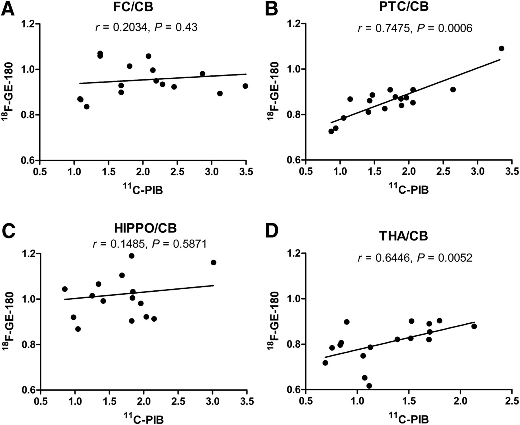
  • FIGURE 3.
    • Download figure
    • Open in new tab
    • Download powerpoint
    FIGURE 3.

    Correlation of the ratios of FC (A), PTC (B), hippocampus (HIPPO) (C), and thalamus (THA) (D) to cerebellum (CB) between 11C-PIB and 18F-GE-180 in transgenic APP23 mice from 17 to 26 mo of age, calculated with Pearson correlation (n = 17).
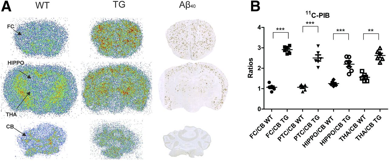
  • FIGURE 4.
    • Download figure
    • Open in new tab
    • Download powerpoint
    FIGURE 4.

    (A) Representative ex vivo autoradiography images of 11C-PIB binding in 26-mo-old transgenic (TG) and WT APP23 mice together with Aβ40-immunostaining (A). (B) Ratios of FC, PTC, hippocampus (HIPPO), and thalamus (THA) to cerebellum (CB). **P < 0.01. ***P < 0.001.
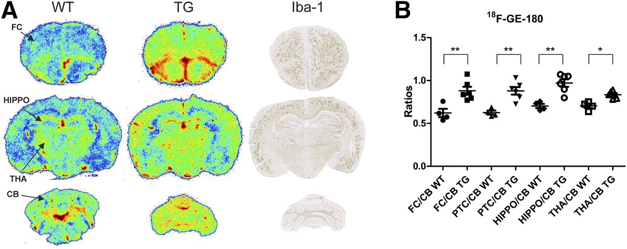
  • FIGURE 5.
    • Download figure
    • Open in new tab
    • Download powerpoint
    FIGURE 5.

    (A) Representative ex vivo brain autoradiography images of 18F-GE-180 binding in 26-mo-old transgenic (TG) and WT APP23 mice together with Iba1 immunostaining. (B) Ratios of FC, PTC, hippocampus (HIPPO), and thalamus (THA) to cerebellum (CB). *P < 0.05. **P < 0.01.

Tables

  • Figures
  • Additional Files
    • View popup
    TABLE 1

    Ratio and Percentage Increase from 17 to 20, 23, and 26 Months in APP23 Transgenic Mice

    11C-PIB18F-GE-180
    Site17–20 mo17–23 mo17–26 mo17–20 mo17–23 mo17–26 mo
    FC/CB0.64 (52%)1.03 (84%)1.72 (140%)0.00 (0%)0.01 (1%)0.02 (2%)
    PTC/CB0.56 (50%)0.83 (75%)1.13 (102%)0.06 (8%)0.08 (10%)0.11 (14%)
    HIPPO/CB0.33 (30%)0.70 (65%)1.07 (100%)0.03 (3%)0.04 (4%)0.01 (1%)
    THA/CB0.16 (17%)0.24 (25%)0.77 (82%)0.03 (4%)0.10 (14%)0.16 (23%)
    • CB = cerebellum; HIPPO = hippocampus; THA = thalamus.

Additional Files

  • Figures
  • Tables
  • Supplemental Data

    Files in this Data Supplement:

    • Supplemental Data
PreviousNext
Back to top

In this issue

Journal of Nuclear Medicine: 59 (3)
Journal of Nuclear Medicine
Vol. 59, Issue 3
March 1, 2018
  • Table of Contents
  • Table of Contents (PDF)
  • About the Cover
  • Index by author
Print
Download PDF
Article Alerts
Sign In to Email Alerts with your Email Address
Email Article

Thank you for your interest in spreading the word on Journal of Nuclear Medicine.

NOTE: We only request your email address so that the person you are recommending the page to knows that you wanted them to see it, and that it is not junk mail. We do not capture any email address.

Enter multiple addresses on separate lines or separate them with commas.
Neuroinflammation Appears Early on PET Imaging and Then Plateaus in a Mouse Model of Alzheimer Disease
(Your Name) has sent you a message from Journal of Nuclear Medicine
(Your Name) thought you would like to see the Journal of Nuclear Medicine web site.
Citation Tools
Neuroinflammation Appears Early on PET Imaging and Then Plateaus in a Mouse Model of Alzheimer Disease
Francisco R. López-Picón, Anniina Snellman, Olli Eskola, Semi Helin, Olof Solin, Merja Haaparanta-Solin, Juha O. Rinne
Journal of Nuclear Medicine Mar 2018, 59 (3) 509-515; DOI: 10.2967/jnumed.117.197608

Citation Manager Formats

  • BibTeX
  • Bookends
  • EasyBib
  • EndNote (tagged)
  • EndNote 8 (xml)
  • Medlars
  • Mendeley
  • Papers
  • RefWorks Tagged
  • Ref Manager
  • RIS
  • Zotero
Share
Neuroinflammation Appears Early on PET Imaging and Then Plateaus in a Mouse Model of Alzheimer Disease
Francisco R. López-Picón, Anniina Snellman, Olli Eskola, Semi Helin, Olof Solin, Merja Haaparanta-Solin, Juha O. Rinne
Journal of Nuclear Medicine Mar 2018, 59 (3) 509-515; DOI: 10.2967/jnumed.117.197608
Twitter logo Facebook logo LinkedIn logo Mendeley logo
  • Tweet Widget
  • Facebook Like
  • Google Plus One
Bookmark this article

Jump to section

  • Article
    • Abstract
    • MATERIALS AND METHODS
    • RESULTS
    • DISCUSSION
    • CONCLUSION
    • DISCLOSURE
    • Footnotes
    • REFERENCES
  • Figures & Data
  • Supplemental
  • Info & Metrics
  • PDF

Related Articles

  • This Month in JNM
  • PubMed
  • Google Scholar

Cited By...

  • Western diet increases brain metabolism and adaptive immune responses in a mouse model of amyloidosis
  • MicroPET evidence for a hypersensitive neuroinflammatory profile of gp120 mouse model of HIV
  • Microglial activation in early Alzheimer trajectory is associated with higher gray matter volume
  • Google Scholar

More in this TOC Section

Neurology

  • Dopamine D1 Receptor Agonist PET Tracer Development: Assessment in Nonhuman Primates
  • Hypermetabolism on Pediatric PET Scans of Brain Glucose Metabolism: What Does It Signify?
  • TauIQ: A Canonical Image Based Algorithm to Quantify Tau PET Scans
Show more Neurology

Basic

  • Dopamine D1 Receptor Agonist PET Tracer Development: Assessment in Nonhuman Primates
  • Optical Navigation of the Drop-In γ-Probe as a Means to Strengthen the Connection Between Robot-Assisted and Radioguided Surgery
  • Synthesis and Preclinical Evaluation of a 68Ga-Labeled Adnectin, 68Ga-BMS-986192, as a PET Agent for Imaging PD-L1 Expression
Show more Basic

Similar Articles

Keywords

  • Alzheimer disease
  • neuroinflammation
  • β-amyloid
  • GE-180
  • PIB
SNMMI

© 2025 SNMMI

Powered by HighWire