


Article Figures & Data
Figures
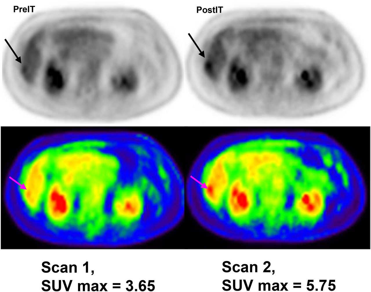
- FIGURE 1.
18F-FDG PET before and 2 wk after starting treatment of HER2 bispecific antibody–armed activated T cells shows inflammatory response.
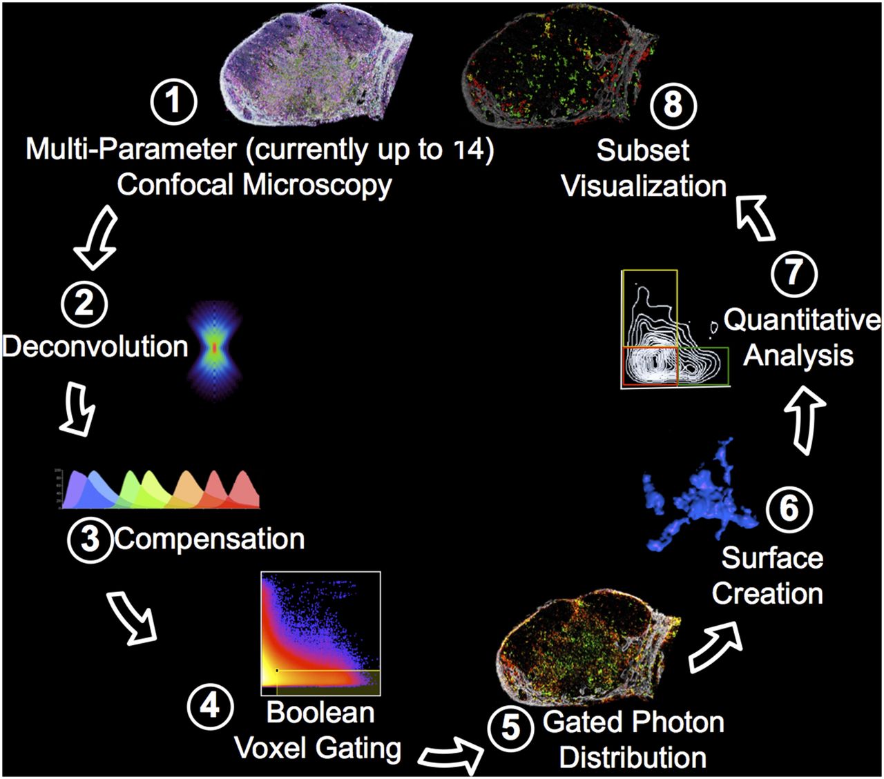
- FIGURE 2.
Histocytometry processing and analysis of tissue sections. (1) Fixed tissue section is simultaneously stained with antibodies directed to as many as 14 determinants and imaged in tiled, high-resolution mode using advanced confocal microscope. (2) Image data are deconvoluted to improve signal-to-noise ratio and spatial resolution. (3) Spillover between fluorescent emissions is corrected (compensated). (4–6) The resulting data are used to create cellular objects based on membrane staining, with defined objects retaining all fluorescence information associated with that object. (7) These data are equivalent to flow cytometry data and can be processed using software for flow data analysis, yielding quantitative information about cellular subset frequency (for example) or intensity of staining for a given determinant. (8) Because data also retain spatial x-y-z information about each object, each gated and defined cellular object can also be placed in its original tissue location.
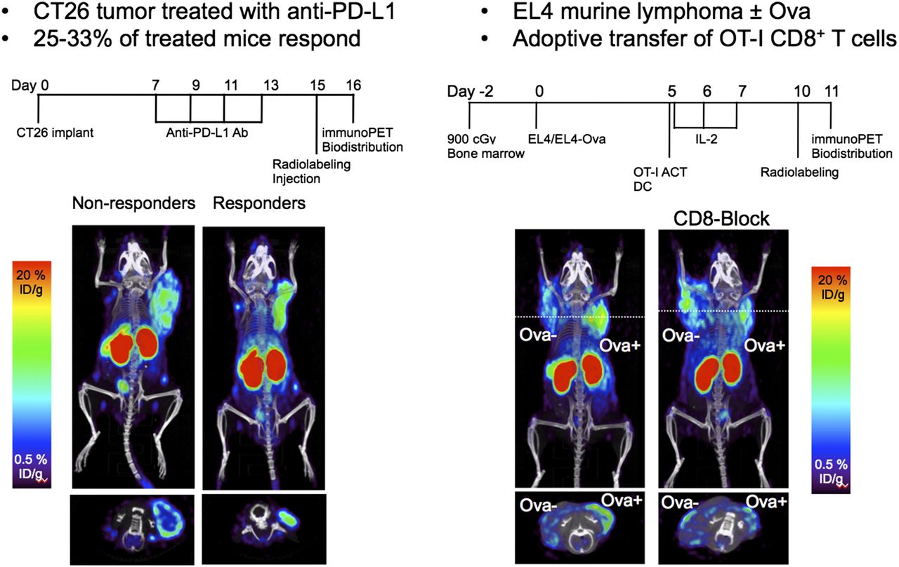
- FIGURE 3.
Imaging CD8 T-cell infiltration in tumor immunotherapy. Anti-CD8 PET was performed 5 d after adoptive transfer of OT-1 ovalbumin-specific T cells in C57BL/6 mice bearing subcutaneous Ova-negative and Ova-positive EL4 tumors. Mouse on right was preblocked with cold anti-CD8 antibody.
- FIGURE 4.
First-in-human imaging with 89Zr-desferrioxamine (Df)-IAb2M anti–prostate-specific membrane antigen minibody in patients with metastatic prostate cancer, compared with 99mTc-methylene diphosphonate (MDP) bone scan and 18F-FDG PET maximum-intensity projection (MIP).
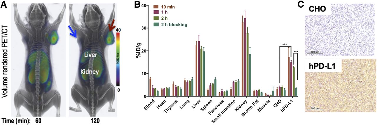
- FIGURE 5.
(A) Volume-rendered PET/CT images at 60 and 120 min after intravenous administration of 5,550 MBq (150 mCi) of 64Cu-WL12 to NSG mice show specific accumulation in hPD-L1 tumor (red arrow) as opposed to CHO tumor (blue arrow). (B) Percentage injected dose per gram of tissue at 10, 30, 60, and 120 min after tracer injection. (C) Photomicrographs illustrating the typical histologic patterns of the two tumor types.


Additional Files
Supplemental Data
Files in this Data Supplement:


Jump to section
- Article
- Abstract
- IMMUNOLOGY OVERVIEW AND CURRENT ROLE OF IMAGING
- T-CELL THERAPIES
- VACCINE THERAPY
- CLARIFYING RESPONSE CRITERIA
- IMAGING CAPABILITIES
- BIOMARKERS OF IMMUNE MODULATION
- IMAGING INFLAMMATION WITH 18F-FDG, 18F-FLT, AND BEYOND
- ANTIBODY IMMUNOTHERAPY IMAGING
- LABELED ANTIBODY FRAGMENTS FOR IMAGING
- IMAGING WITH CHEMOKINES AND SMALL MOLECULES
- BRIDGING THE GAPS TO INVESTIGATIONAL-NEW-DRUG AND NCI SUPPORT
- NEXT STEPS
- CONCLUSION
- DISCLOSURE
- Footnotes
- REFERENCES
- Figures & Data
- Supplemental
- Info & Metrics
Related Articles
Cited By...
- No citing articles found.