


Article Figures & Data
Figures
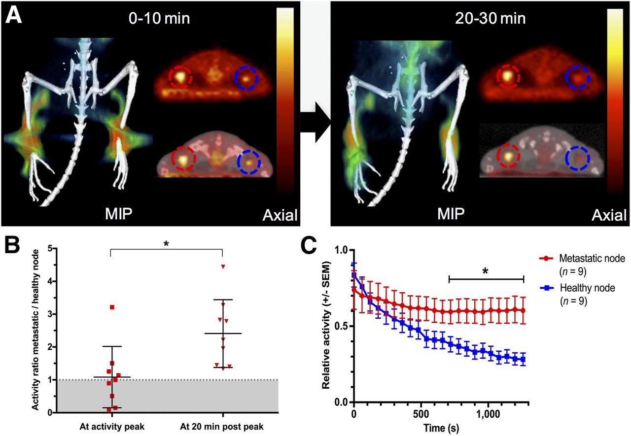
- FIGURE 1.
(A) Coronal maximum-intensity projection (MIP) and corresponding axial PET and fused PET/CT images derived from first 10 and last 10 min of 30-min dynamic 18F-FDG lymphography scan. Prolonged retention of radioactivity is seen in metastatic (red circles) versus healthy (blue circles) popliteal lymph nodes after 18F-FDG lymphography in mice implanted with B16F10 tumors on right hind paw. (B) Relative activity ratios between metastatic and healthy nodes are significantly higher 20 min after intraindividual peak uptake. (C) When displayed over time, longer retention of activity in metastatic nodes is evident, and from minute 13 after intraindividual peak uptake, relative activities remain significantly higher in metastatic nodes than in intraindividual healthy control nodes (P < 0.05).
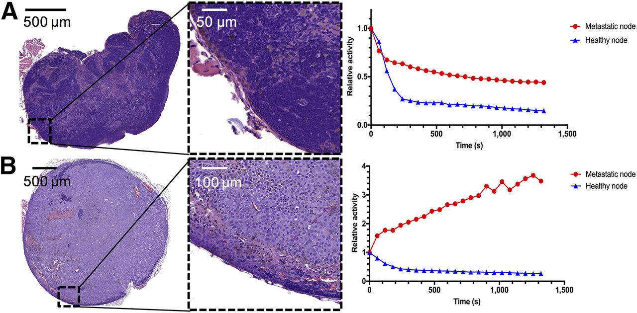
- FIGURE 2.
H&E staining and activity curves. (A) Results in mouse bearing only small melanoma cell infiltrates mainly on lymph node border on metastatic side. Activity curves in metastatic compared with healthy node tended to diverge in later scan minutes, showing retention effect in diseased node. (B) Results in mouse with large popliteal lymph node metastasis. Retention effect in diseased node is much higher, and only residual lymphatic tissue is preserved, as shown in H&E staining.
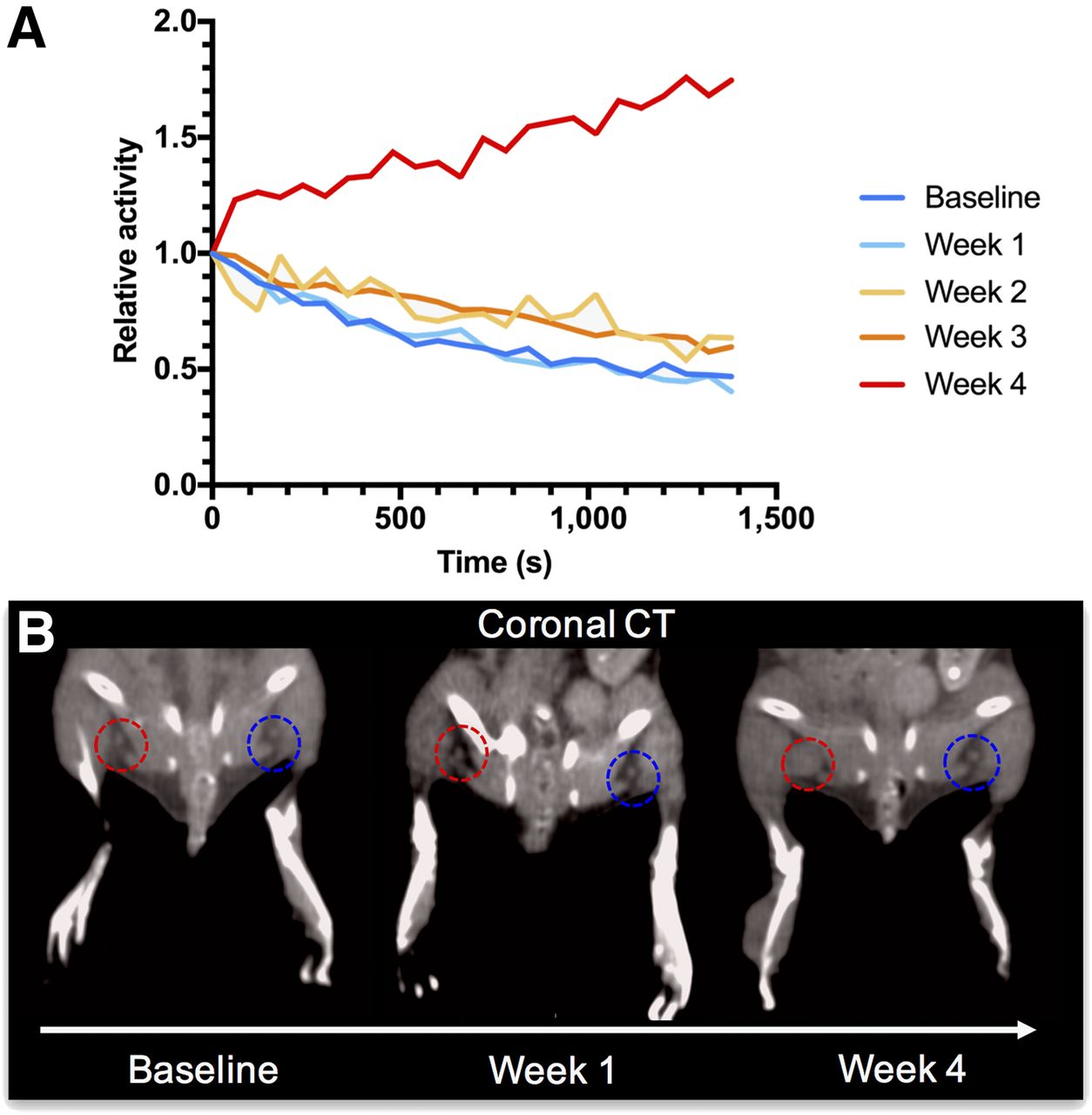
- FIGURE 3.
(A) When repeatedly imaged over 4 wk, alterations in lymphatic drainage become evident with developing metastasis in mouse. Tumor-draining popliteal nodes’ drainage dynamics do not differ from healthy contralateral control at baseline and week 1 after tumor implantation. From second week, subtly elevated activity retention levels can already be detected, whereas markedly prolonged uptake is seen in fourth week after tumor implantation. (B) Fittingly, with growing metastasis in week 4, evidence of lymph node enlargement on tumor-bearing side (red circles) is seen on coronal CT scans, when compared with contralateral healthy control (blue circles). At baseline and in week 1, lymph node sizes do not differ visually between the two sides.
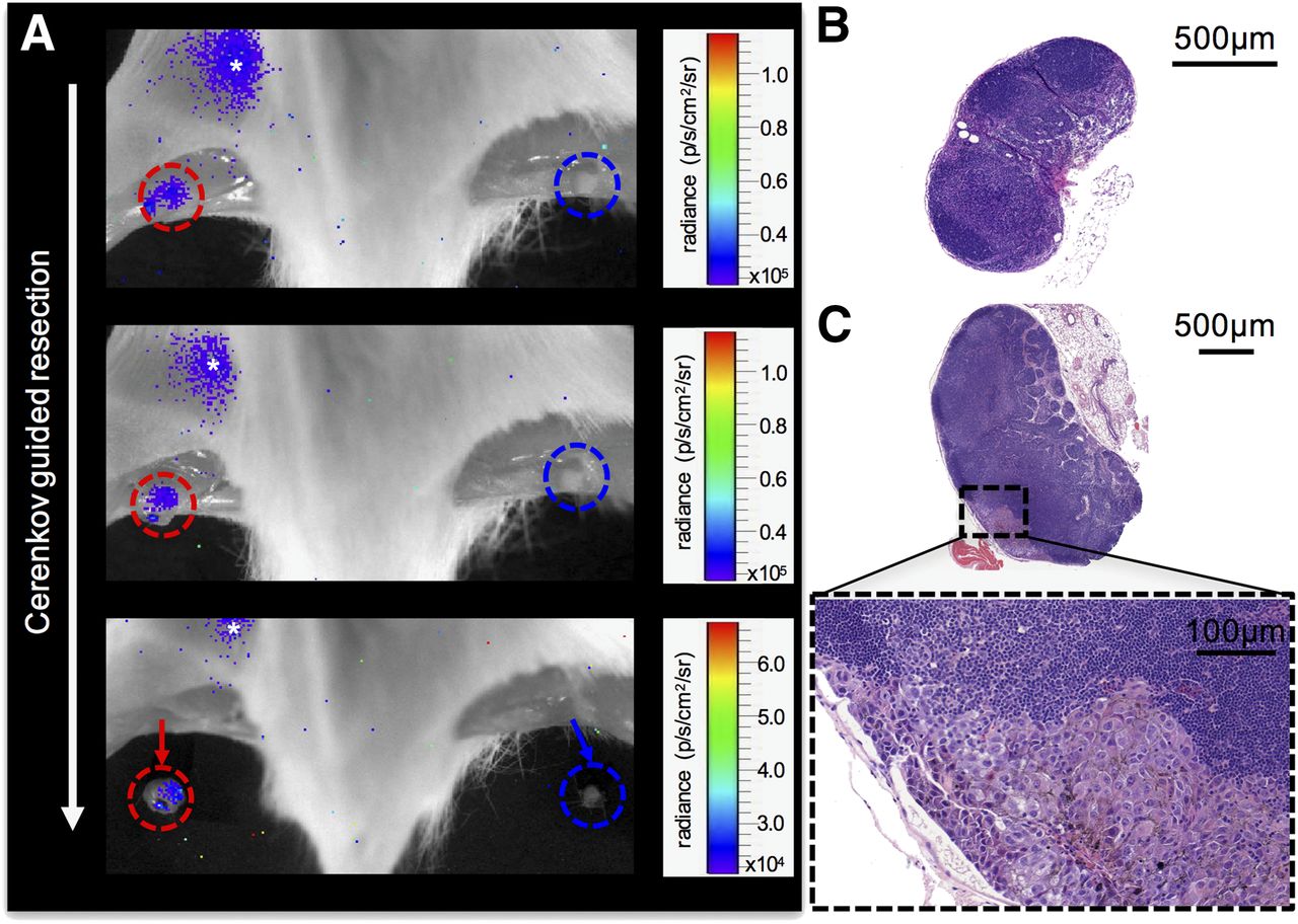
- FIGURE 4.
(A) 18F-FDG lymphography images (tumor not in field of view, mouse positioned prone, fur and skin removed, acquisition time of 5 min) showing stepwise Cerenkov-guided resection of metastatic (red circles) popliteal lymph node and corresponding contralateral healthy control node (blue circles) of mouse implanted with B16F10 tumor on right hind paw. At 1.5 h after bilateral 18F-FDG injection (3.7 MBq per side), metastatic node can still be identified by its subtle Cerenkov signal (mean signal-to-background ratio over time, 1.21 ± 0.14), whereas contralateral healthy control node does not show residual Cerenkov radiation (mean signal-to-background ratio over time, 0.69 ± 0.13), allowing for differentiation between the two and potential selective resection of tumor-bearing nodes. Asterisk indicates 18F-FDG pooling in bladder. (B) Healthy, uninvolved node displayed for size comparison. (C) H&E staining provides evidence of metastatic involvement of tumor side of popliteal lymph node by melanin-containing cells.
Additional Files
Supplemental Data
Files in this Data Supplement:


