


Article Figures & Data
Figures
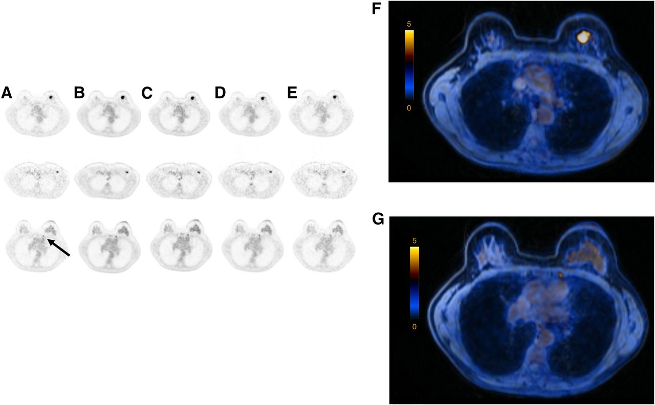
- FIGURE 1.
(A–E) Axial PET images of 57-y-old patient showing invasive left-sided breast cancer (top), axillary lymph node metastasis (middle), and internal mammary lymph node (bottom, arrow) after injection with 180 MBq of 18F-FDG (body weight, 60 kg): 2-min TOF (A), 100% of 18F-FDG dose (B), 36 MBq of 18F-FDG (20% dose) (C), 18 MBq of 18F-FDG (10% dose) (D), and 9 MBq of 18F-FDG (5% dose) (E). For PET/MRI examination with images shown in column D, this patient would receive an estimated radiation burden of 0.36 mSv. (F and G) Axial fused PET/MR images of primary lesion (F) and internal mammary lymph node (G) (PET with 10% dose).

Tables
Grade General image quality + artifact Image sharpness Image noise Lesion detectability 1 Excellent: no artifacts Clear, excellent images Negligible Excellent 2 Good: some diagnostically irrelevant artifacts Diagnostically irrelevant image blurring Diagnostically irrelevant Good 3 Average: diagnostically relevant artifacts Diagnostically relevant image blurring Diagnostically relevant Average 4 Inadequate: marked artifacts Inadequate image with blurring Marked Poor General image quality + artifacts Image sharpness Image noise Lesion detectability Parameter Mean SD Mean SD Mean SD Mean SD 2 min 1.84 0.6 2.28 0.6 2.16 0.6 1.46 0.7 20 min 100% 1.08 0.4 1.12 0.3 1.04 0.2 1.03 0.2 20 min 20% 1.32 0.6 1.64 0.7 1.44 0.5 1.14 0.4 20 min 10% 1.40 0.6 1.76 0.7 1.48 0.6 1.28 0.6 20 min 5% 1.88 0.5 2.60 0.8 2.28 0.5 1.62 0.8 General image quality + artifacts Lesion detectability Parameter Image sharpness Image noise Primary lesion Lymph nodes 20 min 100% <0.001 <0.001 <0.001 0.038 0.001 20 min 20% <0.001 <0.001 <0.001 0.257 0.001 20 min 10% 0.001 0.001 0.001 0.763 0.032 20 min 5% 0.317 0.005 0.180 0.142 0.593
Supplemental Data
Files in this Data Supplement:


Jump to section
Related Articles
Cited By...
- No citing articles found.