


Article Figures & Data
Figures
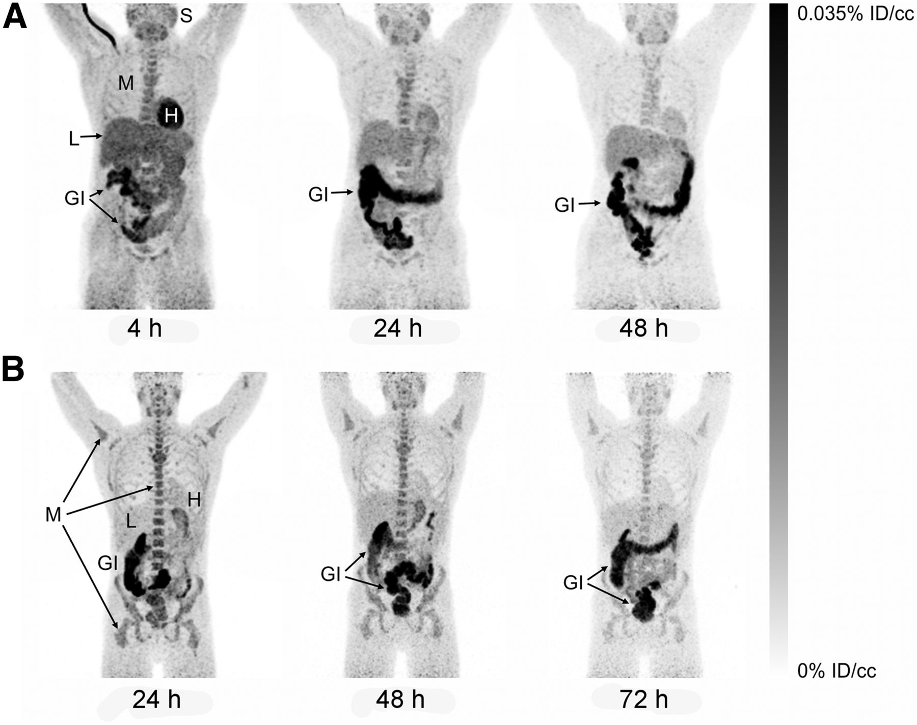
- FIGURE 1.
Maximum-intensity projections of biodistribution at 4–72 h after injection in 2 representative subjects. (A) At 4 h after injection, radiotracer uptake is primarily in heart, liver, salivary tissues, and bone marrow, and gastrointestinal clearance is readily observable. At 24 and 48 h, retention is reduced in all tissues except gastrointestinal tract. (B) At 24–72 h after injection, clearance is seen from most tissues but is slower from bone marrow and salivary tissues. However, signal in gastrointestinal tract is visible through 72 h. %ID/cc = percentage injected dose per cubic centimeter of tissue. GI = gastrointestinal tract; H = heart; L = liver; M = bone marrow; S = salivary tissues.
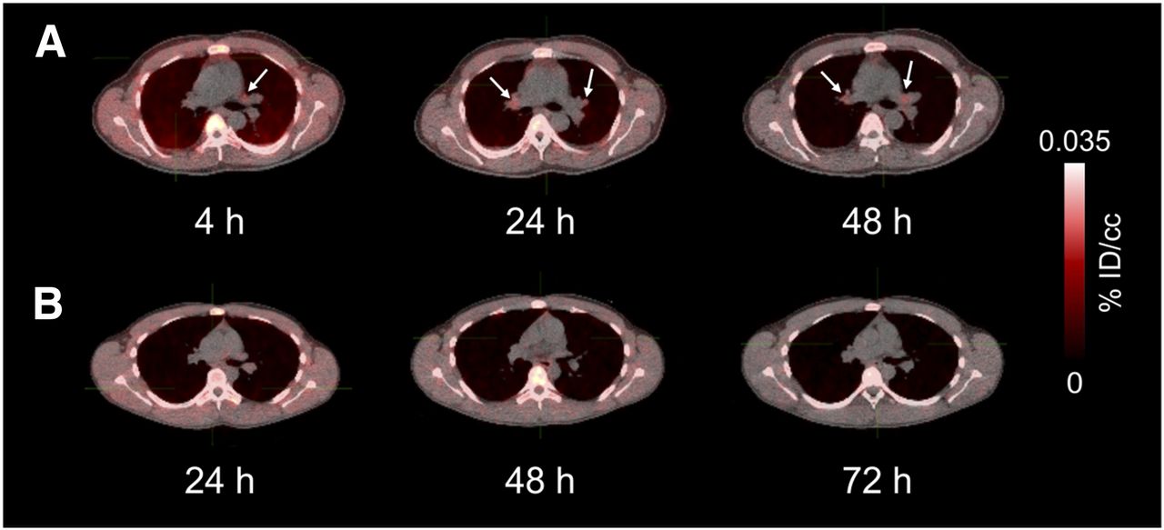
- FIGURE 2.
Biodistribution in lungs and mediastinum 4–72 h after injection as seen on transaxial PET/CT images from subjects A and B of Figure 1. Mediastinal uptake is low in A (arrows) but absent from B. %ID/cc = percentage injected dose per cubic centimeter of tissue.
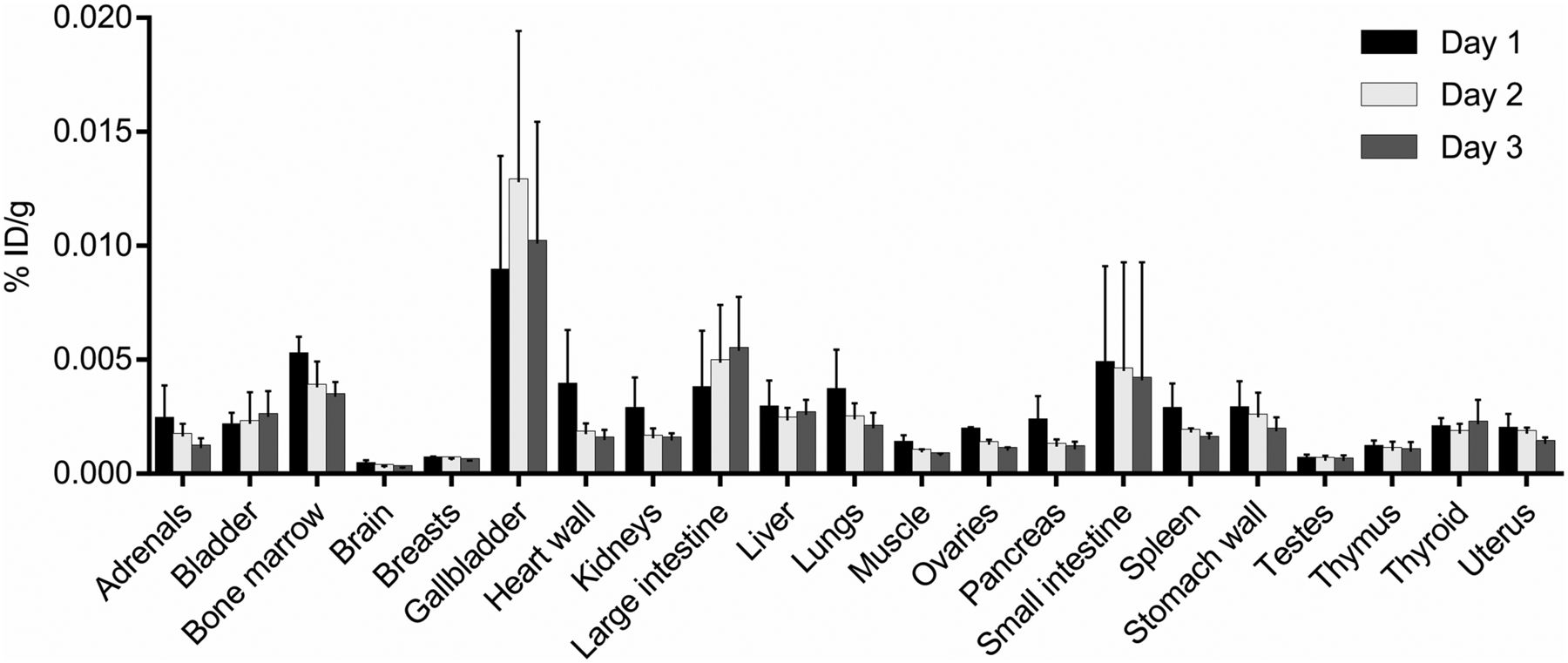
- FIGURE 3.
Mean and SD for physical decay–corrected biodistribution (biodistribution assuming no physical decay of radionuclide) on days 1, 2, and 3. %ID/g = percentage injected dose per cubic centimeter of tissue.
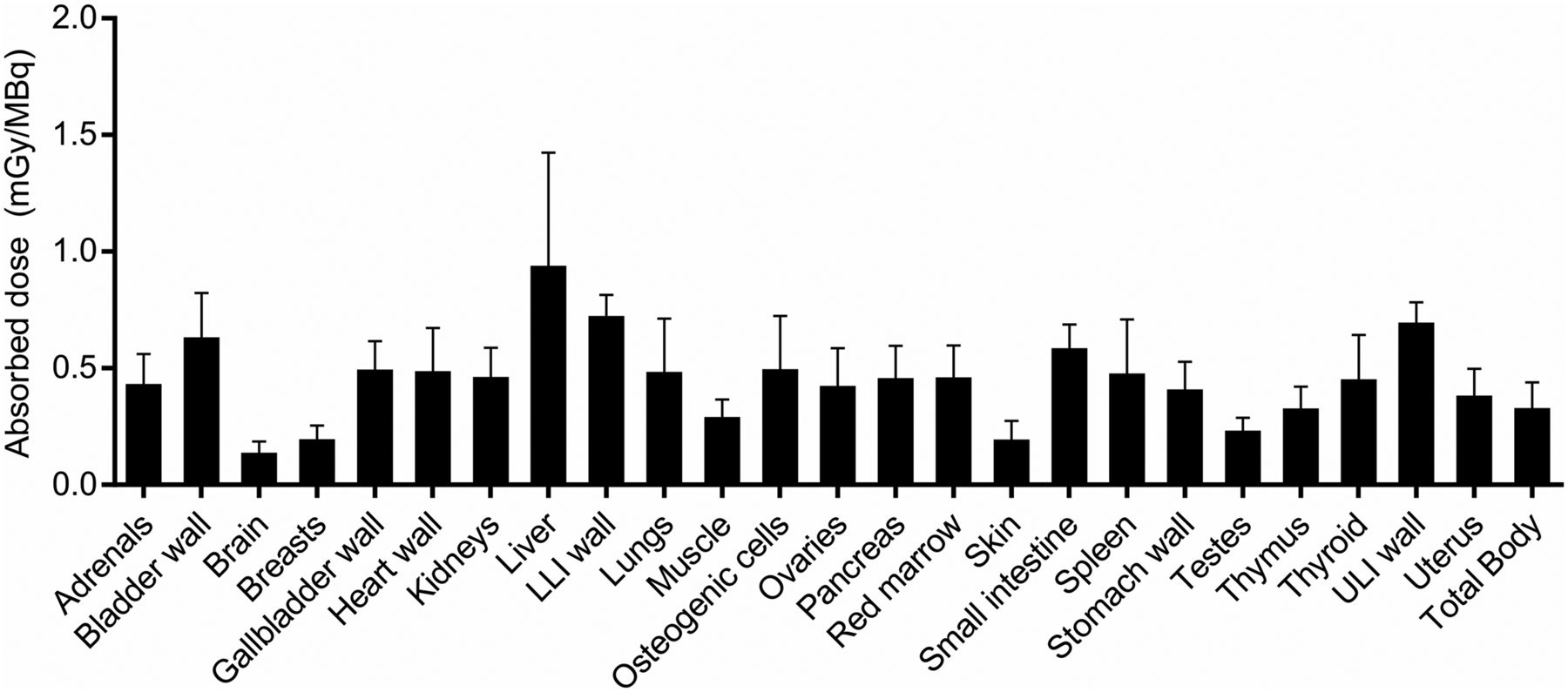
- FIGURE 4.
Mean and SD for organ-absorbed doses. LLI = lower large intestine; ULI = upper large intestine.

Tables
Subject no. Age (y) TSPO genotype Weight (kg) Height (in*) BMI Injected activity (MBq) Injected mass (ng) F1 64 C/T 106.1 62 42.8 69.2 461 F2 43 C/T 97.5 63 38.1 71.6 476 F3 22 C/C 65.8 65 24.1 78.1 520 M1 59 C/T 78.0 73 22.7 71.6 477 M2 28 C/T 76.7 71.5 23.2 69.9 466 M3 22 C/C 71.7 72 21.4 62.4 416 Mean ± SD 40 ± 19 82.6 ± 15.7 68 ± 5 28.7 ± 9.2 70.5 ± 5.1 469 ± 34 ↵* 1 in = 2.54 cm.
BMI = body mass index; C/T = medium-affinity binder; C/C = high-affinity binder.
Site TIAC (h) Brain 0.275 ± 0.136 Lungs 1.647 ± 0.981 Spleen 0.267 ± 0.170 Kidneys 0.386 ± 0.120 Gallbladder 0.073 ± 0.042 Liver 5.766 ± 3.170 Heart wall 0.423 ± 0.175 Bladder 0.738 ± 0.219 Pancreas 0.096 ± 0.041 Red marrow 3.698 ± 1.048 Adrenal glands 0.014 ± 0.008 Small intestine 1.363 ± 0.499 Breasts 0.195 ± 0.054 Testes 0.029 ± 0.015 Thyroid 0.048 ± 0.025 Large intestine 1.727 ± 0.849 Thymus 0.018 ± 0.008 Muscle 15.191 ± 4.921 Stomach wall 0.309 ± 0.212 Uterus 0.083 ± 0.019 Ovaries 0.015 ± 0.010 Whole body 59.844 ± 18.115 Remainder of body 27.676 ± 18.748 Data are mean ± SD.
Site Estimate (mGy/MBq) Adrenals 0.418 ± 0.143 Brain 0.122 ± 0.064 Breasts 0.181 ± 0.074 Gallbladder wall 0.480 ± 0.136 Lower large intestinal wall 0.709 ± 0.105 Small intestine 0.571 ± 0.117 Stomach wall 0.394 ± 0.134 Upper large intestinal wall 0.681 ± 0.102 Heart wall 0.472 ± 0.201 Kidneys 0.448 ± 0.139 Liver 0.924 ± 0.501 Lungs 0.470 ± 0.243 Muscle 0.276 ± 0.090 Ovaries 0.409 ± 0.177 Pancreas 0.442 ± 0.154 Red marrow 0.446 ± 0.151 Osteogenic cells 0.481 ± 0.243 Skin 0.179 ± 0.095 Spleen 0.463 ± 0.246 Testes 0.218 ± 0.069 Thymus 0.312 ± 0.108 Thyroid 0.438 ± 0.204 Urinary bladder wall 0.618 ± 0.204 Uterus 0.367 ± 0.131 Total body 0.315 ± 0.125 Effective dose (mSv/MBq) 0.459 ± 0.127 Data are mean ± SD.
Supplemental Data
Files in this Data Supplement:


Jump to section
Related Articles
Cited By...
- No citing articles found.