


Article Figures & Data
Figures
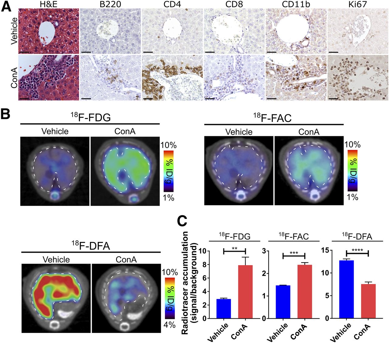
- FIGURE 1.
Hepatic 18F-FDG, 18F-FAC, and 18F-DFA accumulation is affected in mouse model of autoimmune hepatitis. (A) Histochemical and immunohistochemical analyses of liver sections from vehicle- and ConA-treated mice. Scale bars represent 50 μm. (B and C) Transverse PET/CT images (B) and quantification (C) of vehicle- and ConA-treated mice injected with 18F-FDG, 18F-FAC, and 18F-DFA. Livers are outlined. Quantification represents radiotracer accumulation in liver normalized to background organ. **P < 0.01. ***P < 0.001. ****P < 0.0001. H&E = hematoxylin and eosin; ID = injected dose.
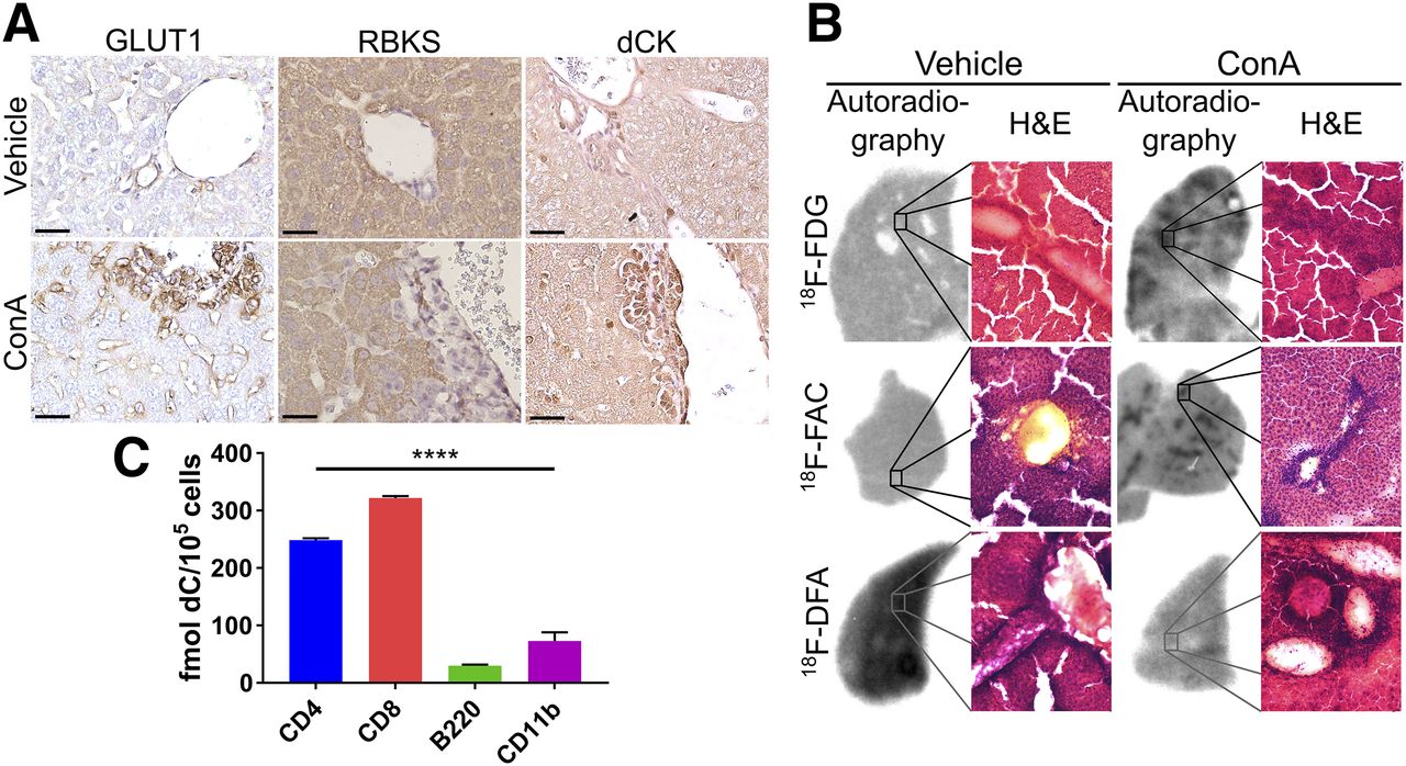
- FIGURE 2.
18F-FDG and 18F-FAC measure liver-infiltrating leukocytes, and 18F-DFA measures hepatocyte inflammation. (A) GLUT1, ribokinase, and dCK immunostained vehicle- and ConA-treated mouse liver sections. Scale bars represent 50 μm. (B) Autoradiographic and histologic analyses of liver sections from vehicle- and ConA-treated mice injected with 18F-FDG, 18F-FAC, or 18F-DFA. (C) Ex vivo accumulation of deoxycytidine in sorted leukocytes from livers of ConA-treated mice. ****P < 0.0001. H&E = hematoxylin and eosin; RBKS = ribokinase.
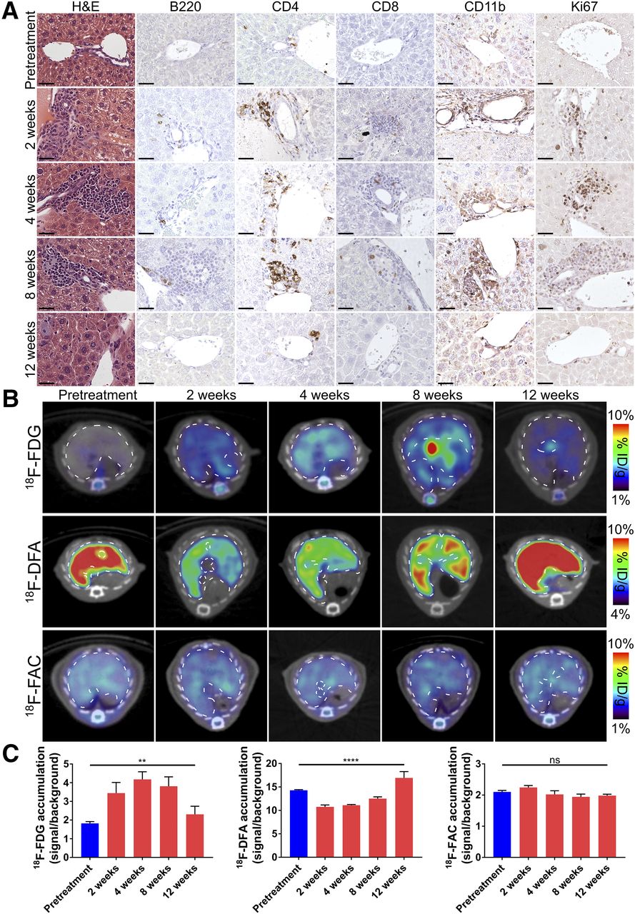
- FIGURE 3.
Hepatic accumulation of 18F-FDG and 18F-DFA but not 18F-FAC is affected in mouse model of viral hepatitis. (A) Histochemical and immunohistochemical analyses of liver sections from pretreatment and adenovirus-treated mice. Scale bars represent 50 μm. (B and C) Transverse PET/CT images (B) and quantification (C) of pretreatment and adenovirus-treated mice injected with 18F-FDG, 18F-DFA, and 18F-FAC. Quantification represents radiotracer accumulation in liver normalized to background organ. **P < 0.01. ****P < 0.0001. H&E = hematoxylin and eosin; ID = injected dose; ns = not significant.
- FIGURE 4.
Changes in hepatic 18F-FAC accumulation can be used to monitor immunosuppressive drug treatments in mouse model of autoimmune hepatitis. (A) Representative hematoxylin- and eosin-stained liver sections of mice treated with vehicle, ConA, or ConA and dexamethasone. Scale bars represent 50 μm. (B) Transverse PET/CT images of hepatic 18F-FAC accumulation in mice treated with vehicle, ConA, or ConA and Dexa. (C) Quantification of hepatic 18F-FAC accumulation in mice treated with vehicle, ConA, or ConA and Dexa. Quantification represents radiotracer accumulation in liver normalized to background organ. ****P < 0.0001. Dexa = dexamethasone; H&E = hematoxylin and eosin; ID = injected dose; ns = not significant.
- FIGURE 5.
Liver-infiltrating leukocytes in patients with autoimmune hepatitis express dCK. Histochemical and immunohistochemical analyses of liver biopsies from patients with autoimmune hepatitis. Scale bars represent 50 μm. H&E = hematoxylin and eosin.


Additional Files
Supplemental Data
Files in this Data Supplement:


