


Abstract
739
Objectives: While 68Ga-PSMA PET-MRI might be superior to PET-CT with regard to soft tissue assessment in prostate cancer evaluation, it is also known to potentially introduce additional PET image artefacts around high-activity structures ("halo artefacts") in current PET-MRI systems. We therefore investigated the impact of PET acquisition duration, attenuation data and scatter correction on halo artefact occurrence in current clinical whole-body PET/MRI.
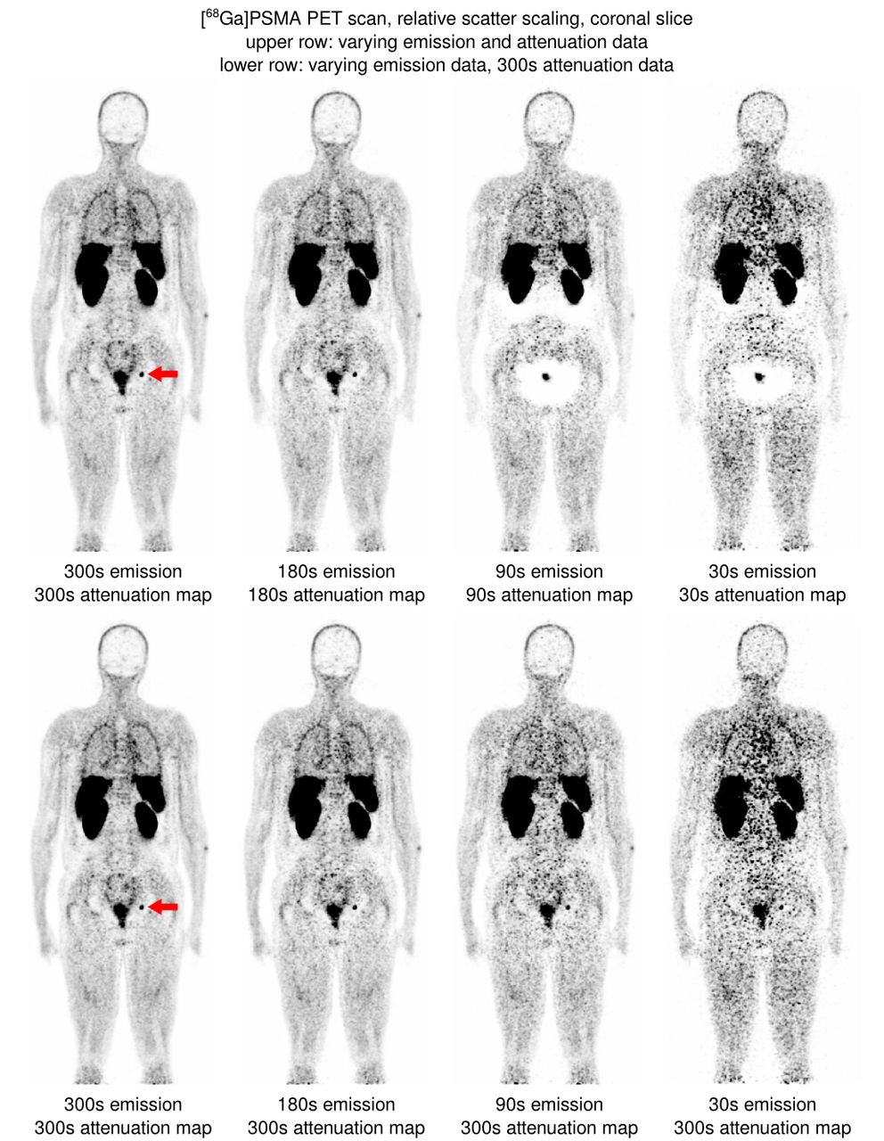
Methods: Whole-body PET list mode data from twelve patients with prostate cancer were acquired one hour after injection of 2 MBq/kg [68Ga]HBED-CC-PSMA on a hybrid PET-MRI system (biograph mMR, Siemens Healthineers, with arms lying next to the body). List mode data were transformed into data sets representing 300s, 180s, 90s and 30s acquisition duration per bed position and were reconstructed without and with scatter correction using both relative and absolute scatter scaling, and additionally a correction for prompt gamma photons. MRI-derived attenuation maps were complemented by emission-based data in areas not covered by MRI (MLAA algorithm). A total of 264 image data sets representing different acquisition durations and attenuation/scatter/prompt gamma corrections were thus reconstructed. PET images were analysed regarding halo artefact occurrence according to the following scoring: 0: no distinct halo visible, 1: halo around either kidneys or bladder, 2: halo around both kidneys and bladder. Attenuation maps were analysed for successful MLAA-based arm completion.
Results: Halo artefacts were not present in non-scatter-corrected images. Decreasing PET acquisition durations resulted in significantly increased incidences of halo artefacts around kidneys and bladder for both relative scatter scaling and prompt gamma correction. Additionally, decreased lesion detectability and markedly lower arm attenuation values were seen. For relative scatter scaling, mean halo scores (± standard deviation) amounted to 0.50 ± 0.67, 0.67 ± 0.78, 1.67 ± 0.78, and 1.67 ± 0.67 for acquisition durations of 300s, 180s, 90s, 30s, respectively, proving a strong dependence of halo occurrences with scanning durations. Prompt gamma correction barely improved this to 0.50 ± 0.67, 0.67 ± 0.78, 1.67 ± 0.78, and 1.25 ± 0.62, respectively, although the areas affected by halos generally seemed smaller than without correction. Absolute scaling resulted in values of 1.42 ± 0.51, 1.42 ± 0.51, 1.42 ± 0.51, and 1.25 ± 0.45, respectively, demonstrating nearly duration-independent artefact occurrences. In case of relative scaling, emission data reconstructed with the 300s attenuation maps restored halo scores to the native 300s level for durations of 180s, 90s, and 30s, while reconstructing 300s emission data with attenuation data of 180s, 90s, and 30s led to halo scores of 0.67 ± 0.78, 1.67 ± 0.78, and 1.67 ± 0.67, respectively. Attenuation maps at 90s and 30s demonstrated markedly lower mean arm attenuation values (0.002 cm-1 at pelvic regions) than those at 300s (0.084 cm-1) and 180s (0.062 cm-1). Additionally, the mean volume of completed arms dropped in the pelvic bed position from 4.6 ± 1.2 L to 4.1 ± 1.3 L, 1.7 ± 0.7 L, and 2.1 ± 0.4 L for 300s, 180s, 90s, and 30s, respectively.
Conclusion: Short acquisition durations of less than 3 minutes per bed position result in unacceptable image artefacts in current clinical protocols of whole-body 68Ga-PSMA PET-MRI when using relative scatter scaling and should be avoided. Imperfections in MLAA-generated attenuation maps seem to be a paramount cause. On the other hand, neither prompt gamma correction nor absolute scaling obviate halo artefacts in the images, highlighting the need for more accurate scatter and attenuation correction methods for 68Ga-PSMA PET-MRI. Research Support: None

In this issue


Jump to section
Related Articles
Cited By...
- No citing articles found.