


Article Figures & Data
Figures
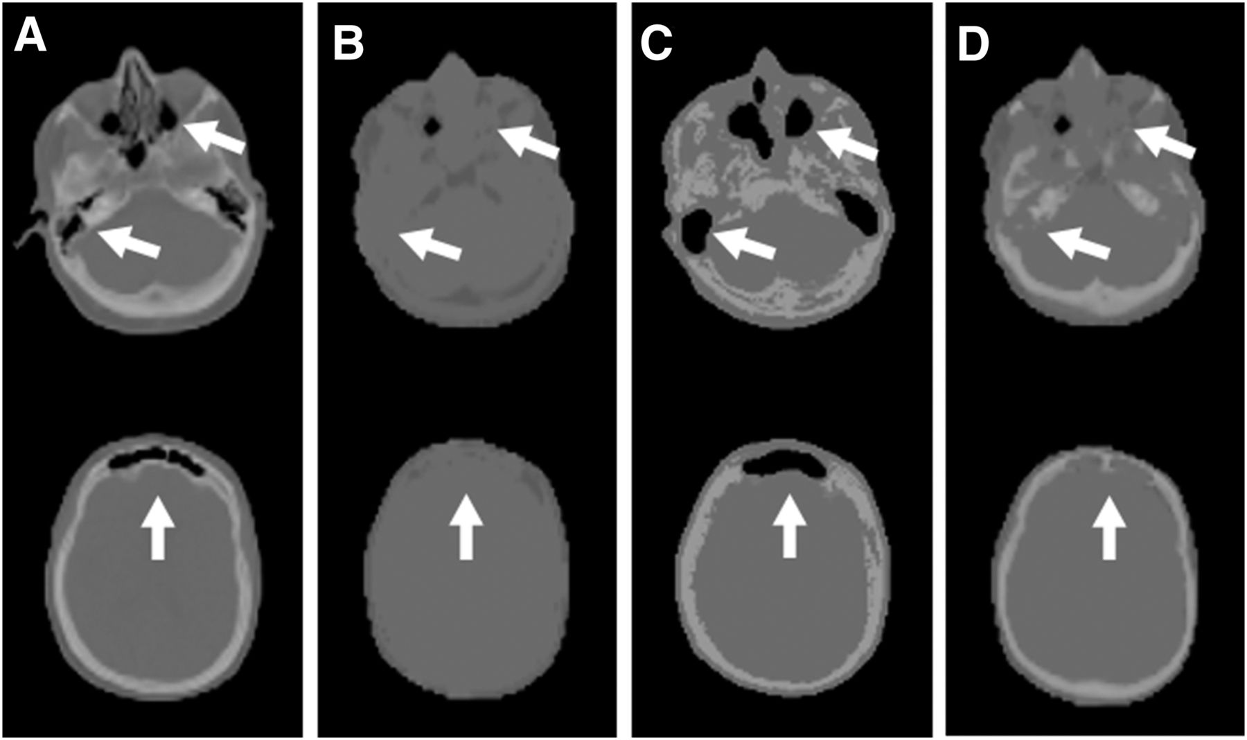
- FIGURE 1.
Example of artifact categories. Axial views at 2 main axial levels of same patient and different AC methods: CT—reference (A), DIXON—artificial filling of air cavities with tissue (B), UTE—overestimation of air cavities due to susceptibility artifacts (C), and BD-based MR-AC—translation of artifacts found in DIXON (D). Arrows indicate artifacts.
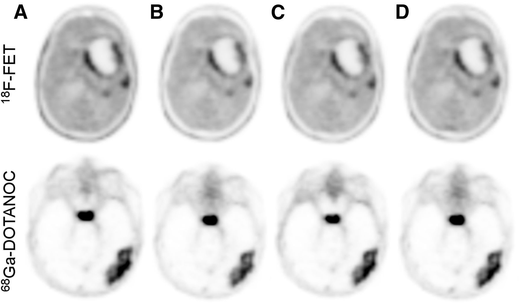
- FIGURE 2.
Axial views of patient with tumor recurrence imaged with 18F-FET (top) and patient with a meningeal tumor formation imaged with 68Ga-DOTANOC (bottom). PET images were reconstructed after AC using: CTref—reference (A), DIXON (B), UTE (C), and BD (D).
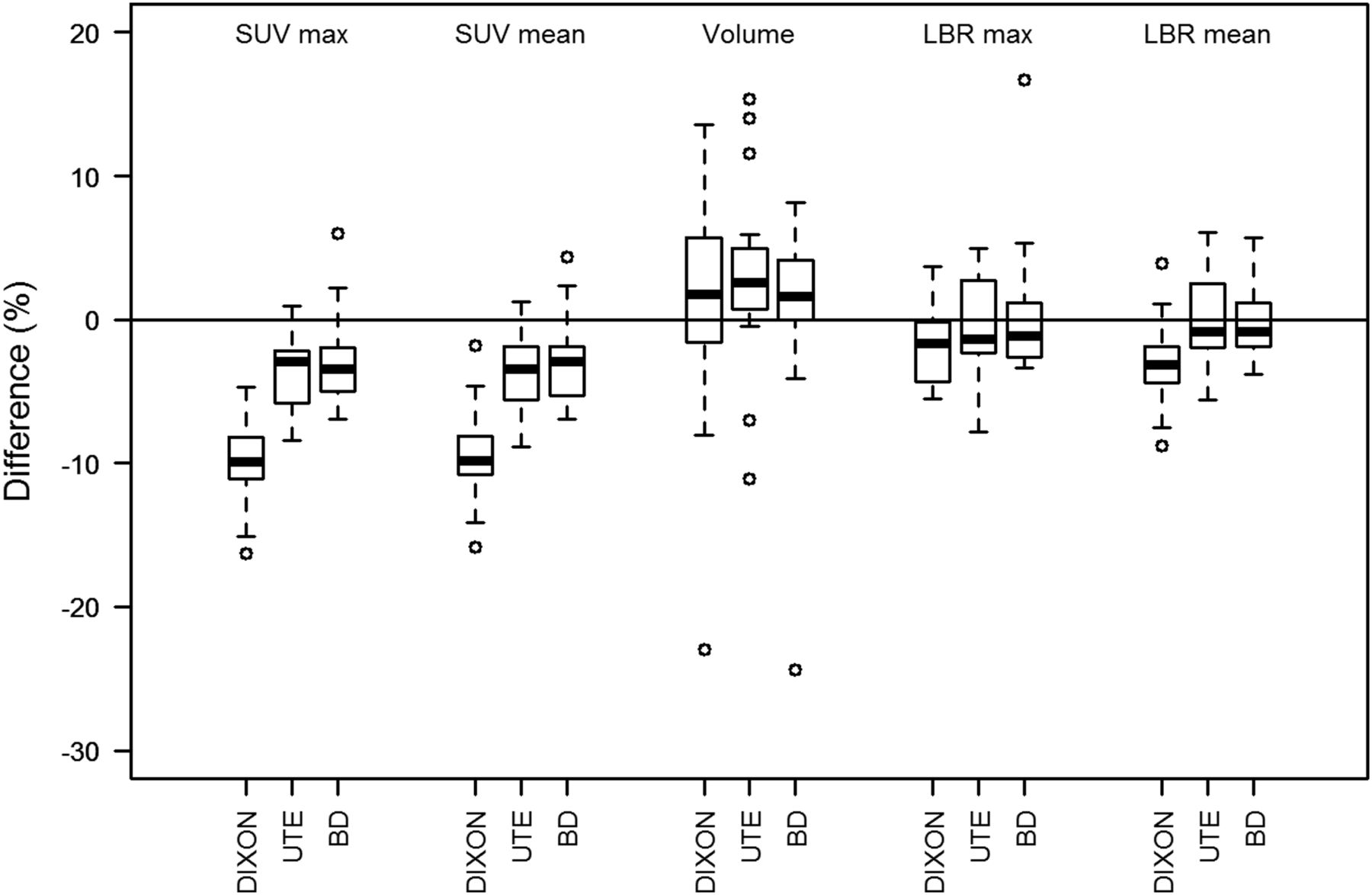
- FIGURE 3.
Box plots of RD of SUVmax and SUVmean, VOI70 (volume), and LBRs of SUVmax (LBRmax) and SUVmean (LBRmean) according to CTref for 18F-FET–avid lesions and 3 different AC methods.
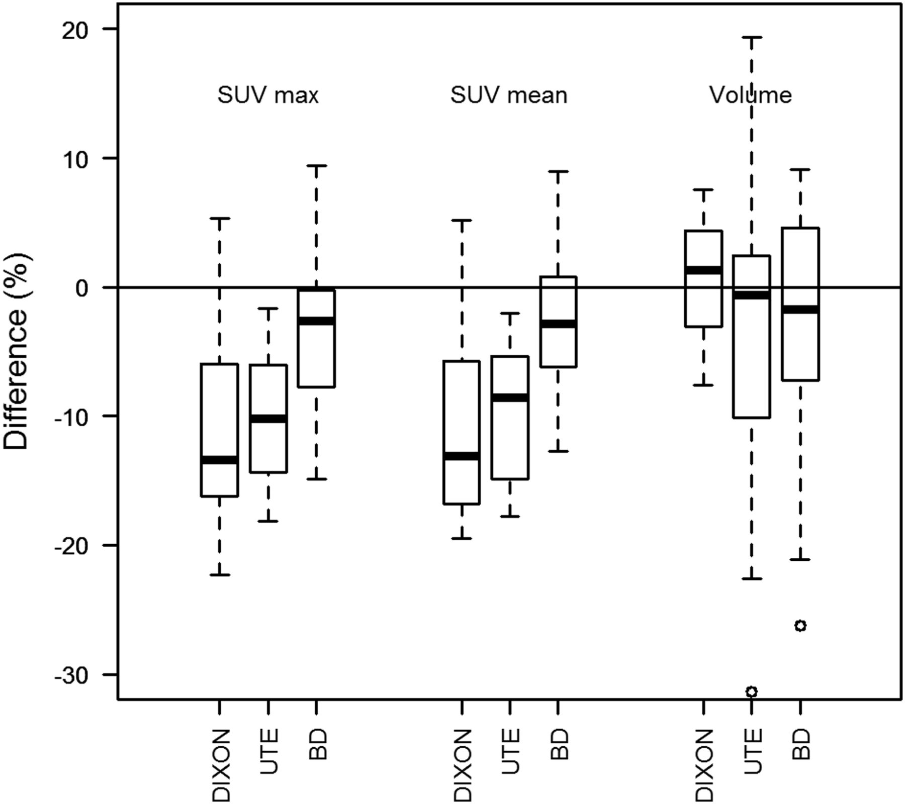
- FIGURE 4.
Box plots of RD of SUVmax and SUVmean and VOI50 (volume) according to CTref for lesions in 68Ga-DOTANOC scans and 3 different AC methods. Data included 11 meningeal tumor formations and for 5 patients additionally pituitary gland (in 2 patients, pituitary gland could not be separated from tumor formation).
- FIGURE 5.
Box plots of RD of SUVmax and SUVmean and VOI50 (volume) according to CTref for evaluated lesions in 68Ga-DOTANOC scans and 3 different AC methods. (A) Findings for lesions and pituitary gland in skull base (n = 9). (B) Results for lesions attached to skull cap (n = 7).
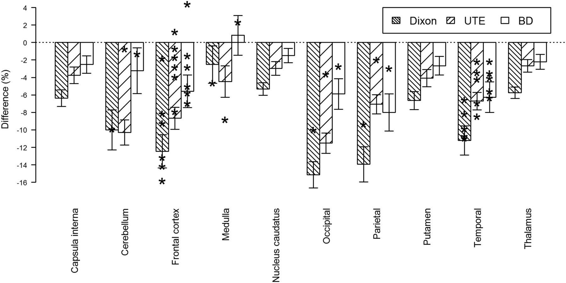
- FIGURE 6.
Average RD of 10 reference regions within brain for 18F-FDG scans of healthy subjects. Error bars correspond to respective RD SD for each reference region. *Corresponding RDs of SUVmean for VOI70 of 18F-FET–avid lesions. Lesions were assigned to reference regions used in 18F-FDG evaluation by experienced nuclear medicine expert.


Tables
- TABLE 1
Mean RDs in Percentage (±SD) of All Investigated Values for 18F-FET and 68Ga-DOTANOC Scans
18F-FET 68Ga-DOTANOC Parameter Dixon UTE BD Dixon UTE BD SUVmax −10 ± 3 −4 ± 3 −3 ± 3 −11 ± 8 −11 ± 8 −3 ± 6 SUVmean −10 ± 4 −4 ± 3 −3 ± 3 −11 ± 7 −11 ± 8 −3 ± 5 Volume +1 ± 9 +2 ± 8 +7 ± 28* +1 ± 4 −4 ± 12 −3 ± 10 LBRmax −2 ± 3 0 ± 3 0 ± 5 — — — LBRmean −3 ± 3 0 ± 3 0 ± 2 — — — ↵* Includes 1 patient with a volume difference of +103%.
LBRmax = LBRs of SUVmax; LBRmean = LBRs of SUVmean.
Images of whole brain, segmented by threshold-based segmentation, were excluded from calculations.
Supplemental Data
Files in this Data Supplement:


