


Article Figures & Data
Figures
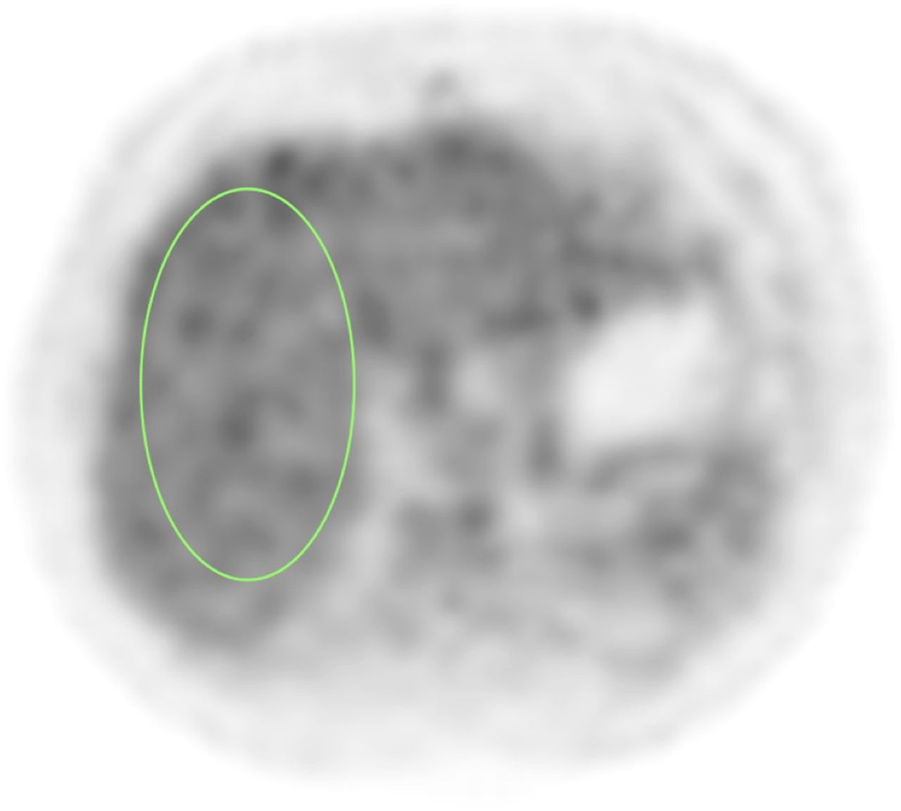
- FIGURE 1.
Image of 1 of 7 adjacent ROIs in liver regions used in body test cases.
- FIGURE 2.
(Left) Typical ROI used for uniformity analysis. (Right) Example of typical plot of mean SUVs per plane in ROI and calculation of maximum (max.) axial deviation (dev.) (MAD).
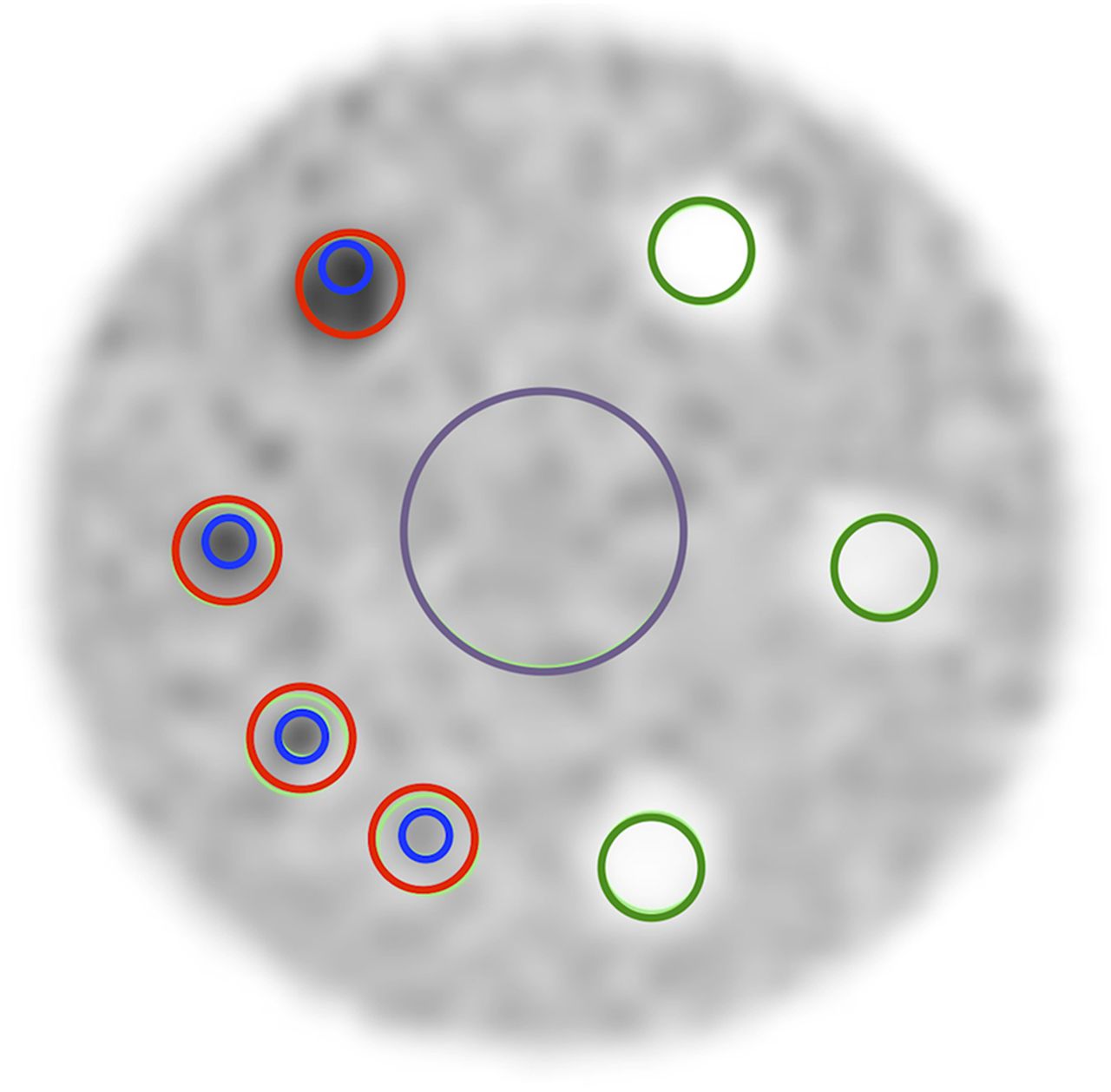
- FIGURE 3.
Image of ACR-approved PET phantom. Red ROIs are used for SUVmax, and blue ROIs are used for SUVpeak. Also shown are large background ROI (purple) and smaller green ROIs for cold cylinders. Latter ROIs were not used in this analysis.
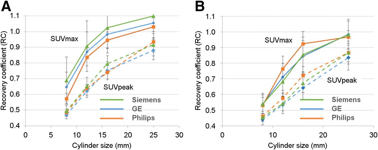
- FIGURE 4.
Recovery coefficients (defined as ratio of measured SUV to true SUV) as function of cylinder diameters for static brain (A) and body (B) acquisitions.


Tables
Manufacturer No. of cases Liver SUV SD GE 62 2.11 0.44 Philips 14 2.06 0.43 Siemens 42 2.42 0.51 T0 T1 T2 Parameter No. Percentage No. Percentage No. Percentage No. of scanners passing first time 25 38 34 87 35 67 No. of scanners passing eventually 39 60 5 13 13 25 Total no. of scanners passing 64 98 39 100 48 92 Total no. of scanners 65 39 52 No. of scanners during: Issue T0 period (65 scanners) T2 period (44 scanners)† Uniformity problem 7 0 SUV outside specifications 14 3 Phantom filling issue 4 3 Reconstruction problem 6 5 Improper acquisition 3 0 Incomplete submission 11 2 Problem with forms 5 1 Total 50 14


