
Main menu

  • Home
  • Content
    • Current
    • Ahead of print
    • Past Issues
    • JNM Supplement
    • SNMMI Annual Meeting Abstracts
    • Continuing Education
    • JNM Podcasts
  • Subscriptions
    • Subscribers
    • Institutional and Non-member
    • Rates
    • Journal Claims
    • Corporate & Special Sales
  • Authors
    • Submit to JNM
    • Information for Authors
    • Assignment of Copyright
    • AQARA requirements
  • Info
    • Reviewers
    • Permissions
    • Advertisers
  • About
    • About Us
    • Editorial Board
    • Contact Information
  • More
    • Alerts
    • Feedback
    • Help
    • SNMMI Journals
  • SNMMI
    • JNM
    • JNMT
    • SNMMI Journals
    • SNMMI

User menu

  • Subscribe
  • My alerts
  • Log in
  • My Cart

Search

  • Advanced search
Journal of Nuclear Medicine
  • SNMMI
    • JNM
    • JNMT
    • SNMMI Journals
    • SNMMI
  • Subscribe
  • My alerts
  • Log in
  • My Cart
Journal of Nuclear Medicine

Advanced Search

  • Home
  • Content
    • Current
    • Ahead of print
    • Past Issues
    • JNM Supplement
    • SNMMI Annual Meeting Abstracts
    • Continuing Education
    • JNM Podcasts
  • Subscriptions
    • Subscribers
    • Institutional and Non-member
    • Rates
    • Journal Claims
    • Corporate & Special Sales
  • Authors
    • Submit to JNM
    • Information for Authors
    • Assignment of Copyright
    • AQARA requirements
  • Info
    • Reviewers
    • Permissions
    • Advertisers
  • About
    • About Us
    • Editorial Board
    • Contact Information
  • More
    • Alerts
    • Feedback
    • Help
    • SNMMI Journals
  • View or Listen to JNM Podcast
  • Visit JNM on Facebook
  • Join JNM on LinkedIn
  • Follow JNM on Twitter
  • Subscribe to our RSS feeds
Research ArticleOncology

Rapid Imaging of Tumor Cell Death In Vivo Using the C2A Domain of Synaptotagmin-I

André A. Neves, Bangwen Xie, Sarah Fawcett, Israt S. Alam, Timothy H. Witney, Maaike M. de Backer, Julia Summers, William Hughes, Sarah McGuire, Dmitry Soloviev, Jodi Miller, William J. Howat, De-en Hu, Tiago B. Rodrigues, David Y. Lewis and Kevin M. Brindle
Journal of Nuclear Medicine June 2017, 58 (6) 881-887; DOI: https://doi.org/10.2967/jnumed.116.183004
André A. Neves
1Cancer Research United Kingdom Cambridge Institute, Li Ka Shing Centre, Cambridge, United Kingdom; and
  • Find this author on Google Scholar
  • Find this author on PubMed
  • Search for this author on this site
Bangwen Xie
1Cancer Research United Kingdom Cambridge Institute, Li Ka Shing Centre, Cambridge, United Kingdom; and
  • Find this author on Google Scholar
  • Find this author on PubMed
  • Search for this author on this site
Sarah Fawcett
1Cancer Research United Kingdom Cambridge Institute, Li Ka Shing Centre, Cambridge, United Kingdom; and
  • Find this author on Google Scholar
  • Find this author on PubMed
  • Search for this author on this site
Israt S. Alam
1Cancer Research United Kingdom Cambridge Institute, Li Ka Shing Centre, Cambridge, United Kingdom; and
  • Find this author on Google Scholar
  • Find this author on PubMed
  • Search for this author on this site
Timothy H. Witney
2Department of Biochemistry, University of Cambridge, Cambridge, United Kingdom
  • Find this author on Google Scholar
  • Find this author on PubMed
  • Search for this author on this site
Maaike M. de Backer
2Department of Biochemistry, University of Cambridge, Cambridge, United Kingdom
  • Find this author on Google Scholar
  • Find this author on PubMed
  • Search for this author on this site
Julia Summers
1Cancer Research United Kingdom Cambridge Institute, Li Ka Shing Centre, Cambridge, United Kingdom; and
  • Find this author on Google Scholar
  • Find this author on PubMed
  • Search for this author on this site
William Hughes
1Cancer Research United Kingdom Cambridge Institute, Li Ka Shing Centre, Cambridge, United Kingdom; and
  • Find this author on Google Scholar
  • Find this author on PubMed
  • Search for this author on this site
Sarah McGuire
1Cancer Research United Kingdom Cambridge Institute, Li Ka Shing Centre, Cambridge, United Kingdom; and
  • Find this author on Google Scholar
  • Find this author on PubMed
  • Search for this author on this site
Dmitry Soloviev
1Cancer Research United Kingdom Cambridge Institute, Li Ka Shing Centre, Cambridge, United Kingdom; and
  • Find this author on Google Scholar
  • Find this author on PubMed
  • Search for this author on this site
Jodi Miller
1Cancer Research United Kingdom Cambridge Institute, Li Ka Shing Centre, Cambridge, United Kingdom; and
  • Find this author on Google Scholar
  • Find this author on PubMed
  • Search for this author on this site
William J. Howat
1Cancer Research United Kingdom Cambridge Institute, Li Ka Shing Centre, Cambridge, United Kingdom; and
  • Find this author on Google Scholar
  • Find this author on PubMed
  • Search for this author on this site
De-en Hu
1Cancer Research United Kingdom Cambridge Institute, Li Ka Shing Centre, Cambridge, United Kingdom; and
  • Find this author on Google Scholar
  • Find this author on PubMed
  • Search for this author on this site
Tiago B. Rodrigues
1Cancer Research United Kingdom Cambridge Institute, Li Ka Shing Centre, Cambridge, United Kingdom; and
  • Find this author on Google Scholar
  • Find this author on PubMed
  • Search for this author on this site
David Y. Lewis
1Cancer Research United Kingdom Cambridge Institute, Li Ka Shing Centre, Cambridge, United Kingdom; and
  • Find this author on Google Scholar
  • Find this author on PubMed
  • Search for this author on this site
Kevin M. Brindle
1Cancer Research United Kingdom Cambridge Institute, Li Ka Shing Centre, Cambridge, United Kingdom; and
2Department of Biochemistry, University of Cambridge, Cambridge, United Kingdom
  • Find this author on Google Scholar
  • Find this author on PubMed
  • Search for this author on this site
  • Article
  • Figures & Data
  • Supplemental
  • Info & Metrics
  • PDF
Loading

Article Figures & Data

Figures

  • Tables
  • Additional Files
  • FIGURE 1.
    • Download figure
    • Open in new tab
    • Download powerpoint
    FIGURE 1.

    Near-infrared fluorescence imaging of cell death. Imaging in vivo of untreated and treated EL4 (A), Colo-205 (B), and Eμ-myc (C) tumors. Images are overlays of bright-field images and 800 nm-channel fluorescence signals, acquired 24 h after C2Am-AF750 administration. AnxV = annexin-V.
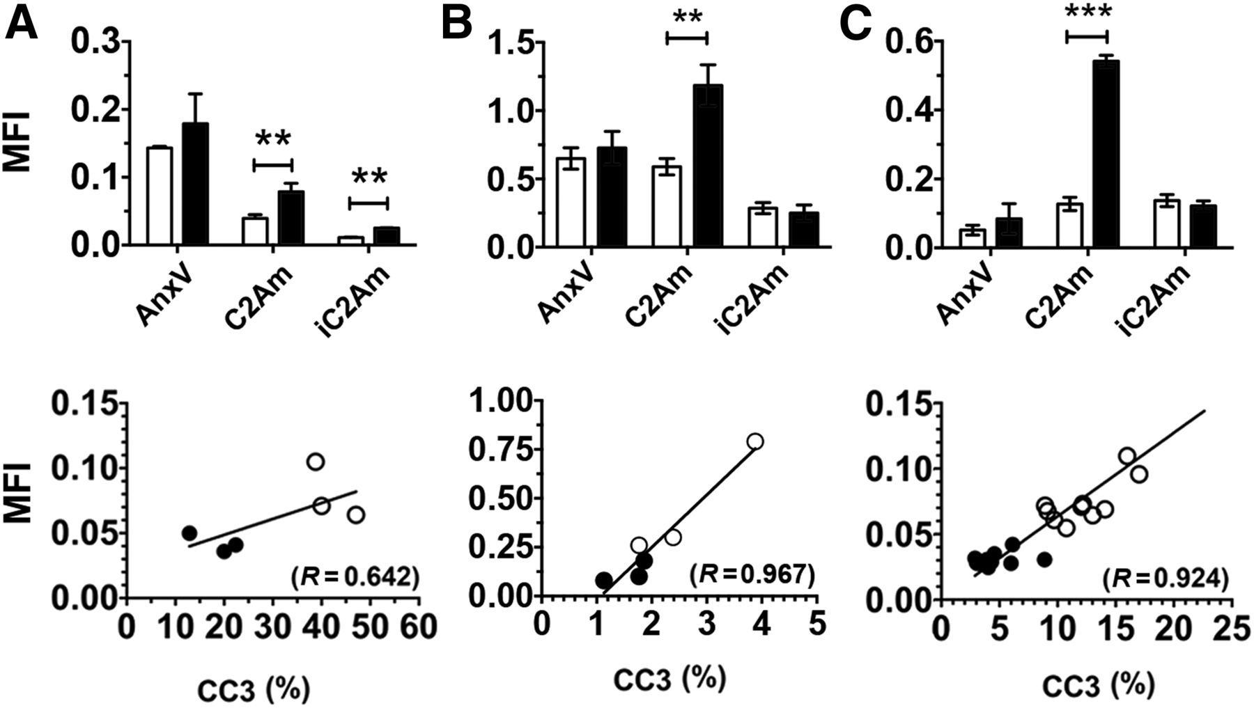
  • FIGURE 2.
    • Download figure
    • Open in new tab
    • Download powerpoint
    FIGURE 2.

    Quantification of tumor mean fluorescence intensity (MFI) for untreated (open bars) and treated (filled bars) EL4 (A), Colo-205 (B), and Eμ-myc (C) tumors. Data for Eμ-myc model was acquired ex vivo, due to skin pigmentation artifacts. (Bottom) Correlation of C2Am whole-tumor MFIs with corresponding CC3 staining, measured in sections of excised EL4 (A), Colo-205 (B), and Eμ-myc (C) tumors. ○ = drug-treated (48 h after treatment); ● = untreated. **P < 0.01, ***P < 0.001, n = 3/group, values are mean ± SD. AnxV = annexin-V.
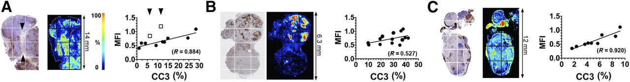
  • FIGURE 3.
    • Download figure
    • Open in new tab
    • Download powerpoint
    FIGURE 3.

    Maps of CC3 staining (left), C2Am-AF750 fluorescence (middle), and correlation (right) of fluorescence intensities of regions of interest (grids indicated on the left), with staining for CC3 in same regions of interest. Tumors were excised 24 h after C2Am-AF750 administration and 48 h after drug treatment. The correlation coefficients (R) of the linear fits to the data (A–C, right) are shown. Arrows in A (left) indicate decellularized regions of tissue. Sections are from EL4 (A), Colo-205 (B), and Eμ-myc (C) tumors. MFI = mean fluorescence intensity.
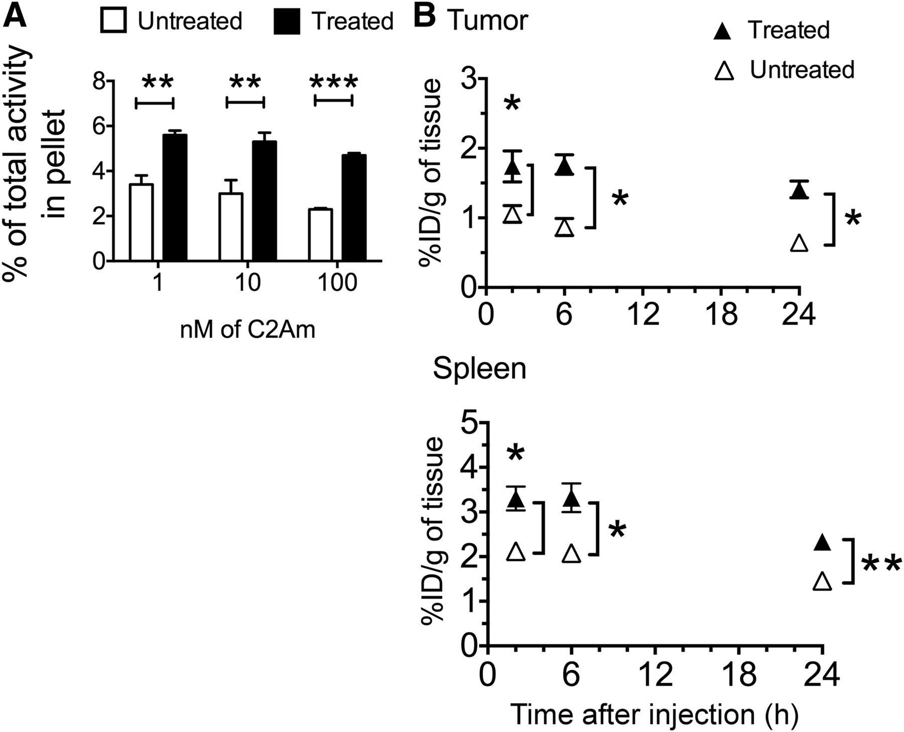
  • FIGURE 4.
    • Download figure
    • Open in new tab
    • Download powerpoint
    FIGURE 4.

    (A) Binding of 99mTc-C2Am to EL4 cells. Labeling of drug-treated and untreated cells is expressed as percentage of total 99mTc activity retained by cell pellets. **P < 0.01, ***P < 0.001, n = 3/group, values are mean ± SD. (B) Retention of 99mTc-C2Am in tumors (top) and spleens (bottom) from EL4 tumor–bearing mice, in untreated (△) and drug-treated (▲) animals, at indicated times after probe administration. Tissue retention is expressed as percentage injected dose per gram (%ID/g) of tissue. *P < 0.05, **P < 0.01, n = 3/group, 2-way ANOVA, with Bonferroni posttest correction, was used for group comparisons. Values are mean ± SD.
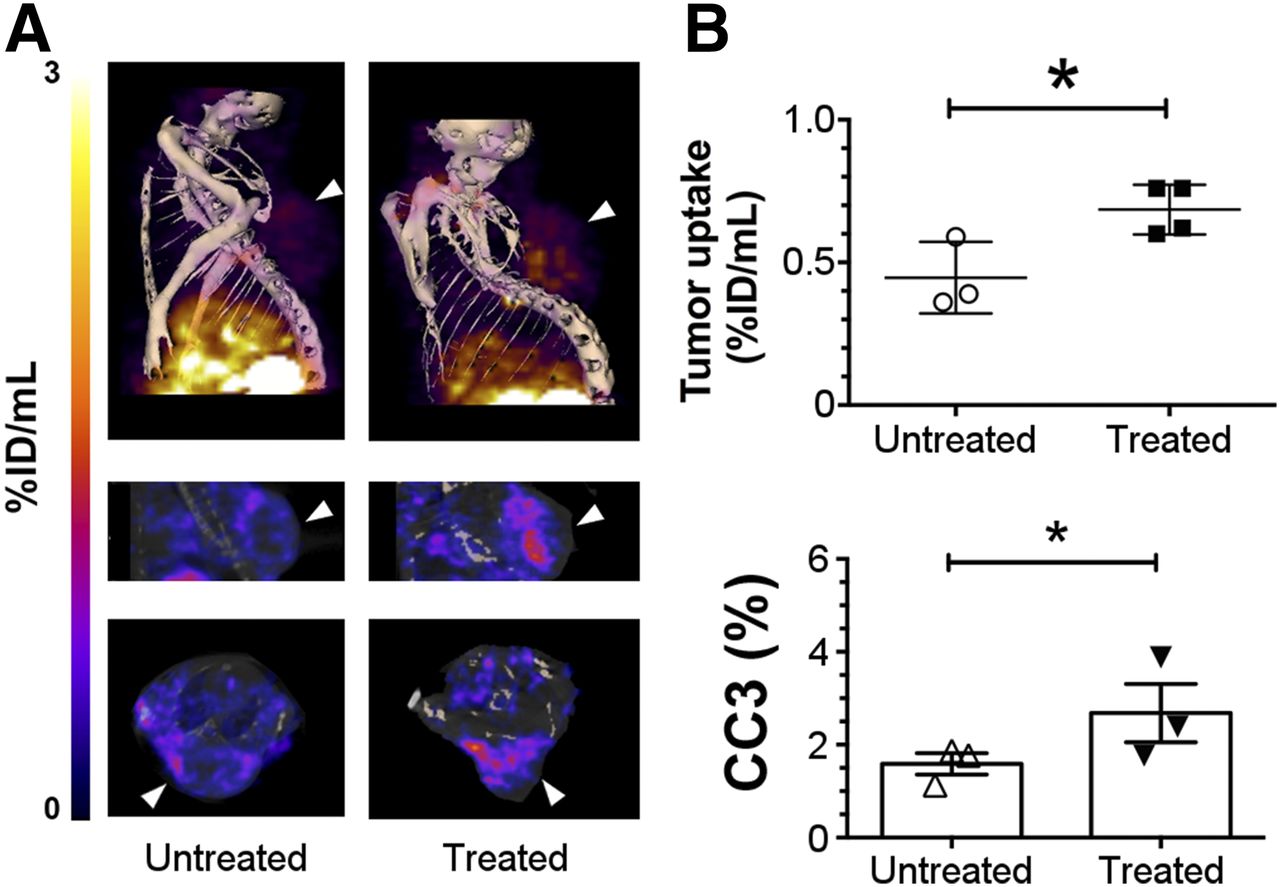
  • FIGURE 5.
    • Download figure
    • Open in new tab
    • Download powerpoint
    FIGURE 5.

    SPECT imaging of cell death in vivo in Colo-205 tumors. Imaging of 111In-labeled C2Am was performed 2 h after probe administration and 24 h after drug treatment. (A) SPECT/CT fusion images of representative untreated Colo-205 tumor–bearing mouse (A, left) and 5-FU–treated animal (A, right), 2 h after administration of 111In-C2Am. Tumor location is indicated by arrowheads. (B) Tumor retention (percentage injected dose [%ID]/mL) (upper) and CC3 staining (lower) in untreated and 5-FU–treated tumors. *P < 0.05, n = 3−4 tumors/group.
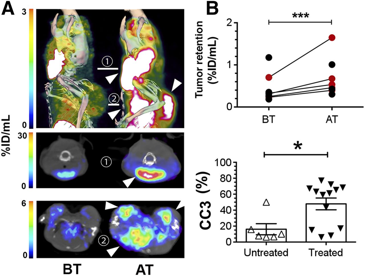
  • FIGURE 6.
    • Download figure
    • Open in new tab
    • Download powerpoint
    FIGURE 6.

    SPECT imaging of cell death in vivo in Eμ-myc tumors. Imaging of 99mTc-labeled C2Am was performed 2 h after probe administration, and 24 h after drug treatment. (A) SPECT/CT fusion images of representative Eμ-myc mice before (left) and after (right) cyclophosphamide treatment. Tumors in neck, axillary region, and chest cavity were visible (arrowheads, top) and in axial sections across cervical (1, middle) and axillary (2, bottom) planes. Red circles in B correspond to percentage injected dose [%ID]/mL retention values for tumors of animals shown in A. CC3 staining (B, bottom) in untreated and drug-treated tumors. *P < 0.05, ***P < 0.0001, n = 6−7 tumors/group (A); n = 6−13 tumors/group (B). AT = after treatment; BT = before treatment. Supplemental materials provide statistical analysis. Three-dimensional rendering of SPECT data are shown in Supplemental Video 1.

Tables

  • Figures
  • Additional Files
    • View popup
    TABLE 1

    Biodistribution of 99mTc-C2Am in Tumor-Bearing EL4 Mice and 111In-C2Am in Tumor-Bearing Colo-205 Mice, 24 Hours After Etoposide and 5-FU Treatment, Respectively

    2 h24 h
    Tissue (%ID/g)MeanSDMeanSD
    Tumor-bearing EL4 mice
     Muscle0.390.050.190.02
     Blood1.020.080.200.01
     Tumor1.740.391.410.21
     Spleen3.300.472.340.29
     Liver6.170.985.140.90
     Kidney194.245.9157.457.0
     Tumor-to-blood1.710.406.961.08
     Tumor-to-muscle4.51.157.31.40
    Tumor-bearing Colo-205 mice
     Muscle0.320.070.180.02
     Blood0.500.220.130.02
     Tumor0.690.090.720.10
     Spleen0.920.171.180.20
     Liver2.010.342.170.53
     Kidney3101227522
     Tumor-to-blood1.380.655.430.96
     Tumor-to-muscle2.160.484.090.88
    • %ID/g = percentage injected dose per gram.

    • n = 3/group.

Additional Files

  • Figures
  • Tables
  • Supplemental Data

    Files in this Data Supplement:

    • Supplemental Data
PreviousNext
Back to top

In this issue

Journal of Nuclear Medicine: 58 (6)
Journal of Nuclear Medicine
Vol. 58, Issue 6
June 1, 2017
  • Table of Contents
  • Table of Contents (PDF)
  • About the Cover
  • Index by author
Print
Download PDF
Article Alerts
Sign In to Email Alerts with your Email Address
Email Article

Thank you for your interest in spreading the word on Journal of Nuclear Medicine.

NOTE: We only request your email address so that the person you are recommending the page to knows that you wanted them to see it, and that it is not junk mail. We do not capture any email address.

Enter multiple addresses on separate lines or separate them with commas.
Rapid Imaging of Tumor Cell Death In Vivo Using the C2A Domain of Synaptotagmin-I
(Your Name) has sent you a message from Journal of Nuclear Medicine
(Your Name) thought you would like to see the Journal of Nuclear Medicine web site.
Citation Tools
Rapid Imaging of Tumor Cell Death In Vivo Using the C2A Domain of Synaptotagmin-I
André A. Neves, Bangwen Xie, Sarah Fawcett, Israt S. Alam, Timothy H. Witney, Maaike M. de Backer, Julia Summers, William Hughes, Sarah McGuire, Dmitry Soloviev, Jodi Miller, William J. Howat, De-en Hu, Tiago B. Rodrigues, David Y. Lewis, Kevin M. Brindle
Journal of Nuclear Medicine Jun 2017, 58 (6) 881-887; DOI: 10.2967/jnumed.116.183004

Citation Manager Formats

  • BibTeX
  • Bookends
  • EasyBib
  • EndNote (tagged)
  • EndNote 8 (xml)
  • Medlars
  • Mendeley
  • Papers
  • RefWorks Tagged
  • Ref Manager
  • RIS
  • Zotero
Share
Rapid Imaging of Tumor Cell Death In Vivo Using the C2A Domain of Synaptotagmin-I
André A. Neves, Bangwen Xie, Sarah Fawcett, Israt S. Alam, Timothy H. Witney, Maaike M. de Backer, Julia Summers, William Hughes, Sarah McGuire, Dmitry Soloviev, Jodi Miller, William J. Howat, De-en Hu, Tiago B. Rodrigues, David Y. Lewis, Kevin M. Brindle
Journal of Nuclear Medicine Jun 2017, 58 (6) 881-887; DOI: 10.2967/jnumed.116.183004
Twitter logo Facebook logo LinkedIn logo Mendeley logo
  • Tweet Widget
  • Facebook Like
  • Google Plus One
Bookmark this article

Jump to section

  • Article
    • Abstract
    • MATERIALS AND METHODS
    • RESULTS
    • DISCUSSION
    • CONCLUSION
    • DISCLOSURE
    • Acknowledgments
    • Footnotes
    • REFERENCES
  • Figures & Data
  • Supplemental
  • Info & Metrics
  • PDF

Related Articles

  • This Month in JNM
  • PubMed
  • Google Scholar

Cited By...

  • Optoacoustic Detection of Early Therapy-Induced Tumor Cell Death Using a Targeted Imaging Agent
  • Google Scholar

More in this TOC Section

Oncology

  • Role of F18-FDG PET/CT in non-cutaneous melanomas.
  • The role of Lymphoscintigraphy in Breast Cancer Reccurence
  • Utility of bone scans in patients with RCC
Show more Oncology

Basic

  • Dopamine D1 Receptor Agonist PET Tracer Development: Assessment in Nonhuman Primates
  • Optical Navigation of the Drop-In γ-Probe as a Means to Strengthen the Connection Between Robot-Assisted and Radioguided Surgery
  • Synthesis and Preclinical Evaluation of a 68Ga-Labeled Adnectin, 68Ga-BMS-986192, as a PET Agent for Imaging PD-L1 Expression
Show more Basic

Similar Articles

Keywords

  • cell death
  • C2A
  • synaptotagmin
  • imaging
  • mouse
  • tumor
SNMMI

© 2025 SNMMI

Powered by HighWire