


Article Figures & Data
Figures
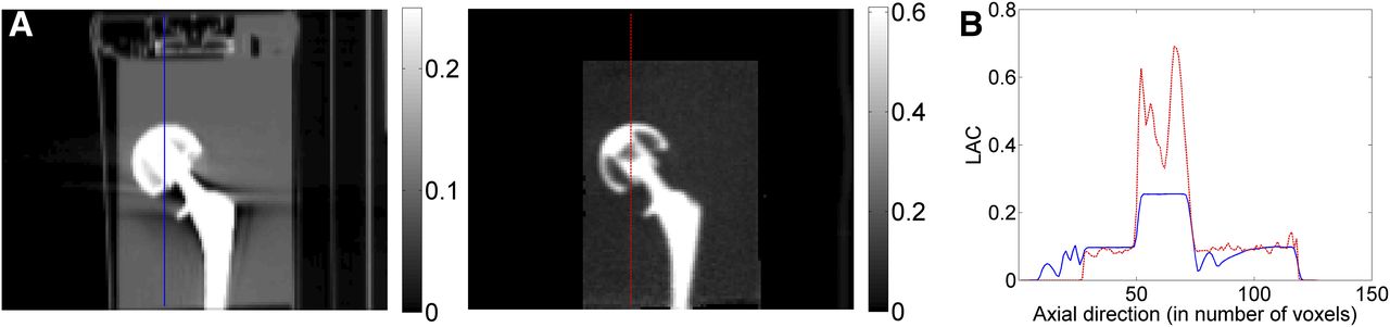
- FIGURE 1.
Uniform 18F-FDG phantom with cobalt–chromium alloy implant. (A) CT μ-map (left) and IPAC μ-map (right) are shown. (B) Corresponding LAC profiles are shown (CT μ-map in blue and IPAC μ-map in red).
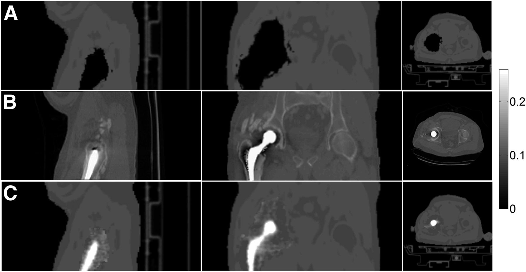
- FIGURE 2.
Patient presenting right hip cobalt–chromium alloy endoprosthesis (patient 1). Dixon (A), CT (B), and IPAC (C) μ-maps are shown. The 3 columns show (from left to right) sagittal, coronal, and axial planes.
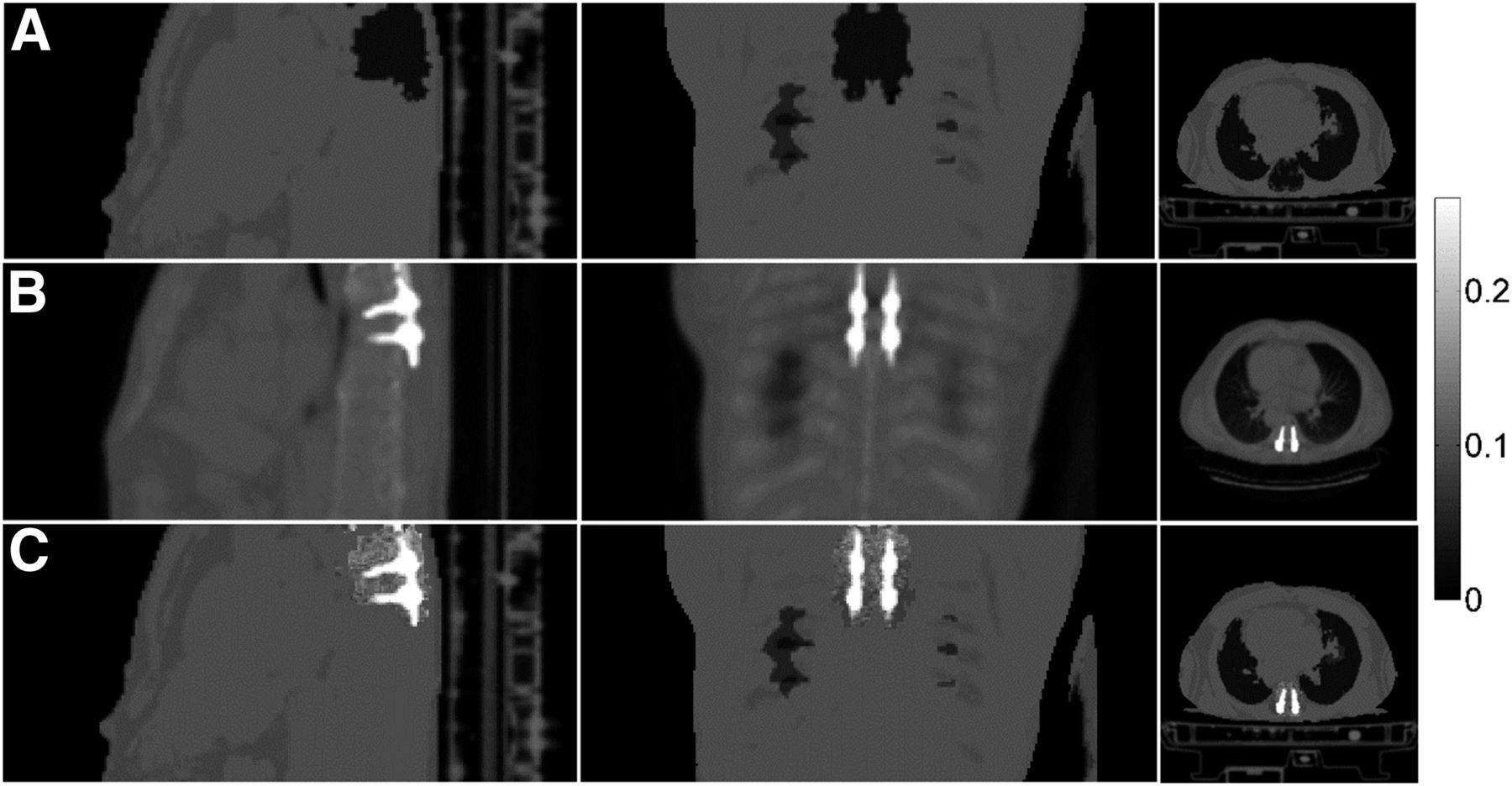
- FIGURE 3.
Patient presenting titanium spinal implant and pedicle screws (patient 1B). Dixon (A), CT (B), and IPAC (C) μ-maps are shown. The 3 columns show (from left to right) sagittal, coronal, and axial planes.
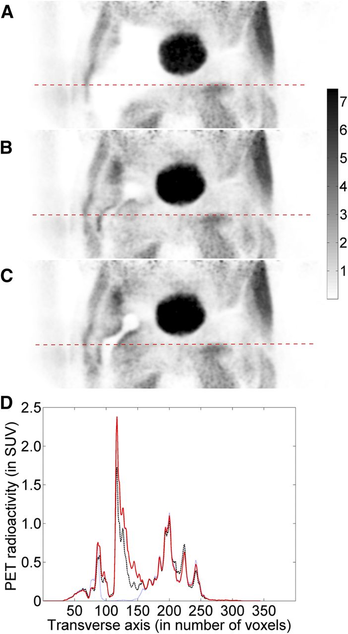
- FIGURE 4.
Reconstructed PET images corrected with the 3 AC methods for the validation subject of Figure 2. Dixon (A), thCT (B), and IPAC (C) μ-maps were, respectively, used for AC during reconstruction. (D) Corresponding radioactivity profiles are shown (Dixon in blue, thCT in black, and IPAC in red).
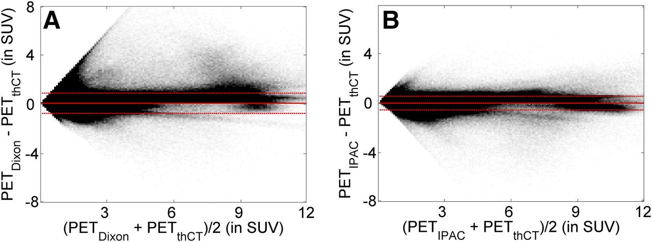
- FIGURE 5.
Bland–Altman plots showing voxel-based comparisons (for all validation subjects) between PETDixon/PETthCT (A) and PETIPAC/PETthCT (B). Continuous red lines show mean, and dashed red lines show the of difference.


Tables
- TABLE 1
Data Acquisition Parameters, Implant Material Specifications, and Results for DSC and Mean LAC Analysis
Patient no. Center Type of implant Material mean LAC Reconstructed mean LAC DSC Total prompts events 1 Martinos Right hip 0.72 0.66 0.89 4.28E+08 2 SDN Napoli Left hip 0.36 0.32 0.80 8.55E+07 3 SDN Napoli Both hips 0.36 0.30 0.75 4.84E+07 4 SDN Napoli Both hips 0.36 0.31 0.75 2.70E+08 5 SDN Napoli Right hip 0.36 0.31 0.69 2.48E+08 6 SDN Napoli Both hips 0.36 0.28 0.69 7.50E+07 7 SDN Napoli Both hips 0.36 0.28 0.79 3.54E+07 8 SDN Napoli Left femur 0.36 0.26 0.65 1.07E+08 1B Martinos Back screws 0.36 0.32 0.72 2.33E+08 2B SDN Napoli Back screws 0.36 0.33 0.66 1.21E+08 9 Martinos Back screws 0.36 0.27 0.65 2.40E+08 10 Martinos Dental — 0.29 0.64 1.80E+08 11 Martinos Dental — 0.28 0.66 1.13E+08 Phantom Martinos Hip 0.71 0.70 0.90 1.92E+08
Supplemental Data
Files in this Data Supplement:


Jump to section
Related Articles
Cited By...
- No citing articles found.