


Article Figures & Data
Figures
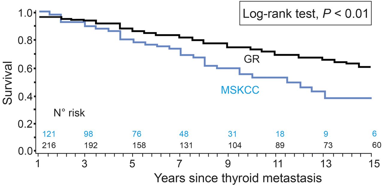
- FIGURE 1.
OS estimated from time of diagnosis of metastases until patient last follow-up or death.
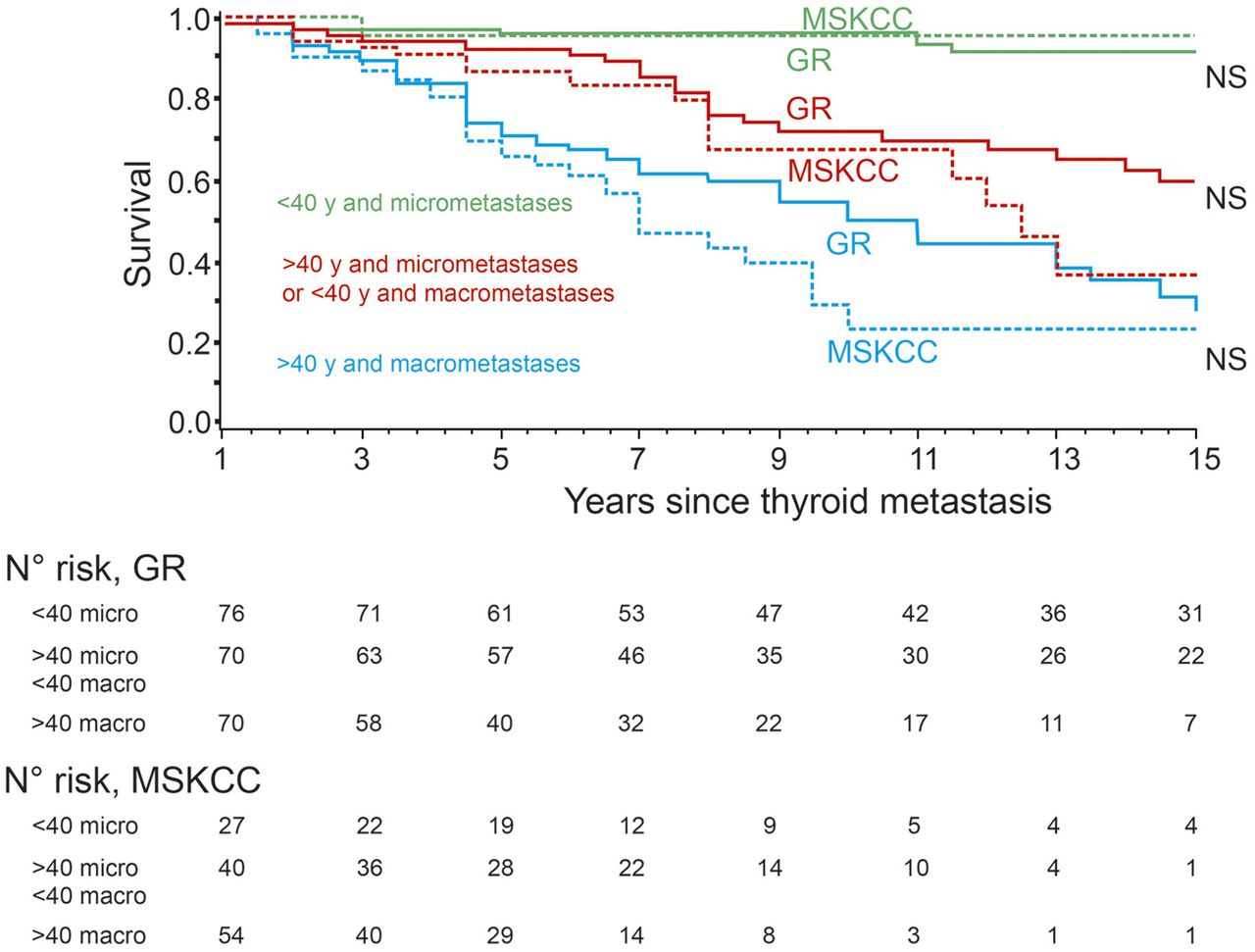
- FIGURE 2.
OS according to age and extent of metastases estimated from time of diagnosis of metastases until patient last follow-up or death.


Tables
Characteristic GR (n = 231) MSKCC (n = 121) GR + MSKCC (n = 352) P Sex (%) Female 153 (66) 68 (56)* 221 (63) 0.09 Primary thyroid tumor Median age at diagnosis (y) 40 (range, 5–90) 49 (range, 7–77) 42 (range, 5–90) 0.03 Histology (%) Follicular thyroid carcinoma 74 (32.0) 32 (26.5) 106 (30.1) Papillary thyroid carcinoma 156 (67.5) 89 (73.5) 245 (69.6) Not significant Other 1 (0.3) 0 1 (0.3) Distant metastases Median age at diagnosis (y) 42 (range, 6–90) 53 (range, 10–80) 46 (range, 6–90) <0.01 Median year of diagnosis 1994 (range, 1981–2007) 2000 (range, 1983–2008) 1997 (range, 1981–2008) <0.0001 Synchronous metastases (%)† 143 (61.9) 67 (55.4) 210 (59.7) Not significant Site (%) Lung 139 (60.2) 66 (54.6) 205 (58.2) Bone 55 (23.8) 29 (24.0) 84 (23.9) Not significant Lung and bone (± others) 37 (16.0) 26 (21.5) 63 (17.9) Metastases size (%) Macronodular lung metastases 37 (21.5) 31 (35.6) 68 (26.3) 0.02 Multiple bone lesions 62 (68.9) 41 (77.4) 103 (72.0) Not significant Extent of metastases‡ Category 1 69 (29.9) 14 (11.6) 83 (23.6) Category 2 66 (28.6) 43 (35.5) 109 (31.0) <0.001 Category 3 96 (41.6) 64 (52.9) 160 (45.4) ↵* Not known, 2 patients.
↵† 6 mo before or after primary tumor diagnosis.
↵‡ According to Durante classification (8).
Category 1 = patients with metastases on 131I WB scan but with normal radiologic imaging; category 2 = patients with micronodular lung metastases defined radiologically < 1 cm or with a single bone metastasis identified radiologically; category 3 = macronodular lung metastases or multiple bone metastases or both bone and lung metastases.
Data in parentheses are percentages unless otherwise indicated.
- TABLE 2
Median Number of 131I Treatments, Median Administered Activity, and Median Cumulative Activity (GBq) According to Patient Group
Group 1 (n = 106) Group 2 (n = 114) Group 3 (n = 132) Parameter GR (n = 79) MSKCC (n = 27) GR (n = 74) MSKCC (n = 40) GR (n = 78) MSKCC (n = 54) 5-y OS 0.96 (95% CI, 0.88–0.99) 0.96 (95% CI, 0.73–0.99) 0.92 (95% CI, 0.83–0.97) 0.87 (95% CI, 0.71–0.94) 0.70 (95% CI, 0.58–0.80) 0.65 (95% CI, 0.50–0.77) 131I treatment Median number 4 (1–14) 2 (1–7) 4 (1–14) 3 (1–9) 4 (1–13) 3 (1–8) Median administered activity (GBq) 3.7 (1.4–5.2) 7.5 (2.7–14.1) 3.7 (1.1–8.7) 8.9 (4.6–18.6) 3.7 (1.8–11.1) 10.6 (3.7–16.7) Median cumulative activity (GBq) 14.8 (3.7–39.2) 17.2 (2.7–56.4) 14.8 (2.2–52.5) 23.1 (5.5–112) 14.8 (1.8–52.5) 29.8 (3.7–83.9) Group 1 = patients aged < 40 y with no visible disease on cross-sectional imaging, micronodular lung metastases or single bone lesion; group 2 = patients aged < 40 y with either macrometastases or patients aged > 40 y with micrometastases; group 3 = patients aged > 40 y with lung macrometastases or multiple bone metastases (8).
Data in parentheses are ranges unless otherwise indicated.
Factor No. of subjects/deaths Crude RR P Adjusted RR P Sex Male 129/57 1 (ref) <0.001 1 (ref) <0.001 Female 221/55 0.42 (0.29–0.62) 0.49 (0.33–0.74) Age at the diagnosis of distant metastasis (y) 0–29 80/3 1 (ref) 1 (ref) 30–39 54/11 6.43 (1.79–23.10) 4.54 (1.24–16.61) 40–49 56/18 9.87 (2.91–33.52) <0.0001 7.65 (2.22–26.32) <0.0001 50–59 70/31 21.57 (6.55–71.10) 11.49 (3.33–39.59) 60–69 57/31 23.60 (7.15–77.95) 13.18 (3.83–45.31) 70–90 35/18 42.88 (12.39–148.37) 23.03 (6.24–85.08) Extent of metastases Category 1 83/8 1 (ref) 1 (ref) Category 2 109/31 3.65 (1.68–7.94) <0.001 2.71 (1.22–6.03) <0.01 Category 3 160/73 8.43 (4.04–17.58) 3.49 (1.56–7.81) Site of metastases Lung 205/51 1 (ref) 1 (ref) Bone 84/32 2.17 (1.38–3.39) <0.001 1.33 (0.77–2.31) 0.24 Lung and bone ± other 63/29 3.58 (2.24–5.72) 1.63 (0.92–2.91) Histology Papillary thyroid carcinoma 245/72 1 (ref) 1 (ref) Follicular thyroid carcinoma 107/40 1.61 (1.09–2.39) 0.02 0.67 (0.42–1.07) 0.09 Center GR 231/68 1 (ref) 1 (ref) MSKCC 121/44 1.77 (1.20–2.62) 0.01 1.41 (0.87–2.29) 0.16 ref = reference; Category 1 = patients with metastases on 131I WB scan but with normal radiologic imaging; category 2 = patients with micronodular lung metastases defined radiologically < 1 cm or with a single bone metastasis identified radiologically; category 3 = macronodular lung metastases or multiple bone metastases or both bone and lung metastases.
Data in parentheses are 95% CIs.


Jump to section
Related Articles
Cited By...
- Appropriate Use Criteria for Nuclear Medicine in the Evaluation and Treatment of Differentiated Thyroid Cancer
- Treatment of refractory thyroid cancer
- Fixed 3.7-GBq 131I Activity for Metastatic Thyroid Cancer Therapy Ignores Science and History
- Reply: Fixed 3.7-GBq 131I Activity for Metastatic Thyroid Cancer Therapy Ignores Science and History
- Prescribed Activity of 131I Therapy in Differentiated Thyroid Cancer
- Reply: Comparison of Empiric Versus Dosimetry-Guided Radioiodine Therapy: The Devil Is in the Details
- Comparison of Empiric Versus Dosimetry-Guided Radioiodine Therapy: The Devil Is in the Details