


Article Figures & Data
Figures
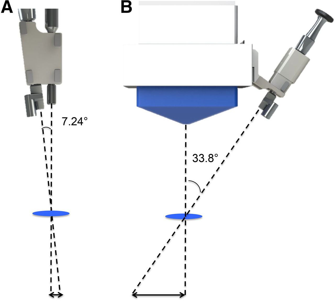
- FIGURE 1.
Schematic overview of VITOM-GP and VITOM-GC combination. (A) VITOM-GP combination. (B) VITOM-GC combination. Because of angle in alignment, devices had to be moved horizontally to compensate for misalignment at distances beyond focal plane.
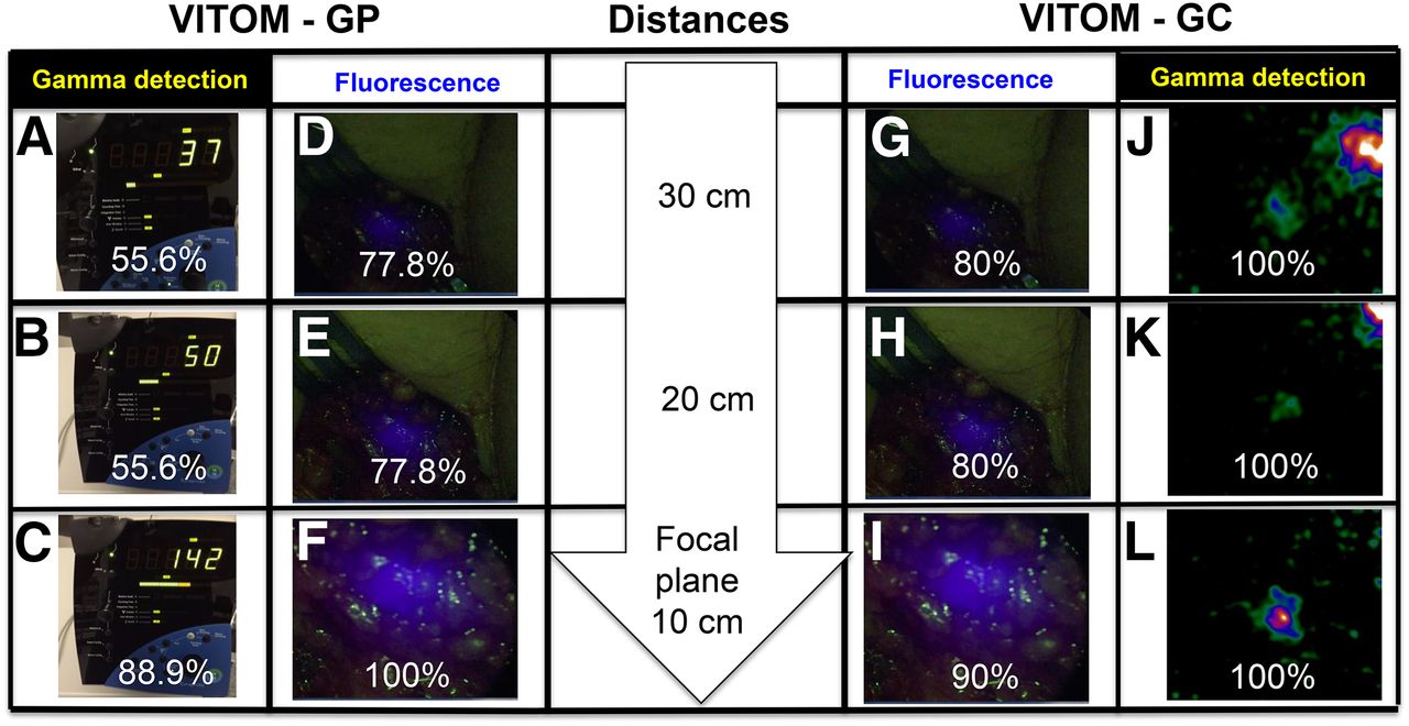
- FIGURE 2.
Fluorescence imaging versus γ-detection. On left side, SN detection rates (%) of VITOM-GP combination are shown in relation to the evaluated working distances (A–F). On right, SN detection rates (%) of VITOM-GC are presented in relation to the different evaluated working distances (G–L).


Tables
Patient no. Clinical TNM stage Injected dose (MBq) No. of SNs on SPECT/CT No. of pursued SNs No. of groins No. of SNs at pathology No. of tumor-positive SNs Patients evaluated with VITOM-GP 1 cT2N0Mx 184.28 4 3 (75%) 1 3 0 2 cT3N0Mx 77.87 2 1 (50%) 1 5 0 3 cT3N0Mx 162.74 2 3 (100%)* 1 3 0 4 cT3N0Mx 84 2 1 (50%) 1 3 0 5 cT2N0Mx 83.69 4 1 (25%) 1 4 1 Total 14 9 5 18 1 Patients evaluated with VITOM-GC 1 cT3N0M0 143.23 3 2 (67%) 1 3 1 2 cT1N0Mx 145.37 1 1 (100%) 1 1 0 3 cT1bN0Mx 144.66 2 2 (100%) 2 2 0 4 cT2N0M0 71.62 2 2 (100%) 2 4 0 5 cT2N0M0 70.37 2 2 (100%) 2 2 0 6 cT1/2N0Mx 87.51 5 1 (20%) 1 7 0 Total 15 10 9 19 1 ↵* Intraoperative additional SN removed.
Supplemental Data
Files in this Data Supplement:


