


Article Figures & Data
Figures
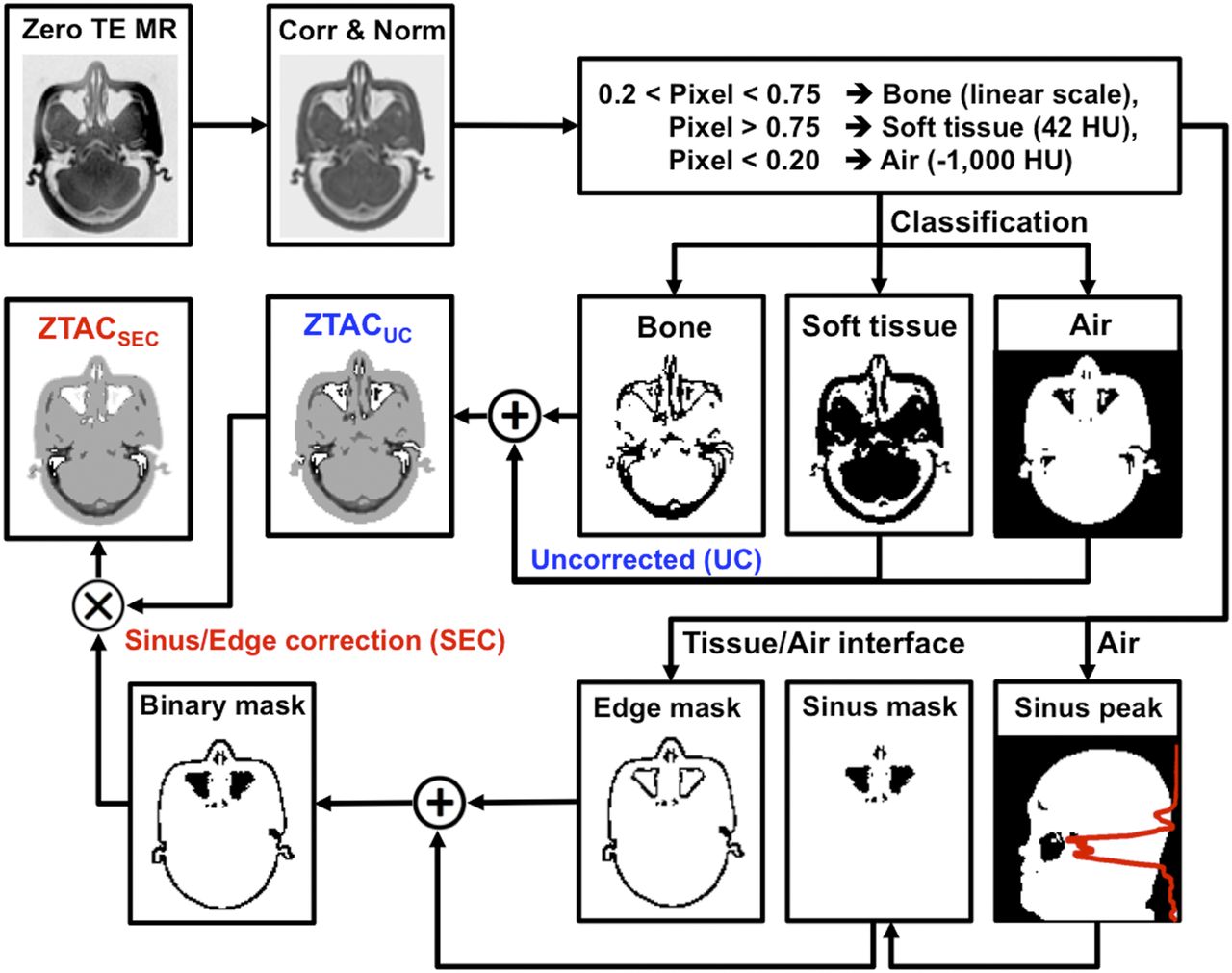
- FIGURE 1.
Workflow of generating pseudo-CT from ZTE images: ZTACSEC and ZTACUC. In the binary mask, false-positive bone pixels were corrected to stay below 42 HU (soft tissue).
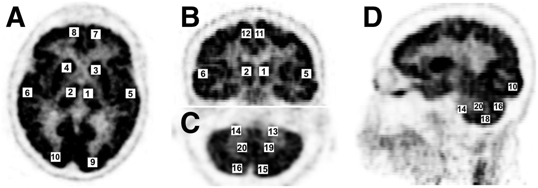
- FIGURE 2.
Localization of 20 VOIs for VOI-based quantification: axial brain (A), coronal brain (B), axial cerebellum (C), sagittal brain with sinus and cerebellum (D); left-right (1–2) thalamus, (3–4) corpus callosum, (5–6) temporal lobe, (7–8) frontal lobe, (9–10) occipital lobe, (11–12) cerebral cortex, (13–20) cerebellum at anterior (13–14), posterior (15–16), inferior (17–18), and center (19–20) positions.
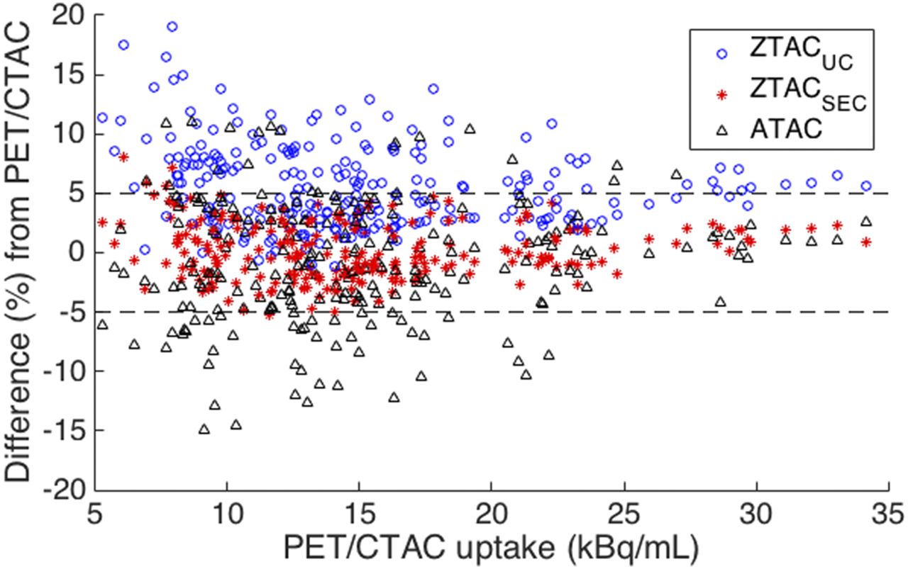
- FIGURE 3.
Bland–Altman plot of 240 VOIs of 12 patients for PET uptake differences (%) with MRAC (ZTACUC, ZTACSEC, ATAC) from PET with CTAC (PET/CTAC). PET/ZTACSEC (red stars) was close to zero mean difference mostly within ±5% boundaries.
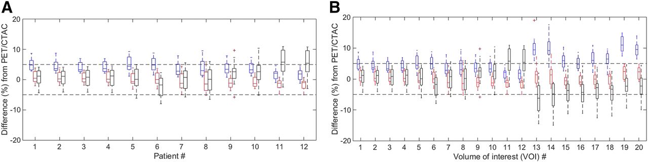
- FIGURE 4.
Box plots of PET uptake differences (%) for 12 patients across 20 VOIs (A) and for 20 VOIs across 12 patients (B) between PET/CTAC and PET/MRAC (left, blue: ZTACUC; middle, red: ZTACSEC; right, black: ATAC). Tops and bottoms of each box are 25th and 75th percentiles of samples, respectively, with median and outlier (+ sign, >1.5 × interquartile range).
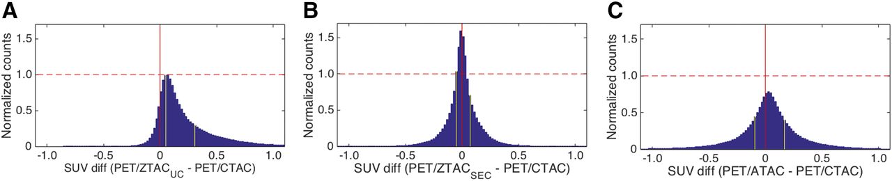
- FIGURE 5.
Histograms of voxel-based PET uptake differences (PET/MRAC − PET/CTAC) across all patients using voxels within SUV range of 0.5–15.0 (g/mL). (A) ZTACUC. (B) ZTACSEC. (C) ATAC. Counts were normalized by maximum count of PET/ZTACUC difference from PET/CTAC. (Left to right) Yellow lines indicate 25th and 75th percentiles.
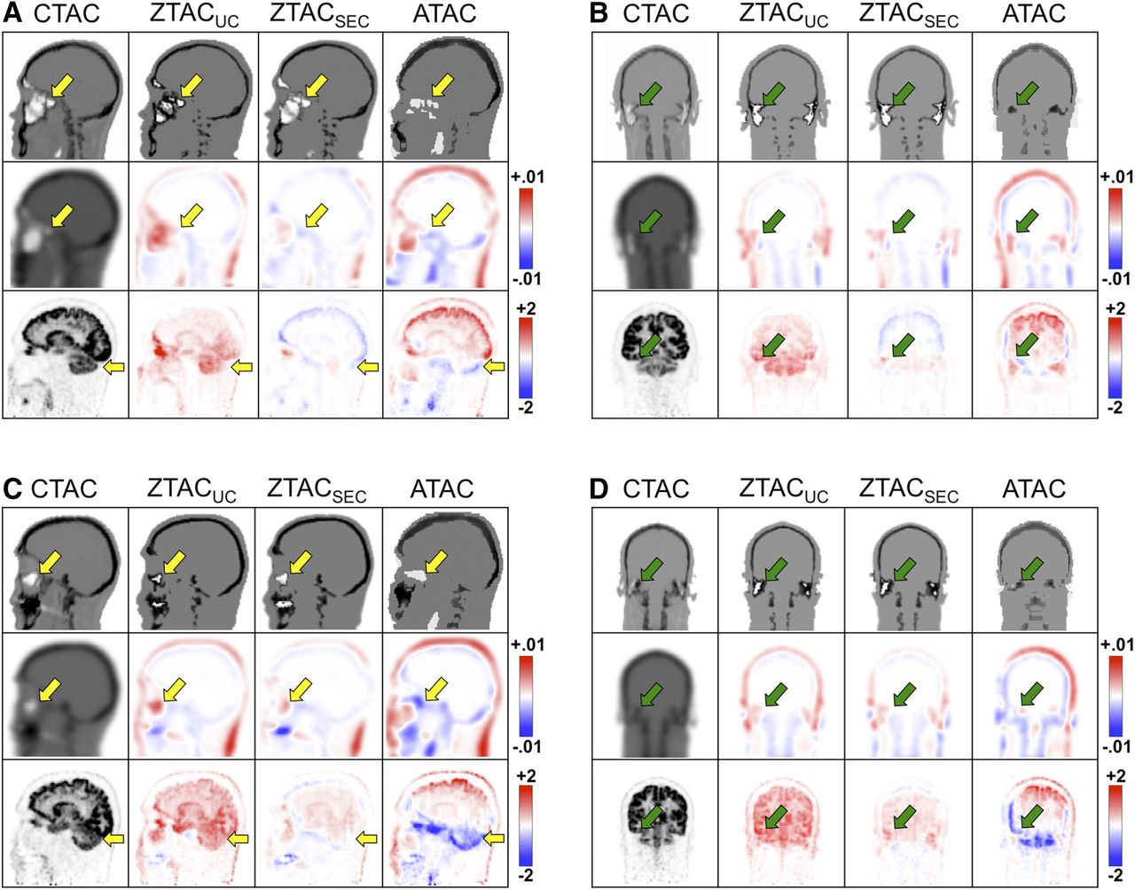
- FIGURE 6.
Patients 5 (A and B) and 8 (C and D) in coronal and sagittal views for CT and pseudo-CTs (first row), attenuation-correction (AC) maps (second row), and corresponding PET images (third row) according to AC methods. Second and third rows illustrate difference images on second through fourth columns for pseudo-CT–derived AC maps from CT-derived AC maps (unit, cm−1) and PET/MRAC from PET/CTAC (unit, SUV), respectively. Arrows indicate sinus, mastoid, and cerebellum.


Tables
- TABLE 1
PET Uptake Differences Between Reference PET/CTAC and PET/MRAC (ZTACUC, ZTACSEC, and ATAC) Across All 240 VOIs
Difference PET/MRAC kBq/mL % P PET/ZTACUC 0.79 ± 0.52 5.6 ± 3.5 <0.01 PET/ZTACSEC 0.03 ± 0.33 0.2 ± 2.4 0.23 PET/ATAC −0.10 ± 0.71 −0.9 ± 5.0 0.03 Differences are mean ± SD. A paired t test was performed for pair of PET/CTAC and PET/MRAC.


Jump to section
Related Articles
Cited By...
- Synthesis of Patient-Specific Transmission Data for PET Attenuation Correction for PET/MRI Neuroimaging Using a Convolutional Neural Network
- Zero-Echo-Time and Dixon Deep Pseudo-CT (ZeDD CT): Direct Generation of Pseudo-CT Images for Pelvic PET/MRI Attenuation Correction Using Deep Convolutional Neural Networks with Multiparametric MRI