


Article Figures & Data
Figures
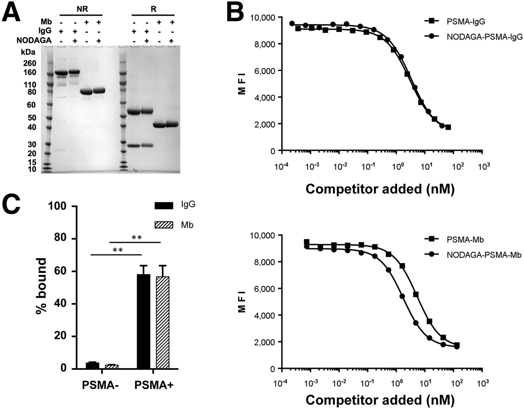
- FIGURE 1.
Characterization of NODAGA-PSMA-IgG and NODAGA-PSMA-Mb. (A) IgG and Mb formats of anti-PSMA huJ591 antibody were analyzed by gel electrophoresis under nonreducing (NR) and reducing (R) conditions before and after conjugation of NODAGA. (B) Competition cell binding experiments demonstrate that conjugation of NODAGA did not significantly disrupt ability of either PSMA-IgG or PSMA-Mb to bind cell surface PSMA. (C) 64Cu-NODAGA-PSMA-IgG and 64Cu-NODAGA-PSMA-Mb bind selectively to PSMA-positive cells.
- FIGURE 2.
CLI with 64Cu-NODAGA-PSMA-IgG and 64Cu-NODAGA-PSMA-Mb. (A) CLI-based evaluation of 64Cu-NODAGA-PSMA-IgG and 64Cu-NODAGA-PSMA-Mb. Luminescence was scaled to allow for direct comparison across time course. (B) Average tumor-to-background ratios across entire time course for PSMA-positive (PSMA+) and PSMA-negative (PSMA–) tumors with both radiotracers. (C) Average radiance in PSMA+ and PSMA– tumors at time of peak uptake for 64Cu-NODAGA-PSMA-IgG and 64Cu-NODAGA-PSMA-Mb.
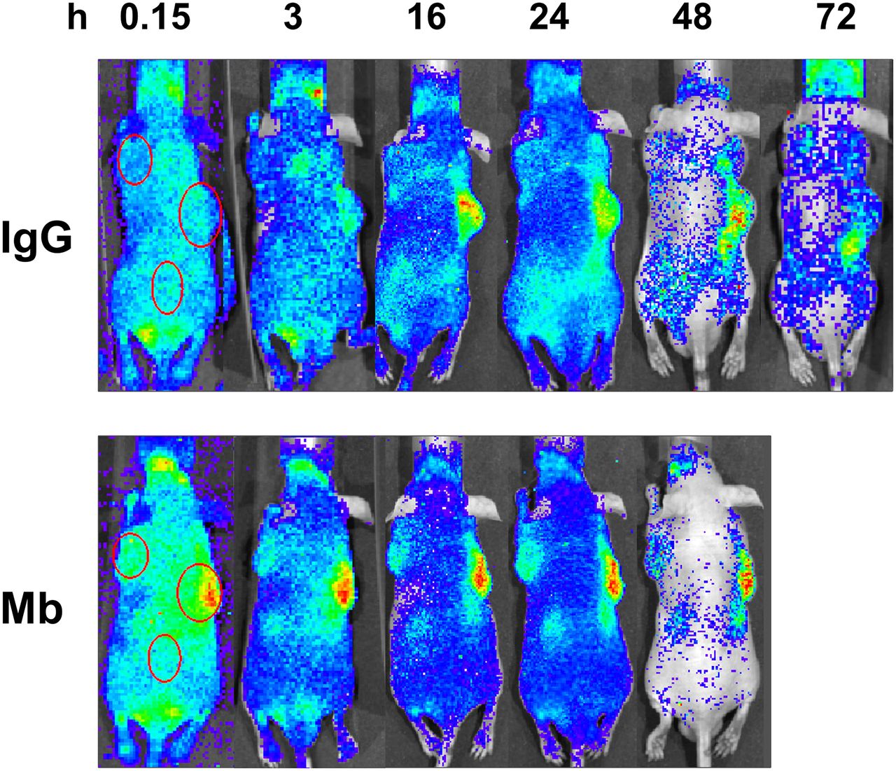
- FIGURE 3.
CLI. Time course analysis of representative mouse imaged with either 64Cu-NODAGA-PSMA-IgG (top) or 64Cu-NODAGA-PSMA-Mb (bottom). Scales were optimized at each time point to provide for maximum contrast between 22Rv1 PSMA-positive (PSMA+) (right shoulder) and PC-3 PSMA-negative (PSMA–) (left shoulder) tumors. Regions of interest over tumor and background areas are shown as ellipses in first panels of each image set.
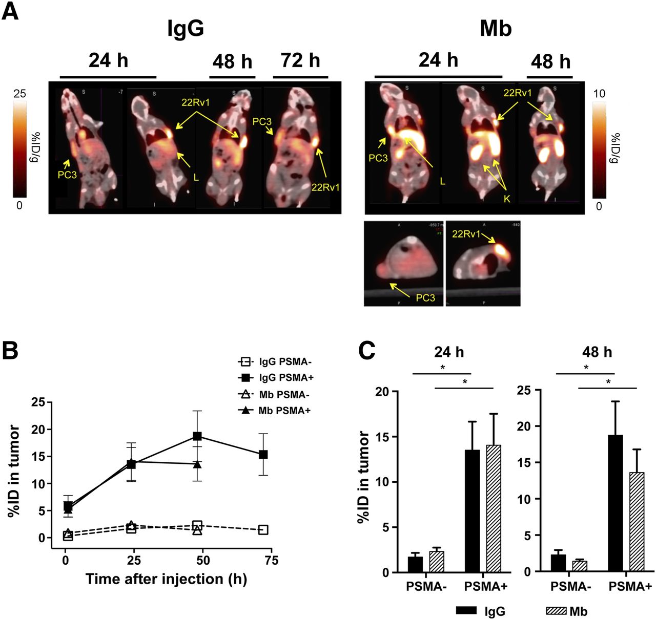
- FIGURE 4.
PET imaging with 64Cu-NODAGA-PSMA-IgG and 64Cu-NODAGA-PSMA-Mb. (A) PET/CT fusion images of representative mice at defined time points. Both 64Cu-NODAGA-PSMA-IgG and 64Cu-NODAGA-PSMA-Mb exhibited selective uptake into PSMA-positive (PSMA+) CWR22Rv1 tumor xenografts. (B) %ID of radiotracers in PSMA+ and PSMA-negative (PSMA–) tumor xenografts over time as quantified by PET imaging. (C) PET-based uptake in PSMA+ and PSMA– tumors at time of peak uptake for 64Cu-NODAGA-PSMA-IgG and 64Cu-NODAGA-PSMA-Mb. K = kidneys; L = liver.
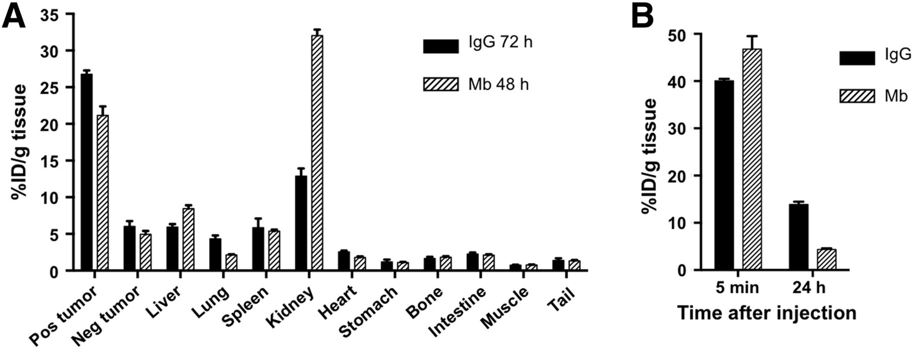
- FIGURE 5.
Biodistribution of 64Cu-NODAGA-PSMA-IgG and 64Cu-NODAGA-PSMA-Mb. (A) Biodistribution of 64Cu-NODAGA-PSMA-IgG and 64Cu-NODAGA-PSMA-Mb radiotracers at 72 and 48 h after injection, respectively, demonstrating statistically significant selective uptake into CWR22Rv1 PSMA-positive (PSMA+) vs. PC-3 PSMA-negative (PSMA–) tumors. (B) 64Cu-NODAGA-PSMA-Mb cleared more rapidly from blood than 64Cu-NODAGA-PSMA-IgG.


Additional Files
Supplemental Data
Files in this Data Supplement:


