


Article Figures & Data
Figures
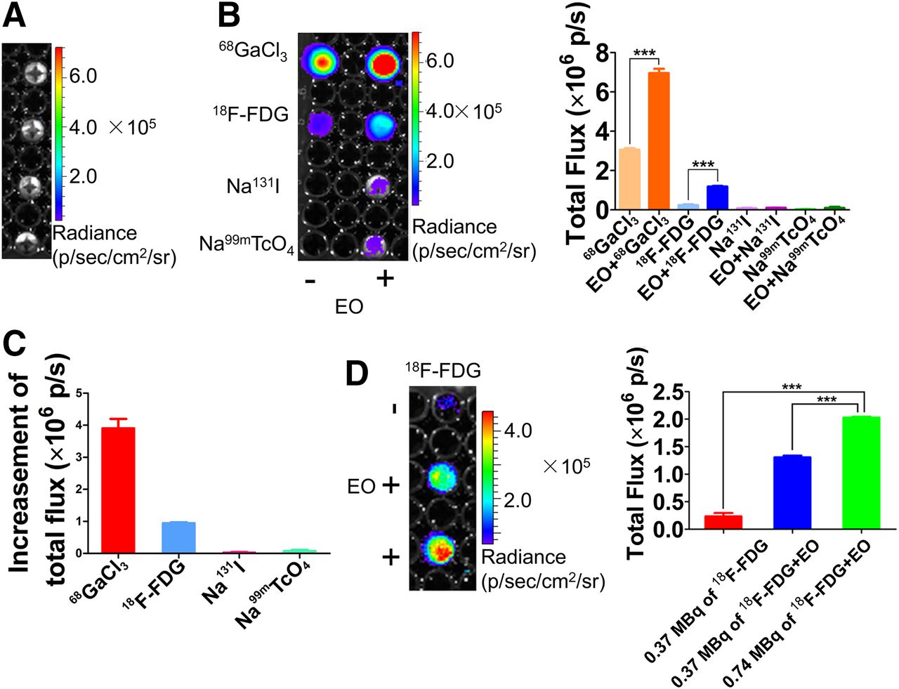
- FIGURE 1.
CLI of radiopharmaceuticals and REFI of mixture of radiopharmaceuticals and EO nanoparticles. (A) Optical image of EO nanoparticles. (B) CL images of radiopharmaceuticals, including Na99mTcO4, 68GaCl3, 18F-FDG, and Na131I, and REF image of mixture of EO nanoparticles and radiopharmaceuticals (left). Quantification analysis of CL signal and REF signal (right). Open field was used for collecting all light. (C) Quantification analysis of optical signal intensity, which was obtained through subtraction of Cerenkov luminescent intensity from optical signal intensity of mixture of radiopharmaceutical and EO nanoparticles. (D) CLI of 18F-FDG and REFI of mixture of 18F-FDG and EO nanoparticles (left) and quantification analysis (right).
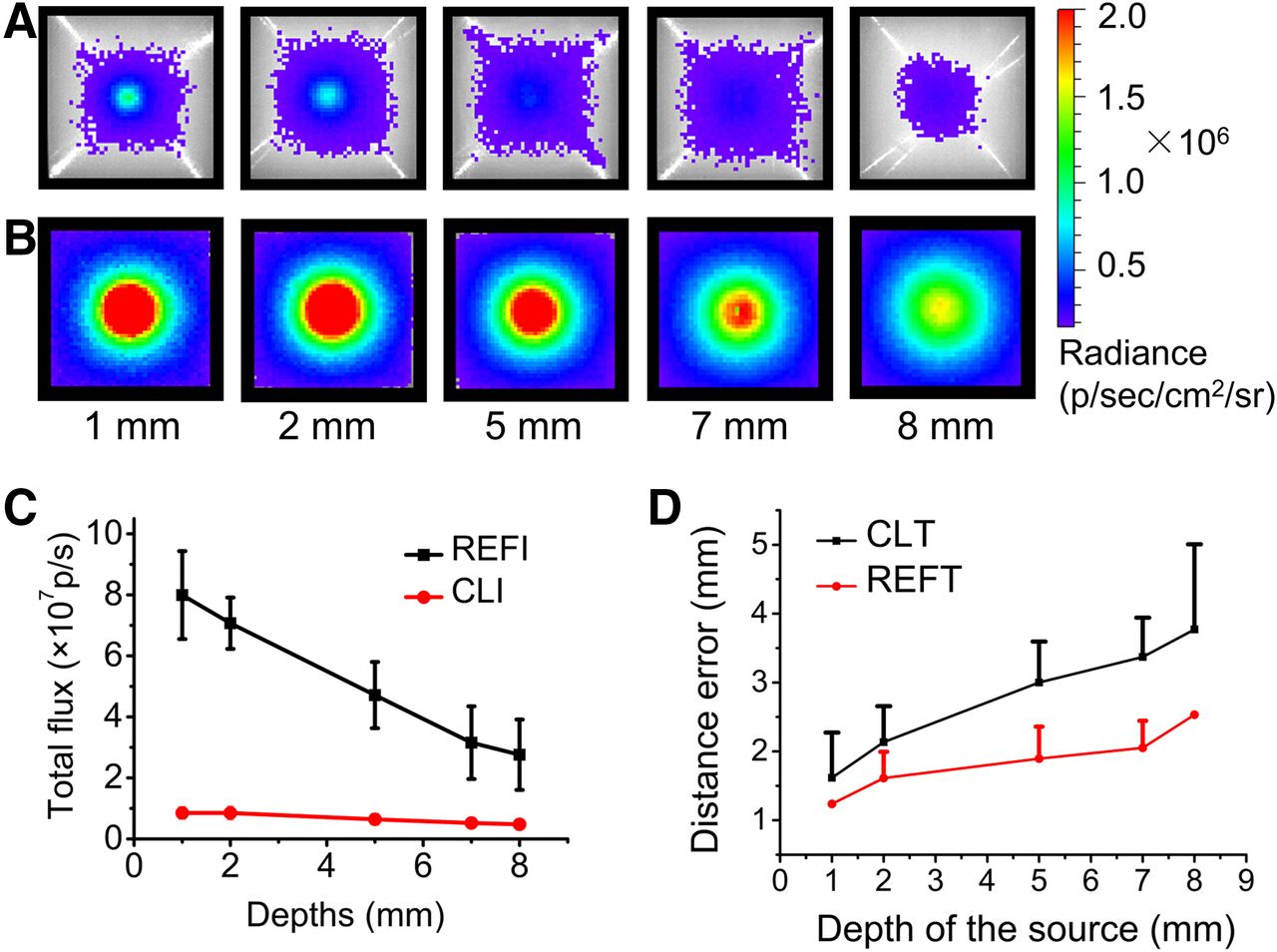
- FIGURE 2.
CLI and REFI of phantoms. (A) Cerenkov luminescence images of phantoms injected with 68Ga with radioactivity of 0.185 MBq. Depths of sources were 1, 2, 5, 7, and 8 mm. (B) Radioluminescent images of 1 mg of EO excited by 68Ga with radioactivity of 0.185 MBq. (C) Quantification comparison results of REFI and CLI. (D) Comparison of 3D reconstruction DEs of CLT and REFT.
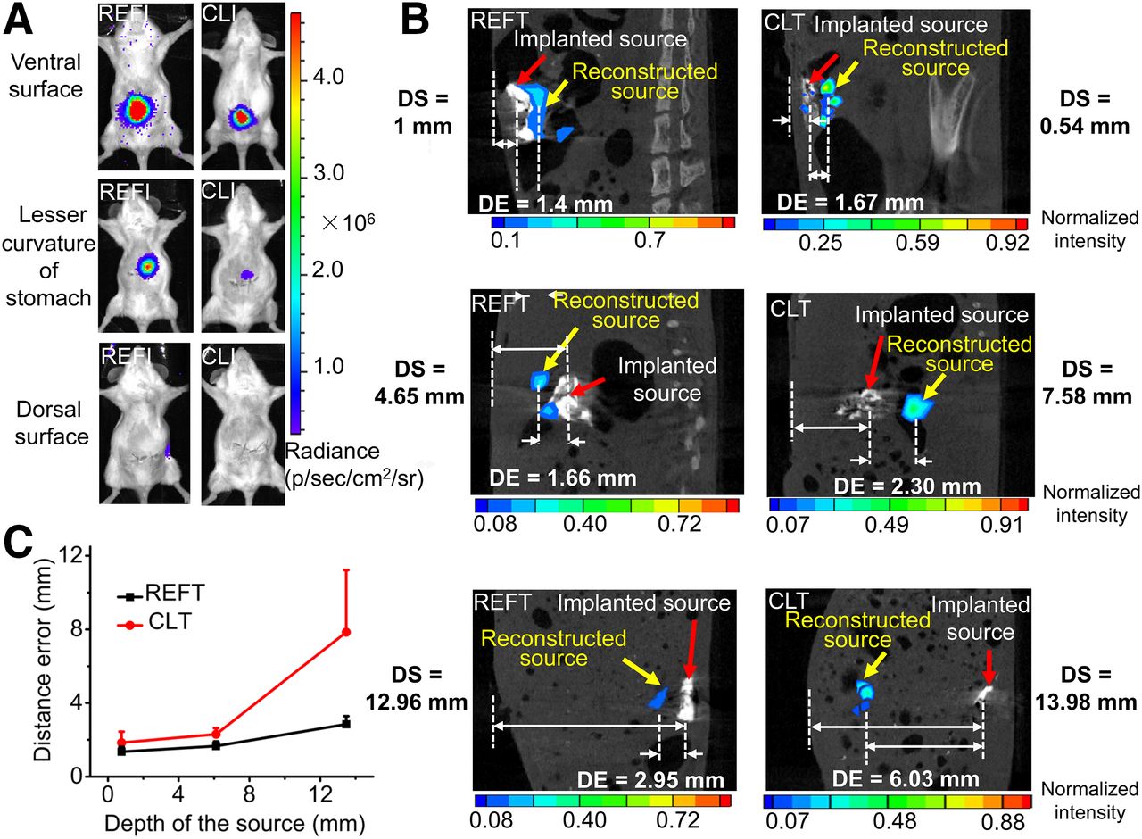
- FIGURE 3.
REFT and CLT of artificial source–implantation mouse models. (A) REFI (left) and CLI (right) of mice implanted with artificial sources. Position of implanted source was close to ventral surface of abdomen (first row), lesser curvature of stomach (second row), and dorsal surface of abdomen of mouse (third row). (B) Comparison of reconstruction results of REFT and CLT. DE = reconstructed distance error, which is defined as distance from real source position to reconstructed source position; DS = source depth from real source position to surface of mouse body. (C) Relationship between DE of REFT or CLT and depth of implanted sources.
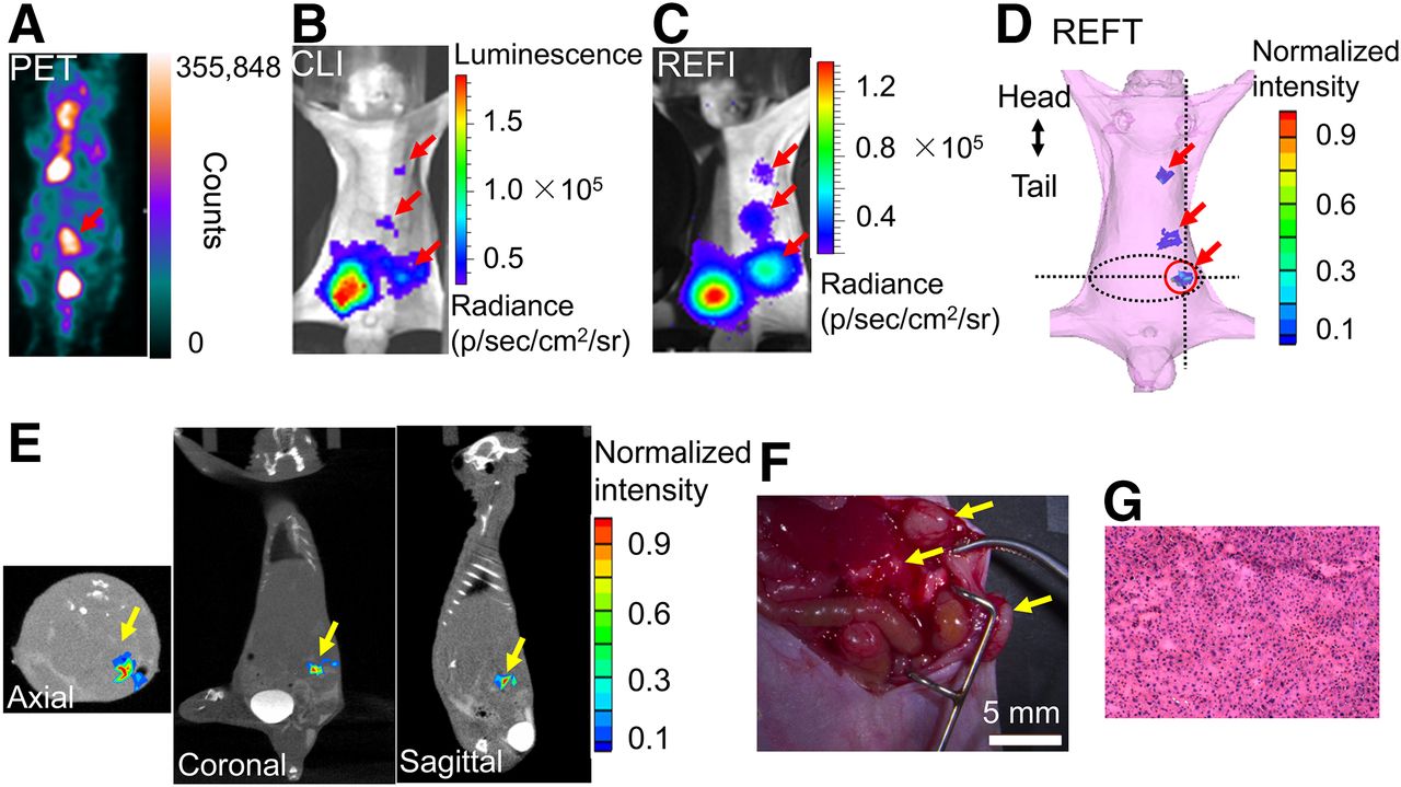
- FIGURE 4.
Small-animal PET, CLI, REFI, and REFT of mouse bearing HCC tumors. (A–D) Small-animal PET, CLI, REFI, and REFT of same mouse. (E) Axial, coronal, and sagittal view of reconstructed results of REFT. (F) Photograph of 3 tumors during operation on mouse (arrows). (G) Hematoxylin and eosin results of tumors.


Tables
Mouse no. Detection rate of REFT (%) Detection rate of PET (%) 1 100 33.3 2 80 60 3 100 50 4 75 25 Mean 88.75 42.08 SD 13.15 15.84
Supplemental Data
Files in this Data Supplement:


Jump to section
Related Articles
Cited By...
- No citing articles found.