


Article Figures & Data
Figures
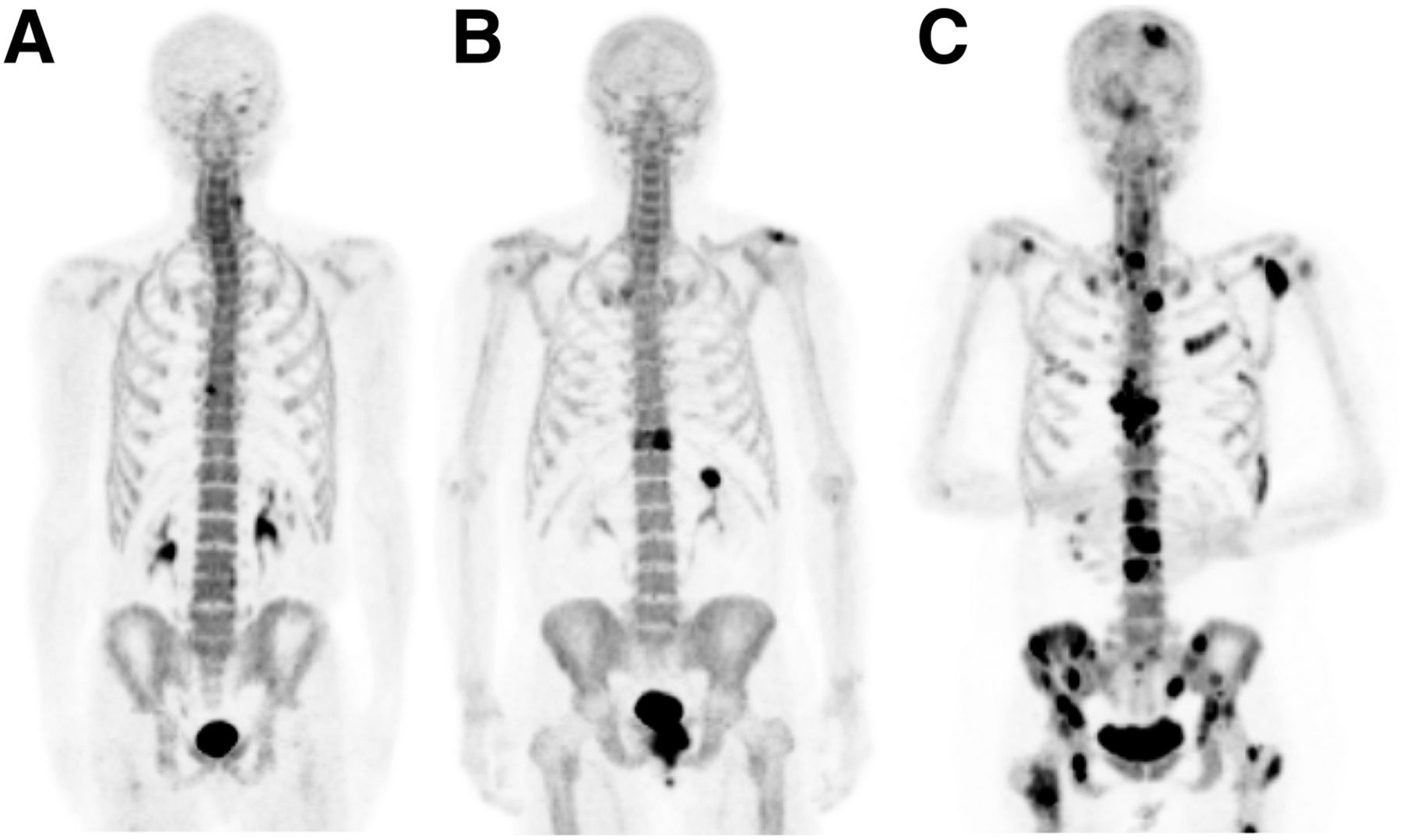
- FIGURE 1.
18F-NaF PET/CT maximum-intensity-projection images illustrating appearance of anterior osteophyte (A), solitary metastasis (B), and multiple metastases (C) in 3 different PC patients.
- FIGURE 2.
73-y-old man with PC. (A and B) 99mTc-MDP bone scan was negative (A), whereas 18F-NaF PET/CT revealed single L4 metastasis (B). (C and D) Axial PET (C) and CT (D) revealed focal uptake in small sclerotic lesion (arrowhead).
- FIGURE 3.
72-y-old man who had PC and was referred for 153Sm-EDTMP treatment of painful bone metastases. 99mTc-MDP bone scan (A) and 153Sm-EDTMP (B) revealed extensive metastases.
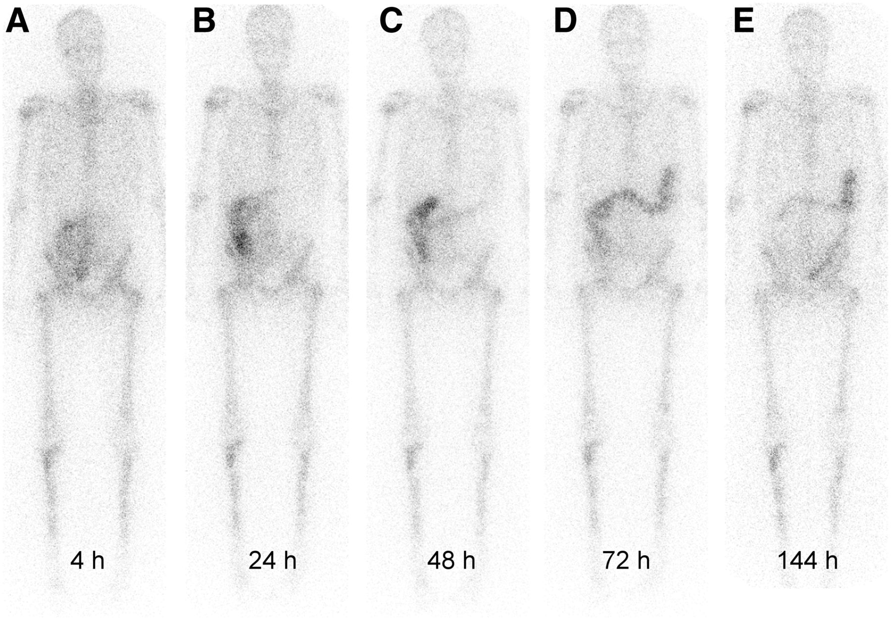
- FIGURE 4.
Biodistribution of 223RaCl2. (Adapted with permission of (47).)


Tables
Radiopharmaceutical Physical half-life (d) Particle type Usual administered activity Typical response time (d) Typical response duration Retreatment interval 89SrCl2 (Metastron; GE Healthcare Ltd.) 50.5 β 148 MBq 14–28 12–26 wk >3 mo 153Sm-EDTMP (Quadramet; IBA-Molecular) 1.9 β 37 MBq/kg 2–7 8 wk >2 mo 223RaCl2 (Xofigo; Bayer Healthcare) 11.4 α 50 kBq/kg × 6 every 4 wk <10 Unknown Unknown


