
Main menu

  • Home
  • Content
    • Current
    • Ahead of print
    • Past Issues
    • JNM Supplement
    • SNMMI Annual Meeting Abstracts
    • Continuing Education
    • JNM Podcasts
  • Subscriptions
    • Subscribers
    • Institutional and Non-member
    • Rates
    • Journal Claims
    • Corporate & Special Sales
  • Authors
    • Submit to JNM
    • Information for Authors
    • Assignment of Copyright
    • AQARA requirements
  • Info
    • Reviewers
    • Permissions
    • Advertisers
  • About
    • About Us
    • Editorial Board
    • Contact Information
  • More
    • Alerts
    • Feedback
    • Help
    • SNMMI Journals
  • SNMMI
    • JNM
    • JNMT
    • SNMMI Journals
    • SNMMI

User menu

  • Subscribe
  • My alerts
  • Log in
  • My Cart

Search

  • Advanced search
Journal of Nuclear Medicine
  • SNMMI
    • JNM
    • JNMT
    • SNMMI Journals
    • SNMMI
  • Subscribe
  • My alerts
  • Log in
  • My Cart
Journal of Nuclear Medicine

Advanced Search

  • Home
  • Content
    • Current
    • Ahead of print
    • Past Issues
    • JNM Supplement
    • SNMMI Annual Meeting Abstracts
    • Continuing Education
    • JNM Podcasts
  • Subscriptions
    • Subscribers
    • Institutional and Non-member
    • Rates
    • Journal Claims
    • Corporate & Special Sales
  • Authors
    • Submit to JNM
    • Information for Authors
    • Assignment of Copyright
    • AQARA requirements
  • Info
    • Reviewers
    • Permissions
    • Advertisers
  • About
    • About Us
    • Editorial Board
    • Contact Information
  • More
    • Alerts
    • Feedback
    • Help
    • SNMMI Journals
  • View or Listen to JNM Podcast
  • Visit JNM on Facebook
  • Join JNM on LinkedIn
  • Follow JNM on Twitter
  • Subscribe to our RSS feeds
Research ArticleBasic Science Investigations

Molecular Imaging and Quantitation of EphA2 Expression in Xenograft Models with 89Zr-DS-8895a

Ingrid J.G. Burvenich, Sagun Parakh, Hui K. Gan, Fook-Thean Lee, Nancy Guo, Angela Rigopoulos, Sze-Ting Lee, Sylvia Gong, Graeme J. O’Keefe, Henri Tochon-Danguy, Masakatsu Kotsuma, Jun Hasegawa, Giorgio Senaldi and Andrew M. Scott
Journal of Nuclear Medicine June 2016, 57 (6) 974-980; DOI: https://doi.org/10.2967/jnumed.115.169839
Ingrid J.G. Burvenich
1Tumour Targeting Laboratory, Ludwig Institute for Cancer Research and Olivia Newton-John Cancer Research Institute, Melbourne, Australia
2School of Cancer Medicine, La Trobe University, Melbourne, Australia
  • Find this author on Google Scholar
  • Find this author on PubMed
  • Search for this author on this site
Sagun Parakh
1Tumour Targeting Laboratory, Ludwig Institute for Cancer Research and Olivia Newton-John Cancer Research Institute, Melbourne, Australia
2School of Cancer Medicine, La Trobe University, Melbourne, Australia
3Department of Medical Oncology, Austin Health, Heidelberg, Melbourne, Australia
  • Find this author on Google Scholar
  • Find this author on PubMed
  • Search for this author on this site
Hui K. Gan
1Tumour Targeting Laboratory, Ludwig Institute for Cancer Research and Olivia Newton-John Cancer Research Institute, Melbourne, Australia
2School of Cancer Medicine, La Trobe University, Melbourne, Australia
3Department of Medical Oncology, Austin Health, Heidelberg, Melbourne, Australia
  • Find this author on Google Scholar
  • Find this author on PubMed
  • Search for this author on this site
Fook-Thean Lee
1Tumour Targeting Laboratory, Ludwig Institute for Cancer Research and Olivia Newton-John Cancer Research Institute, Melbourne, Australia
  • Find this author on Google Scholar
  • Find this author on PubMed
  • Search for this author on this site
Nancy Guo
1Tumour Targeting Laboratory, Ludwig Institute for Cancer Research and Olivia Newton-John Cancer Research Institute, Melbourne, Australia
  • Find this author on Google Scholar
  • Find this author on PubMed
  • Search for this author on this site
Angela Rigopoulos
1Tumour Targeting Laboratory, Ludwig Institute for Cancer Research and Olivia Newton-John Cancer Research Institute, Melbourne, Australia
  • Find this author on Google Scholar
  • Find this author on PubMed
  • Search for this author on this site
Sze-Ting Lee
1Tumour Targeting Laboratory, Ludwig Institute for Cancer Research and Olivia Newton-John Cancer Research Institute, Melbourne, Australia
4Department of Molecular Imaging and Therapy, Austin Health, Melbourne, Australia
  • Find this author on Google Scholar
  • Find this author on PubMed
  • Search for this author on this site
Sylvia Gong
4Department of Molecular Imaging and Therapy, Austin Health, Melbourne, Australia
  • Find this author on Google Scholar
  • Find this author on PubMed
  • Search for this author on this site
Graeme J. O’Keefe
3Department of Medical Oncology, Austin Health, Heidelberg, Melbourne, Australia
4Department of Molecular Imaging and Therapy, Austin Health, Melbourne, Australia
  • Find this author on Google Scholar
  • Find this author on PubMed
  • Search for this author on this site
Henri Tochon-Danguy
4Department of Molecular Imaging and Therapy, Austin Health, Melbourne, Australia
  • Find this author on Google Scholar
  • Find this author on PubMed
  • Search for this author on this site
Masakatsu Kotsuma
5Translational Medicine and Clinical Pharmacology Department, Daiichi-Sankyo Co., Ltd., Tokyo, Japan
  • Find this author on Google Scholar
  • Find this author on PubMed
  • Search for this author on this site
Jun Hasegawa
6Biologics Pharmacology Research Laboratories, Daiichi-Sankyo Co., Ltd., Tokyo, Japan
  • Find this author on Google Scholar
  • Find this author on PubMed
  • Search for this author on this site
Giorgio Senaldi
7Department of Translational Medicine and Clinical Pharmacology, Daiichi-Sankyo Pharma Development, Edison, New Jersey; and
  • Find this author on Google Scholar
  • Find this author on PubMed
  • Search for this author on this site
Andrew M. Scott
1Tumour Targeting Laboratory, Ludwig Institute for Cancer Research and Olivia Newton-John Cancer Research Institute, Melbourne, Australia
3Department of Medical Oncology, Austin Health, Heidelberg, Melbourne, Australia
4Department of Molecular Imaging and Therapy, Austin Health, Melbourne, Australia
8Department of Medicine, University of Melbourne, Melbourne, Australia
  • Find this author on Google Scholar
  • Find this author on PubMed
  • Search for this author on this site
  • Article
  • Figures & Data
  • Info & Metrics
  • PDF
Loading

Article Figures & Data

Figures

  • Tables
  • FIGURE 1.
    • Download figure
    • Open in new tab
    • Download powerpoint
    FIGURE 1.

    (A–C) Lindmo plots showing in vitro binding of 125I-DS-8895a (A), 111In-CHX-A″-DTPA-DS-8895a (B), and 89Zr-Df-Bz-NCS-DS-8895a (C) to increasing concentrations of EphA2-positive MDA-MB-231 cells. (D–F) Scatchard plots showing in vitro binding of 125I-DS-8895a (D), 111In-CHX-A″-DTPA-DS-8895a (E), and 89Zr-Df-Bz-NCS-DS-8895a (F) to MDA-MB-231 cells.

  • FIGURE 2.
    • Download figure
    • Open in new tab
    • Download powerpoint
    FIGURE 2.

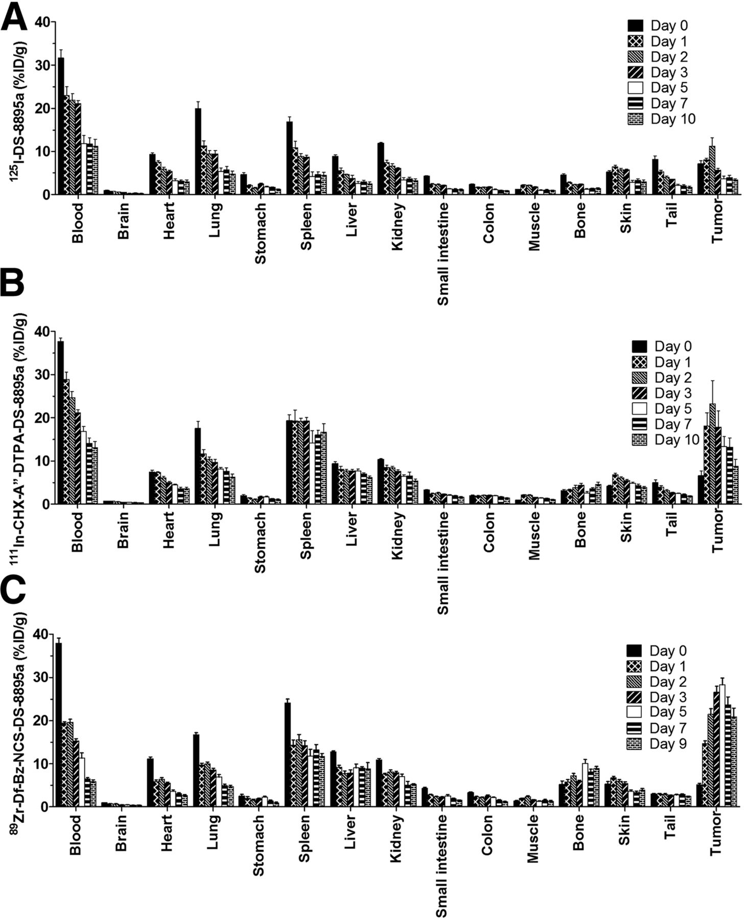
    Biodistribution properties of 125I-DS-8895a (A), 111In-CHX-A″-DTPA-DS-8895a (B), and 89Zr-Df-Bz-NCS-DS-8895a (C) in MDA-MB-231–xenografted BALB/c nu/nu mice. Bars indicate mean ± SD (n = 5).

  • FIGURE 3.
    • Download figure
    • Open in new tab
    • Download powerpoint
    FIGURE 3.

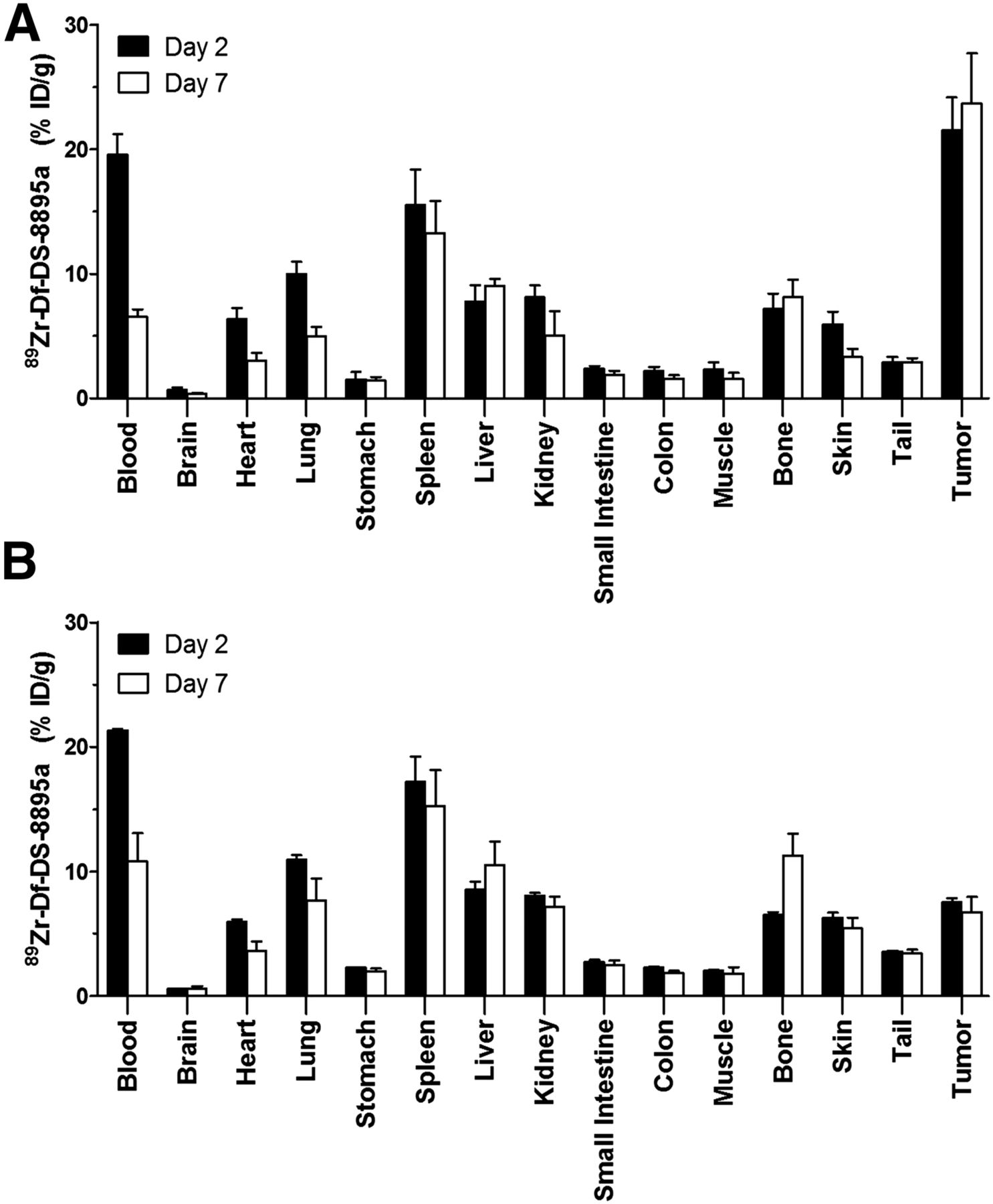
    Biodistribution properties of 89Zr-Df-Bz-NCS-DS-8895a in BALB/c nu/nu mice bearing EphA2-positive MDA-MB-231 breast tumors (A) and EphA2-negative CCRF-CEM human lymphoblastic leukemia (B) on days 2 and 7 after injection. Bars indicate mean ± SD (n = 5).

  • FIGURE 4.
    • Download figure
    • Open in new tab
    • Download powerpoint
    FIGURE 4.

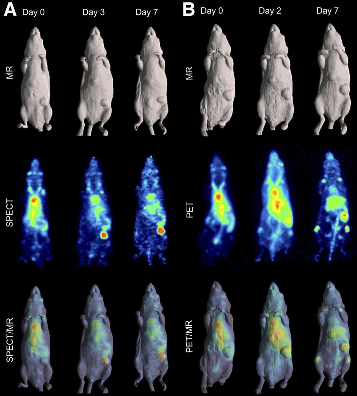
    (A) MR (surface-rendered), SPECT (maximum-intensity projection), and fused SPECT/MR images of MDA-MB-231–xenografted mice at 2 h (day 0), 3 d, and 7 d after injection of 111In-CHX-A″-DTPA-DS-8895a. (B) MR (surface-rendered), PET (maximum-intensity projection), and fused PET/MR images of MDA-MB-231–xenografted mice at 2 h (day 0), 2 d, and 7 d after injection of 89Zr-Df-Bz-NCS-DS-8895a.

  • FIGURE 5.
    • Download figure
    • Open in new tab
    • Download powerpoint
    FIGURE 5.

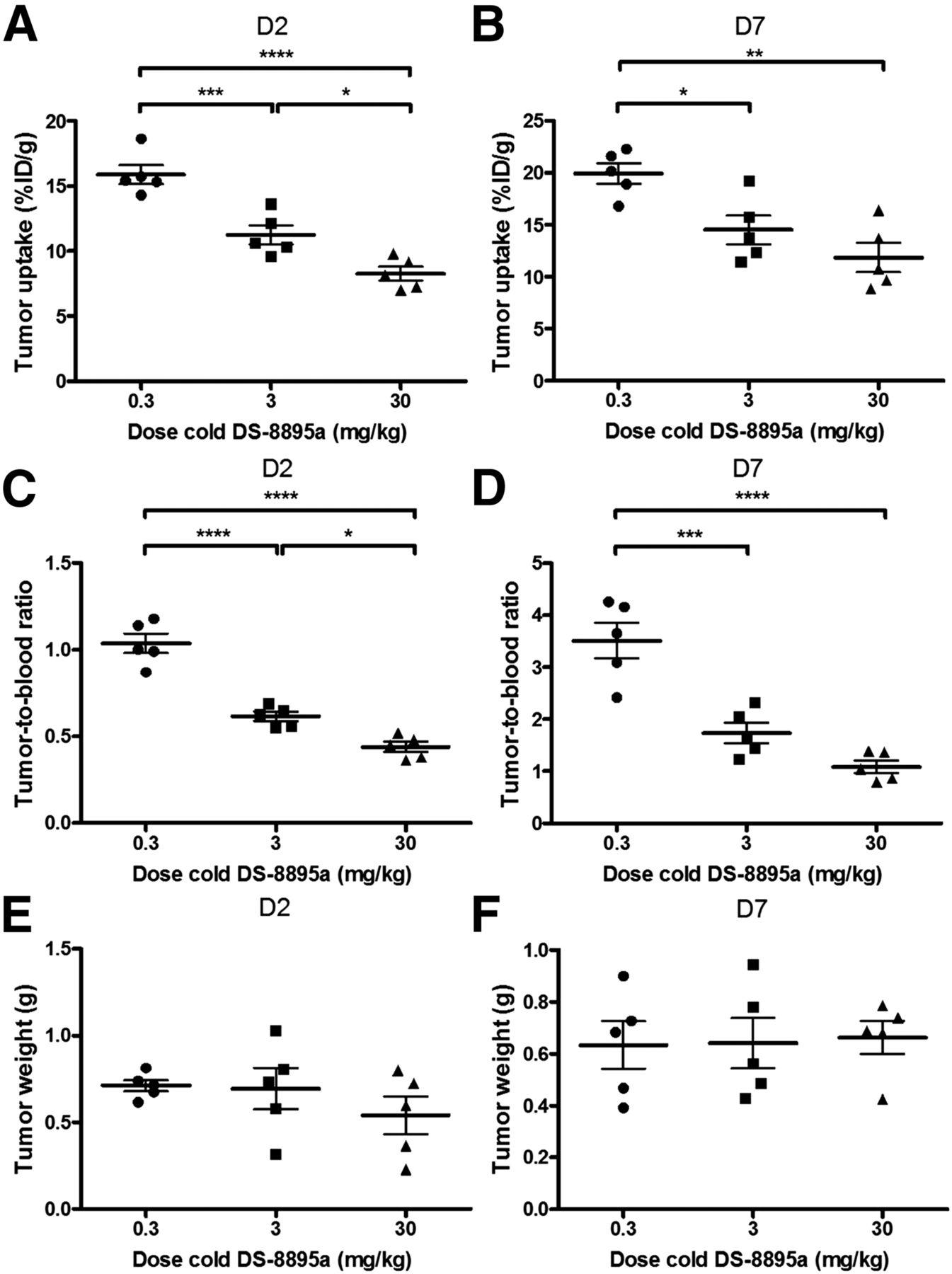
    Influence of cold-dose DS-8895a on tumor uptake of 89Zr-Df-Bz-NCS-DS-8895a in BALB/c nu/nu mice bearing MDA-MB-231 xenografts on days 2 (A) and 7 (B) after injection, and tumor-to-blood ratios on days 2 (C) and 7 (D) after injection. There was no significant difference in average tumor size among the various dose levels on days 2 (E) and 7 (F) after injection. Bars indicate mean ± SD (n = 5). *P < 0.05. **P < 0.005. ***P < 0.0005. ****P < 0.0001.
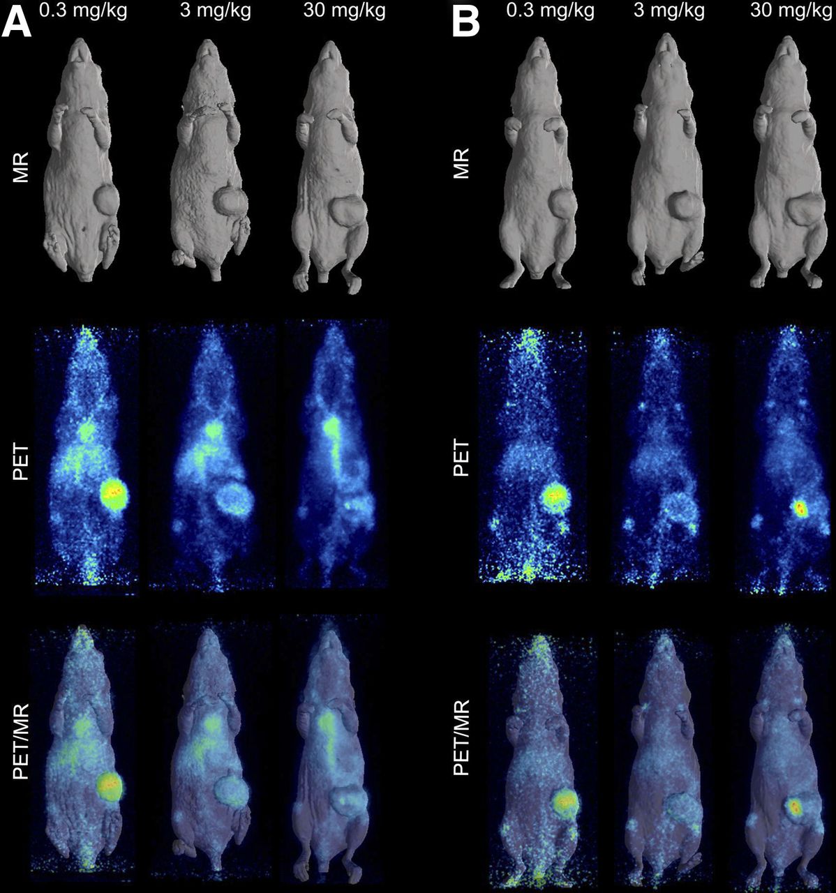
  • FIGURE 6.
    • Download figure
    • Open in new tab
    • Download powerpoint
    FIGURE 6.

    In vivo saturation of 89Zr-labeled DS-8895a as demonstrated by PET/MR on days 2 (A) and 7 (B) after injection. Representative whole-body surface-rendered MR images, maximum-intensity-projection PET images, and fused PET/MR images are shown for each time point with different dose levels of cold DS-8895a (0.3, 3, and 30 mg/kg). Hot spot in tumor on day 7 PET and PET/MR images of mouse receiving 30 mg/kg is due to blood crust.

Tables

  • Figures
    • View popup
    TABLE 1

    Pharmacokinetic Parameters of 89Zr- and 111In-Labeled DS-8895a in MDA-MB-231 Tumor–Bearing Mice

    Parameter89Zr-labeled DS-8895a111In-labeled DS-8895aP
    AUC (h × μg/mL)185.7 ± 26.96417.6 ± 90.600.0033
    t1/2α (h)1.48 ± 0.268.85 ± 4.970.0295
    t1/2β (h)102.4 ± 14.65215.1 ± 72.880.0244
    Cmax (μg/mL)3.03 ± 0.432.07 ± 0.290.0033
    CL (mL/h)0.027 ± 0.0040.012 ± 0.002<0.0001
    Vss (mL)3.90 ± 0.113.59 ± 0.430.1845
    • AUC = area under curve; t1/2α = half-life of initial phase of disposition; t1/2β = half-life of terminal phase of disposition; Cmax = maximum plasma-serum concentration; CL = total serum clearance; Vss = volume of distribution at steady state.

    • Data are mean ± SD (n = 5). P values are result of unpaired t test; Welch correction was used when variances were significantly different.

PreviousNext
Back to top

In this issue

Journal of Nuclear Medicine: 57 (6)
Journal of Nuclear Medicine
Vol. 57, Issue 6
June 1, 2016
  • Table of Contents
  • Table of Contents (PDF)
  • About the Cover
  • Index by author
Print
Download PDF
Article Alerts
Sign In to Email Alerts with your Email Address
Email Article

Thank you for your interest in spreading the word on Journal of Nuclear Medicine.

NOTE: We only request your email address so that the person you are recommending the page to knows that you wanted them to see it, and that it is not junk mail. We do not capture any email address.

Enter multiple addresses on separate lines or separate them with commas.
Molecular Imaging and Quantitation of EphA2 Expression in Xenograft Models with 89Zr-DS-8895a
(Your Name) has sent you a message from Journal of Nuclear Medicine
(Your Name) thought you would like to see the Journal of Nuclear Medicine web site.
Citation Tools
Molecular Imaging and Quantitation of EphA2 Expression in Xenograft Models with 89Zr-DS-8895a
Ingrid J.G. Burvenich, Sagun Parakh, Hui K. Gan, Fook-Thean Lee, Nancy Guo, Angela Rigopoulos, Sze-Ting Lee, Sylvia Gong, Graeme J. O’Keefe, Henri Tochon-Danguy, Masakatsu Kotsuma, Jun Hasegawa, Giorgio Senaldi, Andrew M. Scott
Journal of Nuclear Medicine Jun 2016, 57 (6) 974-980; DOI: 10.2967/jnumed.115.169839

Citation Manager Formats

  • BibTeX
  • Bookends
  • EasyBib
  • EndNote (tagged)
  • EndNote 8 (xml)
  • Medlars
  • Mendeley
  • Papers
  • RefWorks Tagged
  • Ref Manager
  • RIS
  • Zotero
Share
Molecular Imaging and Quantitation of EphA2 Expression in Xenograft Models with 89Zr-DS-8895a
Ingrid J.G. Burvenich, Sagun Parakh, Hui K. Gan, Fook-Thean Lee, Nancy Guo, Angela Rigopoulos, Sze-Ting Lee, Sylvia Gong, Graeme J. O’Keefe, Henri Tochon-Danguy, Masakatsu Kotsuma, Jun Hasegawa, Giorgio Senaldi, Andrew M. Scott
Journal of Nuclear Medicine Jun 2016, 57 (6) 974-980; DOI: 10.2967/jnumed.115.169839
Twitter logo Facebook logo LinkedIn logo Mendeley logo
  • Tweet Widget
  • Facebook Like
  • Google Plus One
Bookmark this article

Jump to section

  • Article
    • Abstract
    • MATERIALS AND METHODS
    • RESULTS
    • DISCUSSION
    • CONCLUSION
    • DISCLOSURE
    • Footnotes
    • REFERENCES
  • Figures & Data
  • Info & Metrics
  • PDF

Related Articles

  • This Month in JNM
  • PubMed
  • Google Scholar

Cited By...

  • PET-Guided Evaluation and Optimization of Internalized Antibody-Drug Conjugates Targeting Erythropoietin-Producing Hepatoma A2 Receptor
  • Google Scholar

More in this TOC Section

  • 11C-Methionine PET of Myocardial Inflammation in a Rat Model of Experimental Autoimmune Myocarditis
  • Counting Rate Characteristics and Image Distortion in Preclinical PET Imaging During Radiopharmaceutical Therapy
  • Design and Fabrication of Kidney Phantoms for Internal Radiation Dosimetry Using 3D Printing Technology
Show more Basic Science Investigations

Similar Articles

Keywords

  • bioimaging
  • DS-8895a
  • EphA2
  • 89Zr
  • cancer
SNMMI

© 2025 SNMMI

Powered by HighWire