


Article Figures & Data
Figures
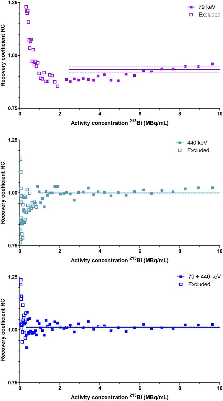
- FIGURE 1.
RCs for syringe initially filled with 86.2 MBq of 213Bi (in 2.0 mL) scanned in 15 frames of 30 min. Results were fitted with a horizontal line when both residuals were normally distributed (D’Agostini–Pearson test) and showed no significant systematic deviation (Runs test). Open symbols indicate excluded RC values. Some data points at low-activity concentrations did not fall within y-axis boundaries.
- FIGURE 2.
RCs for syringe initially filled with 67.69 MBq of 213Bi (in 2.0 mL) scanned in 45 frames of 5 min. Results were fitted with horizontal line when both residuals were normally distributed (D’Agostini–Pearson test) and showed no significant systematic deviation (Runs test). Open symbols indicate excluded RC values. Some data points at low-activity concentrations did not fall within y-axis boundaries. Data for first 13 frames were omitted from graph but were included in averaging.
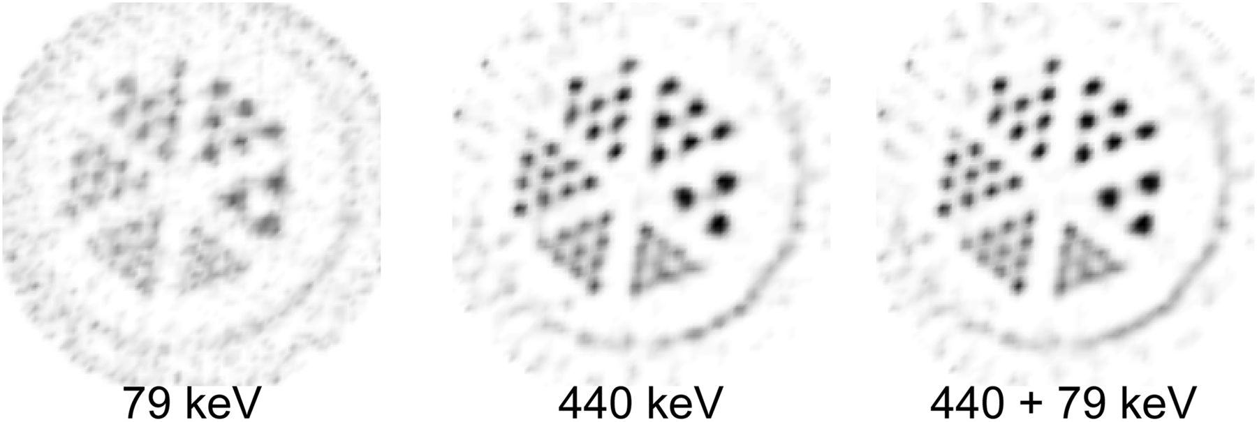
- FIGURE 3.
Resolution phantom images of 213Bi SPECT. Phantom has 6 segments containing capillary diameters of 1.5, 1.2, 1.0, 0.9, 0.8, and 0.7 mm. Images show reconstructions for different energy window settings summed over 5 slices (2 mm in total).
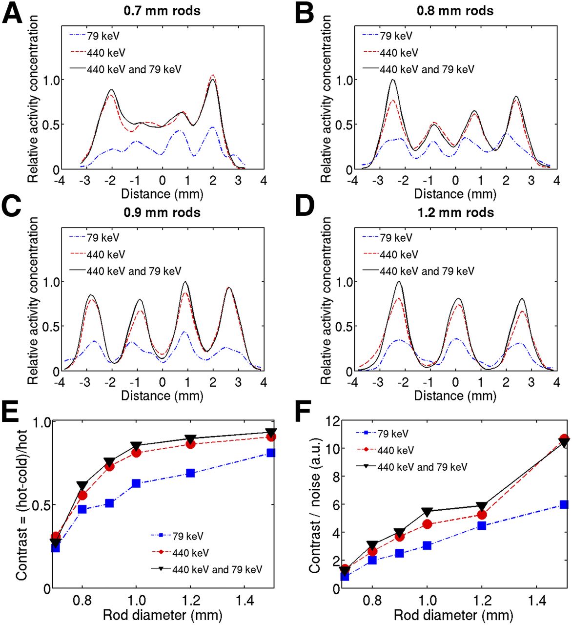
- FIGURE 4.
(A–D) Profiles through 0.7-, 0.8-, 0.9-, and 1.2-mm rods for single 79 and 440 keV and combined 79 keV/440 keV energy windows. (E and F) Contrast and contrast-to-noise curves for different rod sizes.
- FIGURE 5.
Ex vivo image of 3.0 MBq of [213Bi-DOTA,Tyr3]-octreotate injected in nude mouse. (Top to bottom) Maximum-intensity-projection images reconstructed at 79 keV, 440 keV, and both energy windows. Numbers in color table indicate radioactivity concentration in MBq/mL.
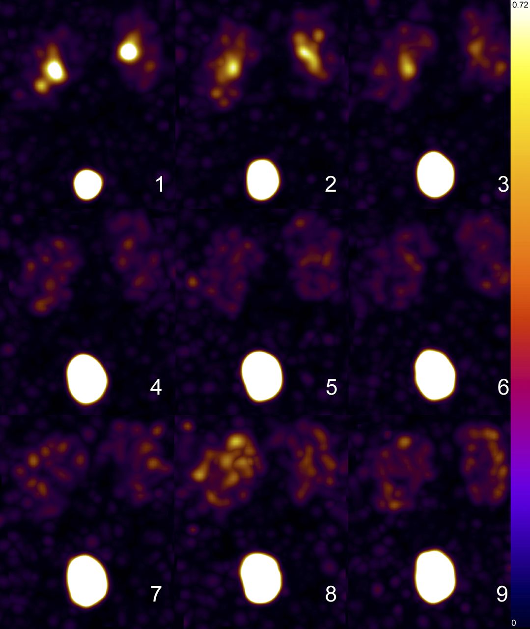
- FIGURE 6.
In vivo mouse maximum-intensity-projection images with 7.4 MBq of 213Bi-DTPA. Images were reconstructed using combined 79 keV/440 keV photopeak setting. In 9 consecutive 5-min frames, kidneys show up in each frame, initially showing ureters at top of image with gradually distribution to renal cortices and filling of bladder. Numbers in color table indicate radioactivity concentration in MBq/mL.
- FIGURE 7.
Kinetic modeling of 213Bi-DTPA in urinary bladder and kidneys. In decay-corrected data (A), bladder activity is fit by single exponential with half-life of 11 ± 2 min (effective half-life [Teff] = 15 min). Kidneys did not show clearance, whereas in uncorrected data (B) renal clearance proceeded with Teff of 52 ± 10 min. Images were reconstructed from combined 79 keV/440 keV photopeaks.


Tables
Energy window 79 keV 440 keV 79 + 440 keV RC 30-min frames (mean ± SE) 1.03 ± 0.007 1.04 ± 0.007 1.04 ± 0.009 Lower limit linearity, 30 min (MBq/mL) 0.94 0.24 0.94 RC 5-min frames (mean ± SE) 0.93 ± 0.007 1.00 ± 0.003 1.01 ± 0.003 Lower limit linearity, 5 min (MBq/mL) 2.33 0.90 0.32 FWHM, 0.7-mm rods (mm) 1.1 ± 0.3 1.4 ± 0.3 1.4 ± 0.3 FWHM, 0.8-mm rods (mm) 1.6 ± 0.5 1.4 ± 0.2 1.4 ± 0.2 FWHM, 0.9-mm rods (mm) 1.4 ± 0.6 1.33 ± 0.14 1.29 ± 0.09 FWHM, 1.0-mm rods (mm) 1.4 ± 0.5 1.34 ± 0.29 1.29 ± 0.12 FWHM, 1.2-mm rods (mm) 1.6 ± 0.6 1.30 ± 0.14 1.25 ± 0.08 FWHM, 1.5-mm rods (mm) 2.0 ± 0.4 1.58 ± 0.14 1.61 ± 0.12
Supplemental Data
Files in this Data Supplement:


