


Article Figures & Data
Figures
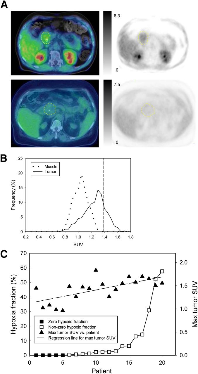
- FIGURE 1.
(A) 18F-FAZA PET images with intensity bars for SUV (left) and fused images (right) obtained from primary pancreatic cancers (dashed yellow lines) showing high (top) and low (bottom) tracer uptake. Biliary stents can be seen in each patient. (B) Histograms of SUV to mean muscle SUV ratios for individual voxels from primary tumor (solid line) and from skeletal muscle (dotted line). HF is derived as percentage of tumor voxels exceeding 3 SDs of the mean value for skeletal muscle, indicated by cursor. (C) Results for tumor SUVmax and HF obtained from 20 patients with static scans, arranged in rank order of HF.
- FIGURE 2.
(A) HF of primary tumor plotted against HF obtained for corresponding normal pancreas. (B) HF plotted against tumor volume assessed by CT.
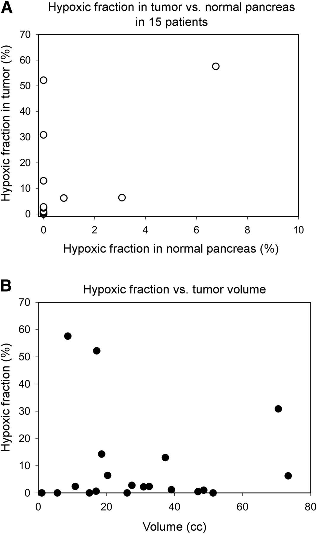
- FIGURE 3.
(A) HFs in rank order, showing tumors that showed no radiologic evidence of metastases and those with overt metastatic disease. (B) Group means of HF for tumors with and without evident metastases. Error bars are SEM; difference is nonsignificant.
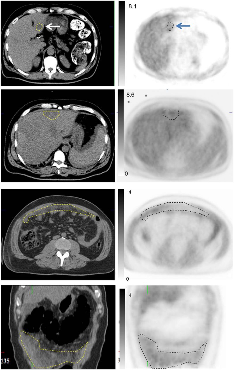
- FIGURE 4.
18F-FAZA uptake at metastatic sites. Upper 4 panels are representative sections showing liver metastases (arrows). High background uptake in normal liver obscures uptake by metastases in PET images, but these could be contoured using CT image, allowing measurement of 18F-FAZA uptake. Lower 4 panels are axial and coronal sections from patient with advanced peritoneal metastases, for which increased 18F-FAZA uptake is easily seen in PET images. SUVs represented in intensity bars.
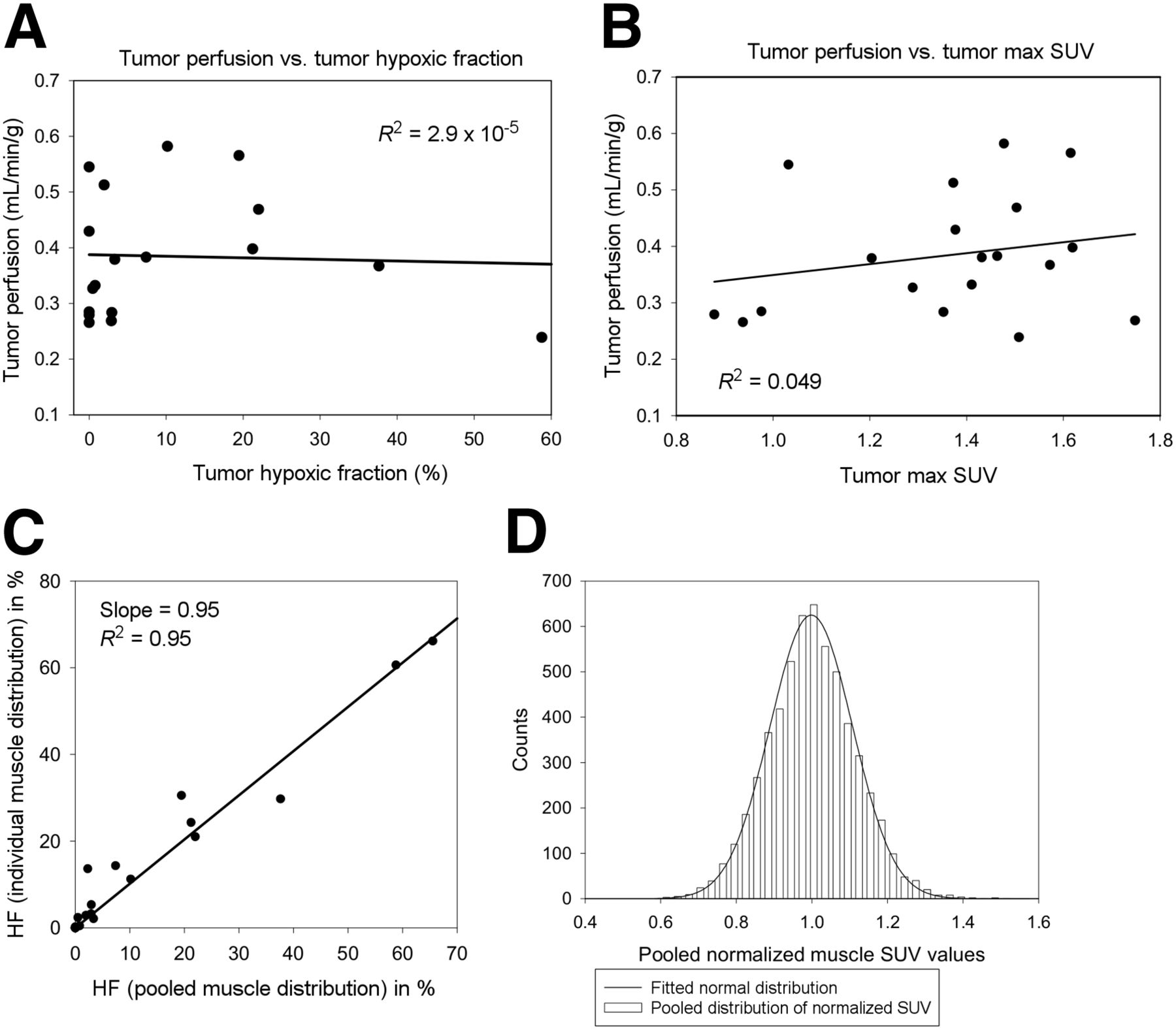
- FIGURE 5.
(A and B) Tumor perfusion, derived from dynamic PET scans, plotted against 18F-FAZA uptake in corresponding static scan, expressed as SUVmax and HF. (C) Correlation between HF values calculated using individual muscle distribution for defining threshold vs. those using pooled muscle distribution. There is strong correlation between the 2 methods (R2 = 0.95). (D) Pooled normalized SUV distribution over 20 patients consisting of 5,705 voxels in total. Data fit normal distribution function.


