


Article Figures & Data
Figures
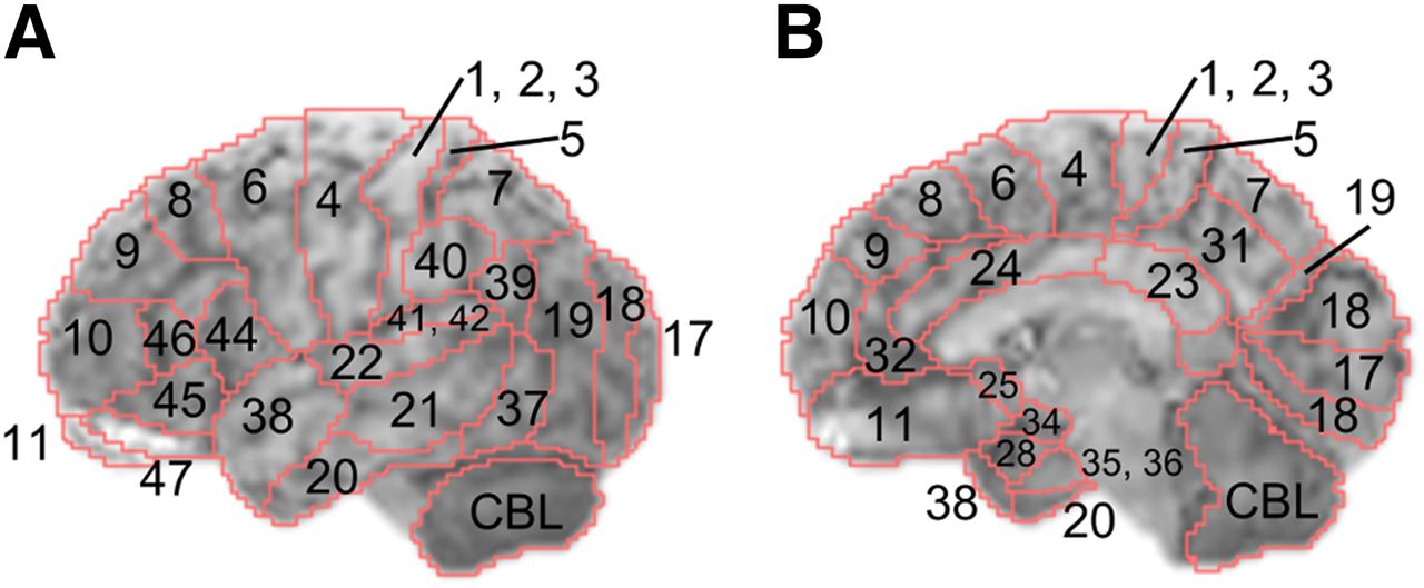
- FIGURE 1.
BAs on 3D-SSP template images based on a previous report (14). Each BA was segmented on brain surface projection atlas (MRI template) of 3D-SSP tools. In this report, right/left hemispheres and lateral/medial cortices were distinguished and analyzed individually. Figures denote left lateral (A) and right medial (B) brain regions.
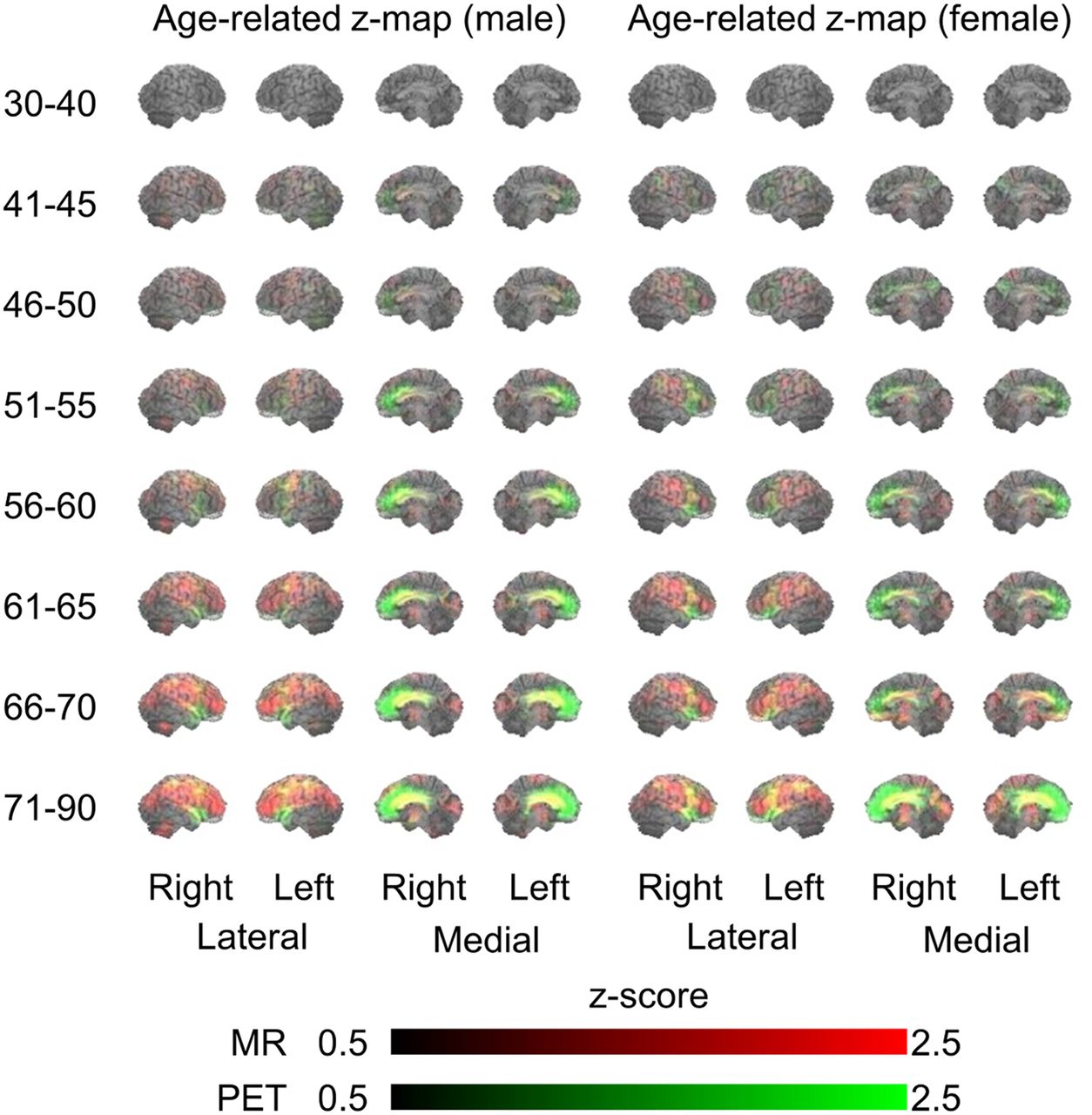
- FIGURE 2.
Age-related changes in 3D-SSP z scores of men and women in different age groups. Red and green indicate reductions in levels of MRI and PET values, respectively. Yellow denotes mixture of MRI (red) and PET (green) values, indicating parallel change.
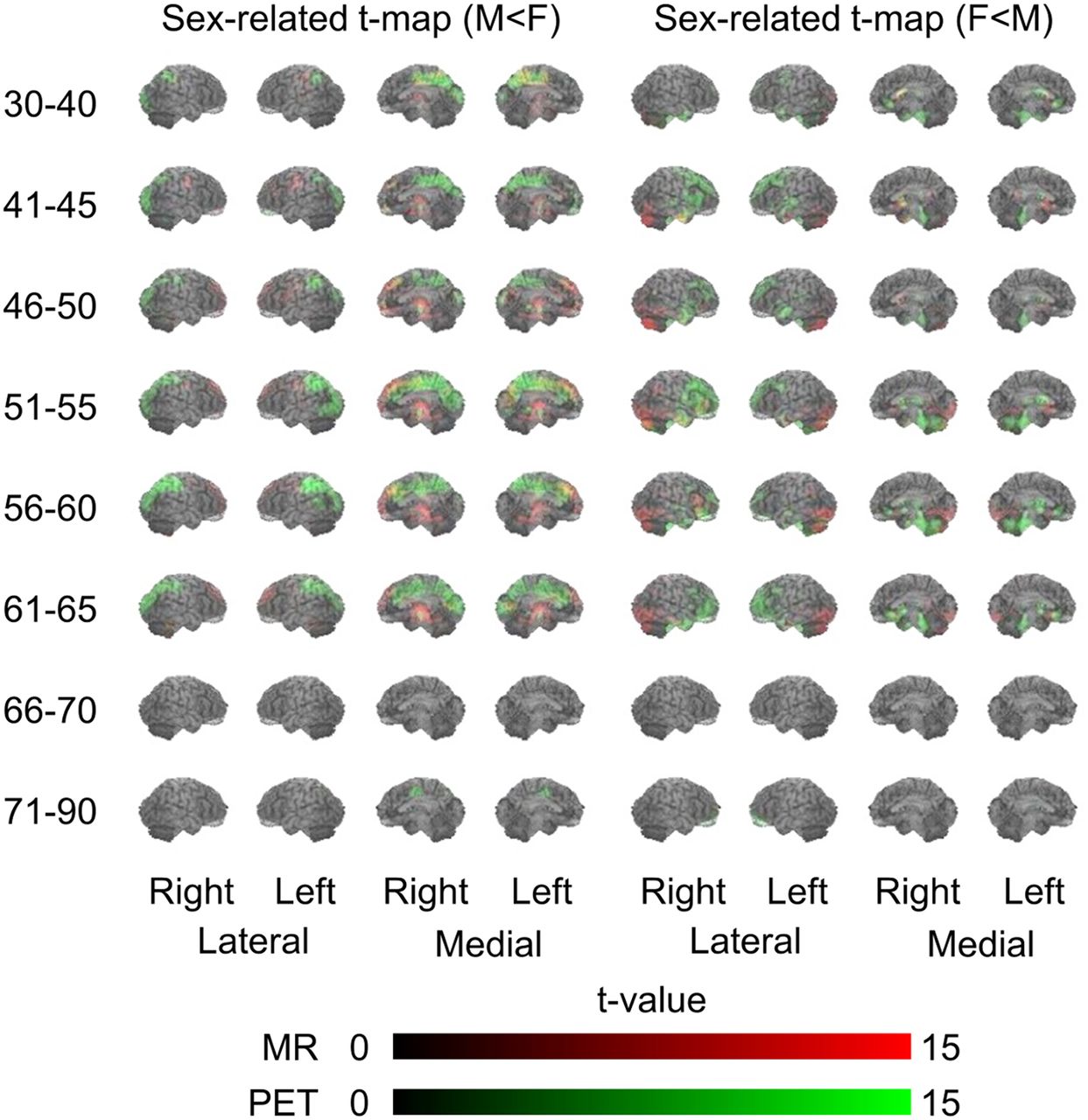
- FIGURE 3.
Sex-specific differences in 3D-SSP t scores (male < female and female < male) in different age groups. Red and green indicate reductions in levels of MRI and PET values, respectively. Yellow denotes mixture of MRI (red) and PET (green) values, indicating parallel change.
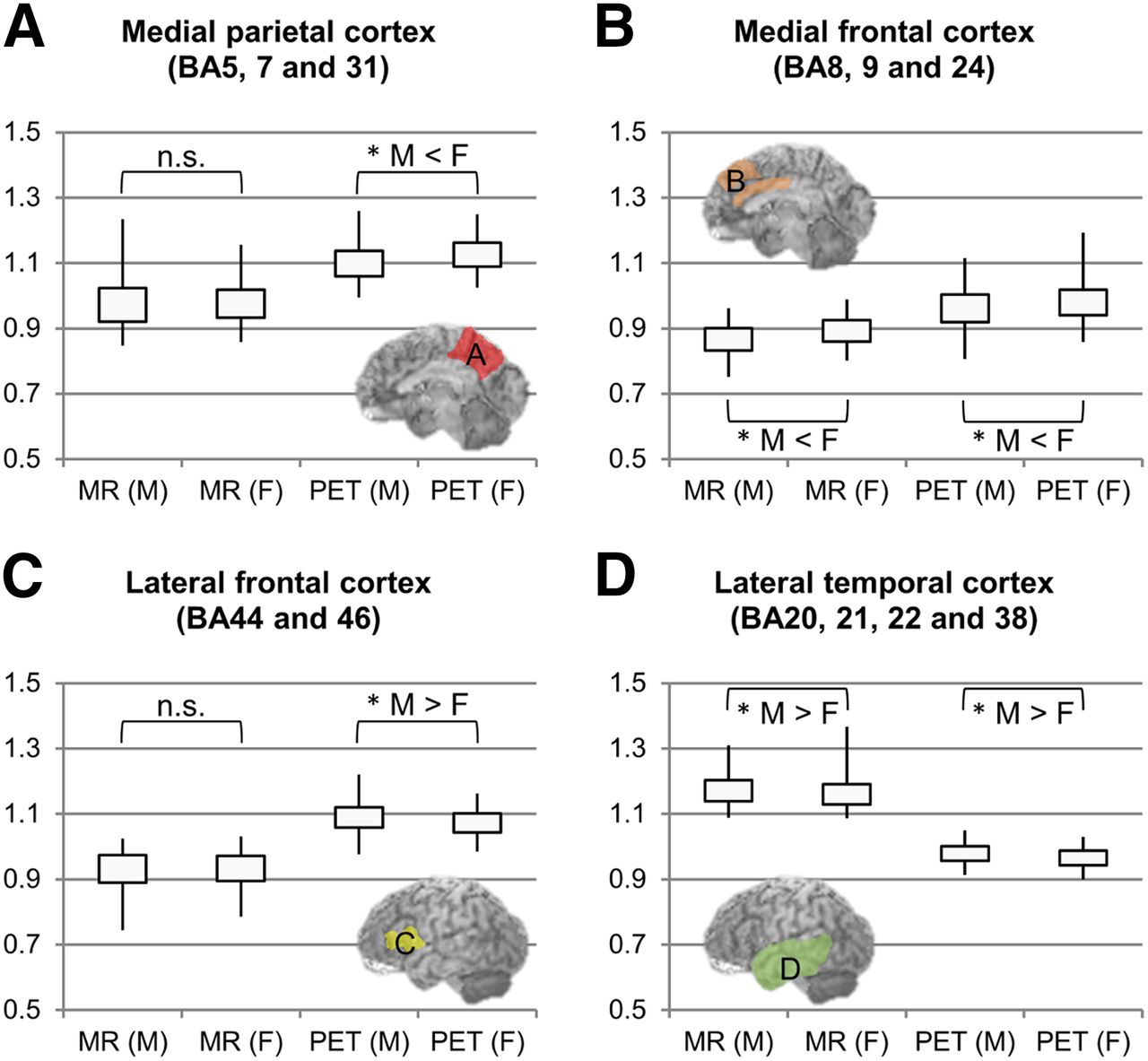
- FIGURE 4.
BA-based analysis between sexes. Vertical axes show pixel value corrected for whole-brain count. (A and C) Asterisks tagged on PET denote dominant changes in metabolism. (B and D) Presence of both asterisks (MRI and PET) denotes parallel changes in morphology and metabolism. Image insets indicate BAs of interest. n.s. = nonsignificant. *P < 0.05.


Tables
Men Women Comorbid disease Age (y) n MMSE n MMSE HT HC DM Insomnia Anxiety OP Other 30–40 40 29.9 ± 0.3 33 29.7 ± 0.5 0 M, 0 F 0 M, 0 F 0 M, 0 F 1 M, 0 F 1 M, 0 F 0 M, 1 F 1 M, 0 F 41–45 85 29.8 ± 0.4 91 29.8 ± 0.4 1 M, 1 F 0 M, 1 F 0 M, 0 F 0 M, 0 F 1 M, 0 F 1 M, 0 F 2 M, 3 F 46–50 75 29.8 ± 0.4 75 29.7 ± 0.4 1 M, 0 F 0 M, 1 F 0 M, 0 F 1 M, 1 F 1 M, 1 F 2 M, 0 F 1 M, 3 F 51–55 81 29.8 ± 0.4 76 29.6 ± 0.5 9 M, 8 F 0 M, 2 F 1 M, 0 F 0 M, 0 F 0 M, 0 F 0 M, 1 F 0 M, 3 F 56–60 72 29.7 ± 0.5 81 29.5 ± 0.6 10 M, 8 F 6 M, 6 F 1 M, 1 F 1 M, 1 F 1 M, 2 F 0 M, 4 F 0 M, 3 F 61–65 70 29.4 ± 0.8 67 29.2 ± 0.8 19 M, 9 F 8 M, 13 F 2 M, 3 F 3 M, 7 F 4 M, 3 F 2 M, 5 F 1 M, 0 F 66–70 38 29.3 ± 0.9 24 29.4 ± 0.9 6 M, 7 F 7 M, 6 F 2 M, 1 F 5 M, 2 F 2 M, 0 F 0 M, 1 F 0 M, 0 F 71–90 32 29.1 ± 1.0 23 28.7 ± 1.4 8 M, 11 F 2 M, 7 F 3 M, 0 F 1 M, 6 F 3 M, 1 F 0 M, 1 F 1 M, 0 F Total 493 29.6 ± 0.6 470 29.5 ± 0.7 54 M, 44 F 23 M, 36 F 9 M, 5 F 12 M, 17 F 13 M, 7 F 5 M, 13 F 6 M, 12 F MMSE = mini-mental state examination; HT = hypertension; HC = hypercholesterolemia; DM = diabetes mellitus; OP = orthopedic illness (arthritis, lumbar pain) treated with analgesics; other = hormonal replacement, pollakiuria, arrhythmia.
Supplemental Data
Files in this Data Supplement:


Jump to section
Related Articles
Cited By...
- The Uniqueness of Human Vulnerability to Brain Aging in Great Ape Evolution
- Nandrolone Decanoate supplementation promotes AMPK activation and divergent brain connectivity rearrangements in adult and aged mice
- Quality control strategies for brain MRI segmentation and parcellation: practical approaches and recommendations - insights from The Maastricht Study
- Time Courses of Cortical Glucose Metabolism and Microglial Activity Across the Life Span of Wild-Type Mice: A PET Study