
Main menu

  • Home
  • Content
    • Current
    • Ahead of print
    • Past Issues
    • JNM Supplement
    • SNMMI Annual Meeting Abstracts
    • Continuing Education
    • JNM Podcasts
  • Subscriptions
    • Subscribers
    • Institutional and Non-member
    • Rates
    • Journal Claims
    • Corporate & Special Sales
  • Authors
    • Submit to JNM
    • Information for Authors
    • Assignment of Copyright
    • AQARA requirements
  • Info
    • Reviewers
    • Permissions
    • Advertisers
  • About
    • About Us
    • Editorial Board
    • Contact Information
  • More
    • Alerts
    • Feedback
    • Help
    • SNMMI Journals
  • SNMMI
    • JNM
    • JNMT
    • SNMMI Journals
    • SNMMI

User menu

  • Subscribe
  • My alerts
  • Log in
  • My Cart

Search

  • Advanced search
Journal of Nuclear Medicine
  • SNMMI
    • JNM
    • JNMT
    • SNMMI Journals
    • SNMMI
  • Subscribe
  • My alerts
  • Log in
  • My Cart
Journal of Nuclear Medicine

Advanced Search

  • Home
  • Content
    • Current
    • Ahead of print
    • Past Issues
    • JNM Supplement
    • SNMMI Annual Meeting Abstracts
    • Continuing Education
    • JNM Podcasts
  • Subscriptions
    • Subscribers
    • Institutional and Non-member
    • Rates
    • Journal Claims
    • Corporate & Special Sales
  • Authors
    • Submit to JNM
    • Information for Authors
    • Assignment of Copyright
    • AQARA requirements
  • Info
    • Reviewers
    • Permissions
    • Advertisers
  • About
    • About Us
    • Editorial Board
    • Contact Information
  • More
    • Alerts
    • Feedback
    • Help
    • SNMMI Journals
  • View or Listen to JNM Podcast
  • Visit JNM on Facebook
  • Join JNM on LinkedIn
  • Follow JNM on Twitter
  • Subscribe to our RSS feeds
Research ArticleClinical Investigations

Evidence of Brain Inflammation in Patients with Human T-Lymphotropic Virus Type 1–Associated Myelopathy (HAM): A Pilot, Multimodal Imaging Study Using 11C-PBR28 PET, MR T1-Weighted, and Diffusion-Weighted Imaging

Rahul Dimber, Qi Guo, Courtney Bishop, Adine Adonis, Aisling Buckley, Agnes Kocsis, David Owen, Nicola Kalk, Rexford Newbould, Roger N. Gunn, Eugenii A. Rabiner and Graham P. Taylor
Journal of Nuclear Medicine December 2016, 57 (12) 1905-1912; DOI: https://doi.org/10.2967/jnumed.116.175083
Rahul Dimber
1Imanova, Centre for Imaging Sciences, London, United Kingdom
  • Find this author on Google Scholar
  • Find this author on PubMed
  • Search for this author on this site
Qi Guo
2Institute of Psychiatry, King’s College London, London, United Kingdom
  • Find this author on Google Scholar
  • Find this author on PubMed
  • Search for this author on this site
Courtney Bishop
1Imanova, Centre for Imaging Sciences, London, United Kingdom
  • Find this author on Google Scholar
  • Find this author on PubMed
  • Search for this author on this site
Adine Adonis
3National Centre for Human Retrovirology, St. Mary's Hospital, Imperial College Healthcare NHS Trust, London, United Kingdom
  • Find this author on Google Scholar
  • Find this author on PubMed
  • Search for this author on this site
Aisling Buckley
4Department of Clinical Health Psychology, St. Mary's Hospital, Imperial College Healthcare NHS Trust, London, United Kingdom; and
  • Find this author on Google Scholar
  • Find this author on PubMed
  • Search for this author on this site
Agnes Kocsis
4Department of Clinical Health Psychology, St. Mary's Hospital, Imperial College Healthcare NHS Trust, London, United Kingdom; and
  • Find this author on Google Scholar
  • Find this author on PubMed
  • Search for this author on this site
David Owen
5Division of Brain Sciences, Imperial College London, London, United Kingdom
  • Find this author on Google Scholar
  • Find this author on PubMed
  • Search for this author on this site
Nicola Kalk
5Division of Brain Sciences, Imperial College London, London, United Kingdom
  • Find this author on Google Scholar
  • Find this author on PubMed
  • Search for this author on this site
Rexford Newbould
1Imanova, Centre for Imaging Sciences, London, United Kingdom
  • Find this author on Google Scholar
  • Find this author on PubMed
  • Search for this author on this site
Roger N. Gunn
1Imanova, Centre for Imaging Sciences, London, United Kingdom
5Division of Brain Sciences, Imperial College London, London, United Kingdom
  • Find this author on Google Scholar
  • Find this author on PubMed
  • Search for this author on this site
Eugenii A. Rabiner
1Imanova, Centre for Imaging Sciences, London, United Kingdom
2Institute of Psychiatry, King’s College London, London, United Kingdom
  • Find this author on Google Scholar
  • Find this author on PubMed
  • Search for this author on this site
Graham P. Taylor
3National Centre for Human Retrovirology, St. Mary's Hospital, Imperial College Healthcare NHS Trust, London, United Kingdom
  • Find this author on Google Scholar
  • Find this author on PubMed
  • Search for this author on this site
  • Article
  • Figures & Data
  • Supplemental
  • Info & Metrics
  • PDF
Loading

Article Figures & Data

Figures

  • Tables
  • Additional Files
  • FIGURE 1.
    • Download figure
    • Open in new tab
    • Download powerpoint
    FIGURE 1.

    Comparison of regional VT (left) and DVR (right) of 11C-PBR28 in HTLV1 subjects compared with healthy volunteers. Error bar is 1 SD. Amy = amygdala; Asym1 = patient 6, asymptomatic; Asym2 = patient 7, asymptomatic; Br.Stem = brain stem; Cau = caudate; Cereb = cerebellum; HAMs1 = patient 1, severe HAM; HAMs2 = patient 2, severe HAM; HAMmi = patient 4, mild HAM; HAMmo = patient 5, moderate HAM; Hip = hippocampus; mid = mid brain; Tha = thalamus.

  • FIGURE 2.
    • Download figure
    • Open in new tab
    • Download powerpoint
    FIGURE 2.

    Spatially normalized VT parametric maps of individual HTLV-1 carriers and population-averaged healthy controls. Asym1 = patient 6, asymptomatic; Asym2 = patient 7, asymptomatic; Avg = average; HAMs1 = patient 1, severe HAM; HAMs2 = patient 2, severe HAM; HAMmi = patient 4, mild HAM; HAMmo = patient 5, moderate HAM.
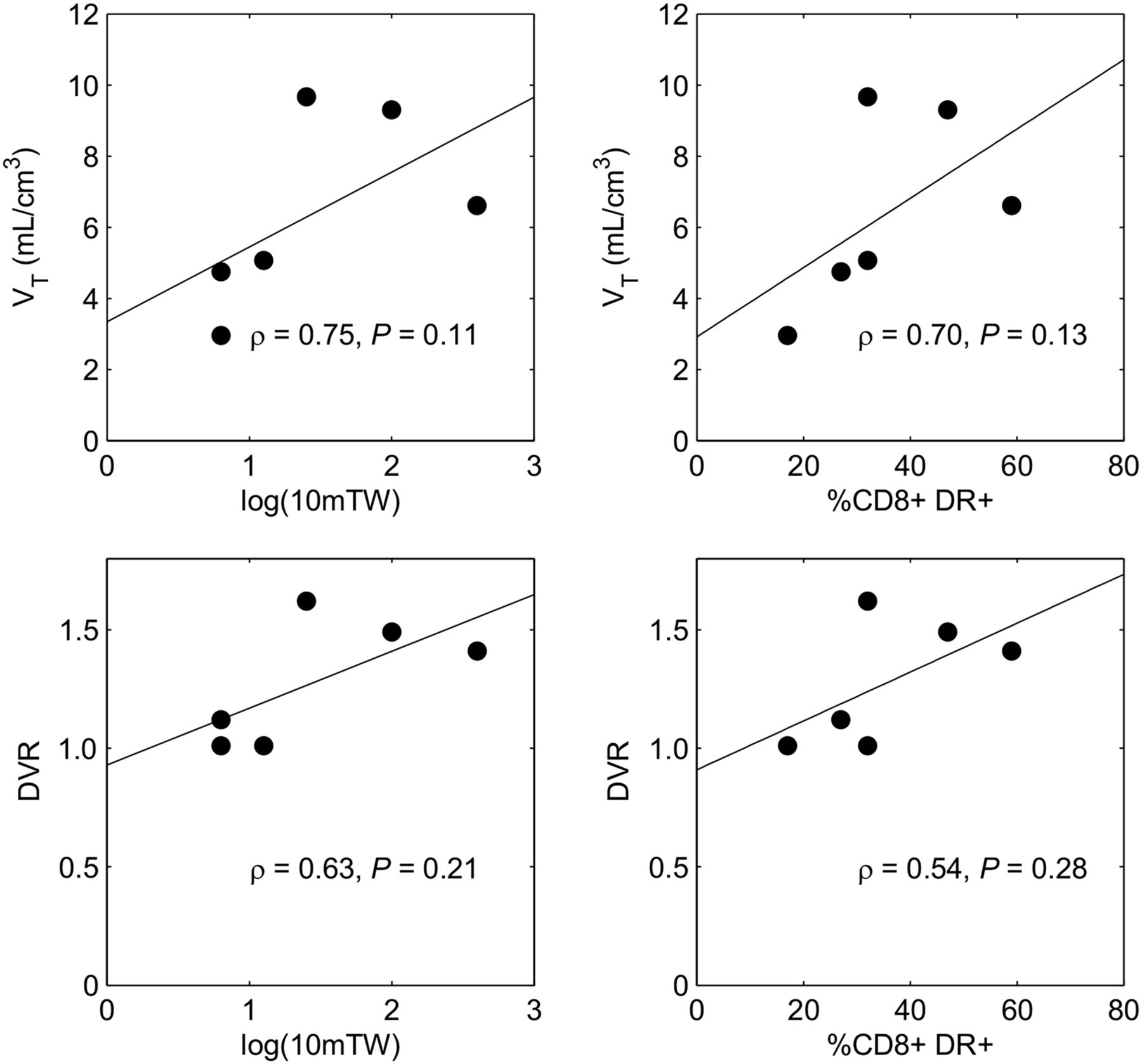
  • FIGURE 3.
    • Download figure
    • Open in new tab
    • Download powerpoint
    FIGURE 3.

    Spearman rank correlation analysis of in vivo thalamus PET outcome measures in HTLV-1–infected subjects with laboratory and clinical assessments. Top (left to right): correlation between VT and 10-m walk time in log scale and CD8+DR+ cell counts. Bottom (left to right): correlation between DVR and 10-m walk time in log scale and CD8+DR+ cell counts. TW = timed walk.

  • FIGURE 4.
    • Download figure
    • Open in new tab
    • Download powerpoint
    FIGURE 4.

    VBM analysis: small clusters of altered GM topography in HTLV-1–infected patients compared with controls.
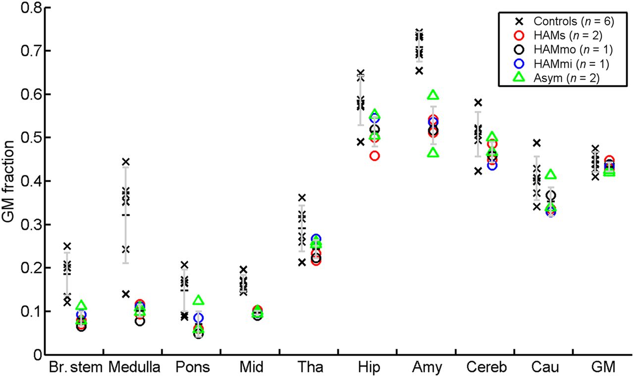
  • FIGURE 5.
    • Download figure
    • Open in new tab
    • Download powerpoint
    FIGURE 5.

    Regional GM fractions in HTLV-1 patients compared with healthy volunteers. Error bar is 1 SD.
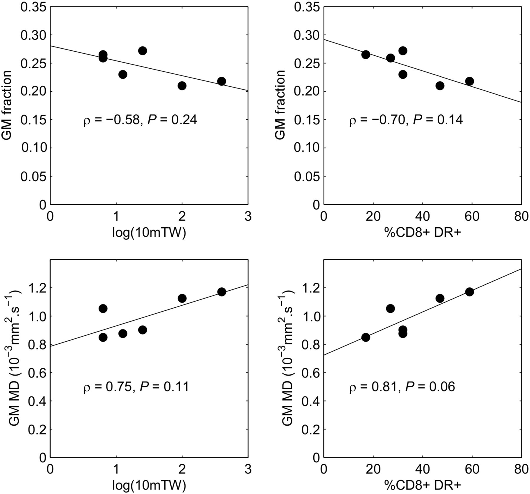
  • FIGURE 6.
    • Download figure
    • Open in new tab
    • Download powerpoint
    FIGURE 6.

    Correlations of left thalamus GM fraction (top) and right thalamus MD (bottom) with (left to right) clinical (10-m timed walk) and laboratory (% circulating CD8+ lymphocytes expressing HLA-DR) scores.

Tables

  • Figures
  • Additional Files
    • View popup
    TABLE 1

    Demographics, Neurology, HTLV-1 Virology, and Immunology

    Subject no. in analysisAge (y)SexEthnicityDuration disease (y)Mobility aid10-m TW (s)PETMRIDWIHTLV-1* proviral loadCD8 DRDisease
    #1 HAMs155FemaleCaucasian19Frame36655 min✓✓4.959Severe HAM progressing
    #2 HAMs248FemaleCaucasian5Frame109✓✓✓17.447Severe HAM progressing
    #3 HAMmo66FemaleAfro-Caribbean81 stick14.50 min✓x3.842Moderate HAM stable
    #4 HAMmi59FemaleAfro-Caribbean2Unaided11.5✓✓✓7.932Mild HAM stable
    #5 HAMmo54MaleAfro-Caribbean13Elbow crutches24.7✓✓✓10.532Moderate HAM slow progression
    #6 Asym157FemaleAfro-CaribbeanN/AUnaided6.5✓✓✓6.427Asymptomatic
    #7 Asym257MaleAfro-CaribbeanN/AUnaided6.6✓✓✓2.617Asymptomatic
    • ↵* HTLV-1 DNA copies per 100 PBMCs.

    • 10-m TW = time taken to walk 10 m on the flat; CD8 DR = % CD8 lymphocytes expressing human leukocyte antigen–antigen D related; HAMs1 = patient 1, severe HAM; HAMs2 = patient 2, severe HAM; HAMmo = patient 5, moderate HAM; HAMmi = patient 4, mild HAM; Asym1 = patient 6, asymptomatic; Asym 2 = patient 7, asymptomatic.

    • View popup
    TABLE 2

    PET Regional VT Data Analysis: z Scores of Individual Patient Regional VT Values Compared with Healthy Volunteers

    Subject codeHAMs1HAMs2HAMmoHAMmiAsym1Asym2
    Brain stem−0.301.942.11−0.06−0.19−1.30
    Medulla−0.331.882.270.25−0.28−0.68
    Pons−0.482.011.30−0.10−0.17−1.50
    Mid brain−0.051.382.98−0.20−0.25−1.24
    Thalamus0.602.452.71−0.47−0.16−1.93
    Hippocampus−0.551.671.76−0.33−0.19−1.34
    Amygdala−0.621.220.95−0.73−0.28−1.35
    Cerebellum−0.221.951.540.27−0.17−1.56
    Caudate−0.071.072.081.32−0.27−2.10
    Gray matter−0.161.291.080.12−0.13−1.61
    Brain−0.171.381.120.25−0.13−1.57
    • HAMs1 = patient 1, severe HAM; HAMs2 = patient 2, severe HAM; HAMmo = patient 5, moderate HAM; HAMmi = patient 4, mild HAM; Asym1 = patient 6, asymptomatic; Asym2 = patient 7, asymptomatic.

    • View popup
    TABLE 3

    PET Regional DVR Data Analysis: z Scores of Individual Patient Regional DVR Values Compared with Healthy Volunteers

    Subject codeHAMs1HAMs2HAMmoHAMmiAsym1Asym2
    Brain stem−0.403.084.23−0.29−0.99−0.11
    Medulla−0.292.032.920.52−0.370.78
    Pons−0.802.481.46−0.36−0.70−0.93
    Mid brain0.431.356.26−0.83−1.600.33
    Thalamus4.726.168.84−3.33−1.17−3.33
    Hippocampus−1.572.443.52−1.53−1.600.31
    Amygdala−1.490.940.77−2.42−2.30−0.31
    Cerebellum−0.273.352.790.79−1.660.17
    Caudate0.05−0.301.071.42−1.07−0.81
    Gray matter0.413.394.010.17−2.080.00
    Brain0.416.135.944.44−2.482.58
    • HAMs1 = patient 1, severe HAM; HAMs2 = patient 2, severe HAM; HAMmo = patient 5, moderate HAM; HAMmi = patient 4, mild HAM; Asym1 = patient 6, asymptomatic; Asym2 = patient 7, asymptomatic.

Additional Files

  • Figures
  • Tables
  • Supplemental Data

    Files in this Data Supplement:

    • Supplemental Data
PreviousNext
Back to top

In this issue

Journal of Nuclear Medicine: 57 (12)
Journal of Nuclear Medicine
Vol. 57, Issue 12
December 1, 2016
  • Table of Contents
  • Table of Contents (PDF)
  • About the Cover
  • Index by author
Print
Download PDF
Article Alerts
Sign In to Email Alerts with your Email Address
Email Article

Thank you for your interest in spreading the word on Journal of Nuclear Medicine.

NOTE: We only request your email address so that the person you are recommending the page to knows that you wanted them to see it, and that it is not junk mail. We do not capture any email address.

Enter multiple addresses on separate lines or separate them with commas.
Evidence of Brain Inflammation in Patients with Human T-Lymphotropic Virus Type 1–Associated Myelopathy (HAM): A Pilot, Multimodal Imaging Study Using 11C-PBR28 PET, MR T1-Weighted, and Diffusion-Weighted Imaging
(Your Name) has sent you a message from Journal of Nuclear Medicine
(Your Name) thought you would like to see the Journal of Nuclear Medicine web site.
Citation Tools
Evidence of Brain Inflammation in Patients with Human T-Lymphotropic Virus Type 1–Associated Myelopathy (HAM): A Pilot, Multimodal Imaging Study Using 11C-PBR28 PET, MR T1-Weighted, and Diffusion-Weighted Imaging
Rahul Dimber, Qi Guo, Courtney Bishop, Adine Adonis, Aisling Buckley, Agnes Kocsis, David Owen, Nicola Kalk, Rexford Newbould, Roger N. Gunn, Eugenii A. Rabiner, Graham P. Taylor
Journal of Nuclear Medicine Dec 2016, 57 (12) 1905-1912; DOI: 10.2967/jnumed.116.175083

Citation Manager Formats

  • BibTeX
  • Bookends
  • EasyBib
  • EndNote (tagged)
  • EndNote 8 (xml)
  • Medlars
  • Mendeley
  • Papers
  • RefWorks Tagged
  • Ref Manager
  • RIS
  • Zotero
Share
Evidence of Brain Inflammation in Patients with Human T-Lymphotropic Virus Type 1–Associated Myelopathy (HAM): A Pilot, Multimodal Imaging Study Using 11C-PBR28 PET, MR T1-Weighted, and Diffusion-Weighted Imaging
Rahul Dimber, Qi Guo, Courtney Bishop, Adine Adonis, Aisling Buckley, Agnes Kocsis, David Owen, Nicola Kalk, Rexford Newbould, Roger N. Gunn, Eugenii A. Rabiner, Graham P. Taylor
Journal of Nuclear Medicine Dec 2016, 57 (12) 1905-1912; DOI: 10.2967/jnumed.116.175083
Twitter logo Facebook logo LinkedIn logo Mendeley logo
  • Tweet Widget
  • Facebook Like
  • Google Plus One
Bookmark this article

Jump to section

  • Article
    • Abstract
    • MATERIALS AND METHODS
    • RESULTS
    • DISCUSSION
    • CONCLUSION
    • DISCLOSURE
    • Acknowledgments
    • Footnotes
    • REFERENCES
  • Figures & Data
  • Supplemental
  • Info & Metrics
  • PDF

Related Articles

  • This Month in JNM
  • PubMed
  • Google Scholar

Cited By...

  • Imaging Spectrum of HTLV-1-Related Neurologic Disease: A Pooled Series and Review
  • Google Scholar

More in this TOC Section

  • Cardiac Presynaptic Sympathetic Nervous Function Evaluated by Cardiac PET in Patients with Chronotropic Incompetence Without Heart Failure
  • Validation and Evaluation of a Vendor-Provided Head Motion Correction Algorithm on the uMI Panorama PET/CT System
  • Left Ventricular Strain from Myocardial Perfusion PET Imaging: Method Development and Comparison to 2-Dimensional Echocardiography
Show more Clinical Investigations

Similar Articles

Keywords

  • HTLV1
  • HAM
  • neuroinflammation
  • MRI
  • DWI
  • PET
  • [11C] PBR28
SNMMI

© 2025 SNMMI

Powered by HighWire