


Article Figures & Data
Figures
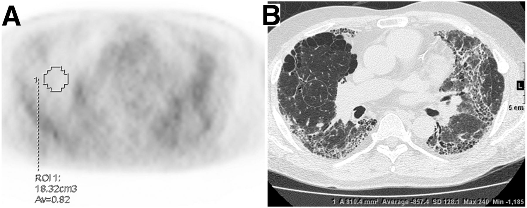
- FIGURE 1.
Representative images of VOIs and region of interest for SUVmean and CTmean. (A) VOI of 18 cm3 was manually placed on background lung field of PET image, and SUVmean was automatically calculated as 0.82 on a workstation. (B) Corresponding region of interest was manually placed on HRCT image, and CTmean was automatically calculated as –857.
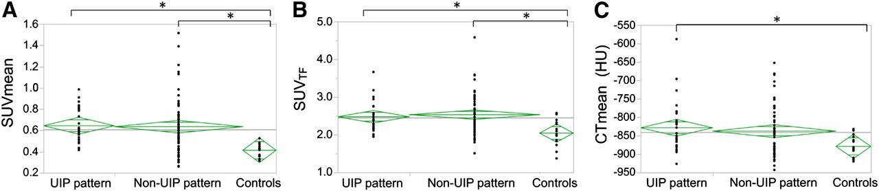
- FIGURE 2.
SUVmean (A), SUVTF (B), and CTmean (C) of ILD patients with UIP pattern, non-UIP pattern, and healthy controls. Horizontal bars in each rhombus represent 25th, 50th, and 75th percentiles from top. *P < 0.016 (Bonferroni adjustment).
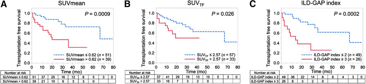
- FIGURE 3.
Kaplan–Meier analysis of SUVmean (≤0.62 vs. >0.62) (A), SUVTF (≤2.57 vs. >2.57) (B), and ILD-GAP index (<3 vs. ≥3) (C) as predictors of TFS in patients with ILD. Patients with higher SUVmean (>0.62), higher SUVTF (>2.57), and higher ILD-GAP index (≥3) had significantly poorer prognosis.
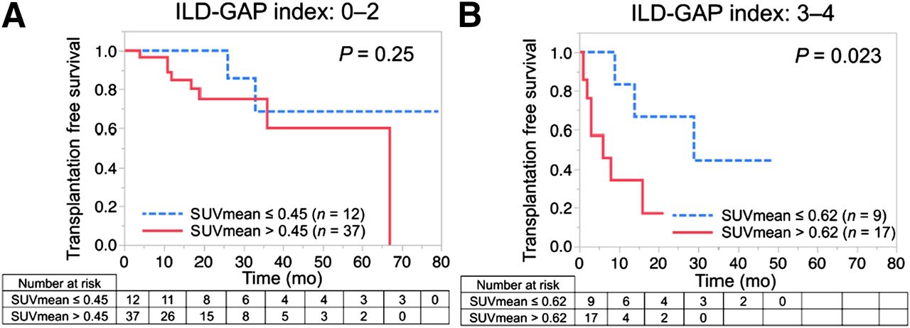
- FIGURE 4.
Kaplan–Meier analysis of SUVmean as predictor of TFS in groups of patients with ILD-GAP index of 0–2 (A) and 3–4 (B). In patients with ILD-GAP index of 0–2, there was no difference in TFS between patients with higher (>0.45) and lower (≤0.45) SUVmean. In group with moderate mortality risk (ILD-GAP index 3–4), however, patients with higher SUVmean (>0.62) had significantly poorer TFS than those with lower SUVmean (≤0.62).


Tables
Characteristic Mean age ± SD (y) 55.4 ± 11.0 Sex (male:female) 51:39 (56.7%:43.3%) Lung transplantation (Yes:No) 18:72 (20.0%:80.0%) Type of ILD IPF 24 (26.7%) Unclassifiable ILD 7 (7.8%) CT-ILD/idiopathic NSIP 55 (61.1%) Chronic HP 4 (4.4%) PFT Mean %FVC ± SD (n = 75; %)* 58.1 ± 22.1 Mean %DLco ± SD (n = 61; %)* 29.9 ± 15.7 ILD-GAP index (n = 75)* 0 9 (12.0%) 1 15 (20.0%) 2 25 (33.3%) 3 19 (25.3%) 4 7 (9.3%) 5–8 0 Median observation period (d) 395 (range, 2–2,392) ↵* Because data were not acquired during designated period, numbers do not total 90.
NSIP = nonspecific interstitial pneumonia; HP = hypersensitivity pneumonitis.
Image parameters Values Mean SUVmax ± SD (n = 90) 2.46 ± 0.76 Mean SUVmean ± SD (n = 90) 0.60 ± 0.24 Mean SUVTF ± SD (n = 90) 2.44 ± 0.50 Mean CTmean ± SD (HU) (n = 83) −833 ± 68 TF = tissue fraction.
Image parameters %FVC %DLco KL-6 SP-D CRP LDH SUVmax NS NS 0.29 (0.014) NS 0.22 (0.043) NS SUVmean −0.45 (<.0001) −0.46 (0.0002) 0.56 (<.0001) 0.36 (0.0098) NS NS SUVTF NS −0.29 (0.022) 0.41 (0.0003) 0.29 (0.040) NS 0.066 (0.040) CTmean −0.50 (<.0001) −0.40 (0.0028) 0.29 (0.019) 0.40 (0.0060) NS NS NS = not significant; TF = tissue fraction.
Rho (ρ) values are demonstrated. P values are in parentheses.
Variable n HR 95% CI P Age 90 1.00 0.96–1.03 0.81 Sex 90 3.47 1.44–9.65 0.0047* UIP or non-UIP pattern 90 0.70 0.25–1.71 0.45 %FVC 75 0.97 0.95–0.99 0.0087* %DLco 61 0.93 0.88–0.97 0.0002* ILD-GAP index 75 2.28 1.44–3.82 0.0003* KL-6 73 1.00 1.00–1.00 0.049* SP-D 49 1.00 1.00–1.01 0.050 CRP 85 0.81 0.52–1.05 0.13 LDH 83 1.00 1.00–1.00 0.87 SUVmax 90 0.97 0.58–1.62 0.91 SUVmean 90 51.0 8.24–306 <0.0001* SUVTF 90 3.08 1.42–6.26 0.0055* CTmean 84 1.00 1.00–1.01 0.059 ↵* P < 0.05.
HR = hazard ratio; CI = confidence interval; TF = tissue fraction.
Supplemental Data
Files in this Data Supplement:


