
Main menu

  • Home
  • Content
    • Current
    • Ahead of print
    • Past Issues
    • JNM Supplement
    • SNMMI Annual Meeting Abstracts
    • Continuing Education
    • JNM Podcasts
  • Subscriptions
    • Subscribers
    • Institutional and Non-member
    • Rates
    • Journal Claims
    • Corporate & Special Sales
  • Authors
    • Submit to JNM
    • Information for Authors
    • Assignment of Copyright
    • AQARA requirements
  • Info
    • Reviewers
    • Permissions
    • Advertisers
  • About
    • About Us
    • Editorial Board
    • Contact Information
  • More
    • Alerts
    • Feedback
    • Help
    • SNMMI Journals
  • SNMMI
    • JNM
    • JNMT
    • SNMMI Journals
    • SNMMI

User menu

  • Subscribe
  • My alerts
  • Log in
  • My Cart

Search

  • Advanced search
Journal of Nuclear Medicine
  • SNMMI
    • JNM
    • JNMT
    • SNMMI Journals
    • SNMMI
  • Subscribe
  • My alerts
  • Log in
  • My Cart
Journal of Nuclear Medicine

Advanced Search

  • Home
  • Content
    • Current
    • Ahead of print
    • Past Issues
    • JNM Supplement
    • SNMMI Annual Meeting Abstracts
    • Continuing Education
    • JNM Podcasts
  • Subscriptions
    • Subscribers
    • Institutional and Non-member
    • Rates
    • Journal Claims
    • Corporate & Special Sales
  • Authors
    • Submit to JNM
    • Information for Authors
    • Assignment of Copyright
    • AQARA requirements
  • Info
    • Reviewers
    • Permissions
    • Advertisers
  • About
    • About Us
    • Editorial Board
    • Contact Information
  • More
    • Alerts
    • Feedback
    • Help
    • SNMMI Journals
  • View or Listen to JNM Podcast
  • Visit JNM on Facebook
  • Join JNM on LinkedIn
  • Follow JNM on Twitter
  • Subscribe to our RSS feeds
Research ArticleBasic Science Investigations

Remodeling the Vascular Microenvironment of Glioblastoma with α-Particles

Katja Behling, William F. Maguire, Valentina Di Gialleonardo, Lukas E.M. Heeb, Iman F. Hassan, Darren R. Veach, Kayvan R. Keshari, Philip H. Gutin, David A. Scheinberg and Michael R. McDevitt
Journal of Nuclear Medicine November 2016, 57 (11) 1771-1777; DOI: https://doi.org/10.2967/jnumed.116.173559
Katja Behling
1Department of Radiology, Memorial Sloan Kettering Cancer Center, New York, New York
  • Find this author on Google Scholar
  • Find this author on PubMed
  • Search for this author on this site
William F. Maguire
2Department of Molecular Pharmacology, Memorial Sloan Kettering Cancer Center, New York, New York
  • Find this author on Google Scholar
  • Find this author on PubMed
  • Search for this author on this site
Valentina Di Gialleonardo
1Department of Radiology, Memorial Sloan Kettering Cancer Center, New York, New York
  • Find this author on Google Scholar
  • Find this author on PubMed
  • Search for this author on this site
Lukas E.M. Heeb
1Department of Radiology, Memorial Sloan Kettering Cancer Center, New York, New York
  • Find this author on Google Scholar
  • Find this author on PubMed
  • Search for this author on this site
Iman F. Hassan
1Department of Radiology, Memorial Sloan Kettering Cancer Center, New York, New York
  • Find this author on Google Scholar
  • Find this author on PubMed
  • Search for this author on this site
Darren R. Veach
1Department of Radiology, Memorial Sloan Kettering Cancer Center, New York, New York
  • Find this author on Google Scholar
  • Find this author on PubMed
  • Search for this author on this site
Kayvan R. Keshari
1Department of Radiology, Memorial Sloan Kettering Cancer Center, New York, New York
  • Find this author on Google Scholar
  • Find this author on PubMed
  • Search for this author on this site
Philip H. Gutin
3Department of Neurosurgery, Memorial Sloan Kettering Cancer Center, New York, New York
4Department of Neurological Surgery, Weill Cornell Medical College, New York, New York
  • Find this author on Google Scholar
  • Find this author on PubMed
  • Search for this author on this site
David A. Scheinberg
2Department of Molecular Pharmacology, Memorial Sloan Kettering Cancer Center, New York, New York
5Department of Pharmacology, Weill Cornell Medical College, New York, New York; and
  • Find this author on Google Scholar
  • Find this author on PubMed
  • Search for this author on this site
Michael R. McDevitt
1Department of Radiology, Memorial Sloan Kettering Cancer Center, New York, New York
6Department of Medicine, Weill Cornell Medical College, New York, New York
  • Find this author on Google Scholar
  • Find this author on PubMed
  • Search for this author on this site
  • Article
  • Figures & Data
  • Supplemental
  • Info & Metrics
  • PDF
Loading

Article Figures & Data

Figures

  • Additional Files
  • FIGURE 1.
    • Download figure
    • Open in new tab
    • Download powerpoint
    FIGURE 1.

    Histologic changes in glioblastoma microenvironment 10 d after 225Ac-E4G10 treatment. (A) Confocal immunofluorescence imaging of tumor sections co-stained for anti-CD31, anti-desmin, and anti–collagen IV. Overlay of all stains is presented, with colocalization of endothelial cells (red) and pericytes (green) in yellow and counterstaining with DAPI (blue). Scale bars are 20 μm. (B and C) Quantification of pericyte density (desmin normalized by DAPI) (B) and pericyte coverage of blood vessels (desmin normalized by CD31) (C). (D) Immunohistochemical staining for regulatory T cells. Arrows indicate FoxP3-positive cells. (E) Quantification of FoxP3-positive regulatory T cells. Scale bars are 100 μm (full panels) and 20 μm (inset panels). Representative images of treatment and control groups are shown in A and D; positively stained area was counted and calculated as percentage of whole-tumor section for quantifications shown in B, C, and E. Data are mean ± SEM. HSA = human serum albumin.
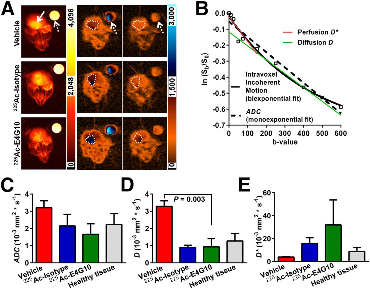
  • FIGURE 2.
    • Download figure
    • Open in new tab
    • Download powerpoint
    FIGURE 2.

    Diffusion-weighted MRI study of vessel functionality after 225Ac-E4G10 treatment. (A) Representative MR images on day 10 after treatment: axial T2-weighted fast spin echo (left); corresponding echoplanar at b = 50 (predominantly perfusion) (middle); and echoplanar at b = 600 (predominantly diffusion) (right). Scale bars indicate relative intensities; dotted outline, tumor margin; solid arrow, tumor; and dashed arrows, water-filled phantom (measurement control). (B) Representative logarithmic plot of signal intensities of whole-tumor ROIs—plotted normalized to intensity at b = 0 and as natural logarithm (ln(Sb/Sb=0))—vs. measured b-values. Biexponential decay fitting of data (intravoxel incoherent motion, straight line) results in 2 slopes, D* (red line) and D (green line). ADC is derived by monoexponential decay fitting of data (dashed line). (C–E) Quantification of coefficients: ADC (C), D (D), and D* (E). Data are mean ± SEM.
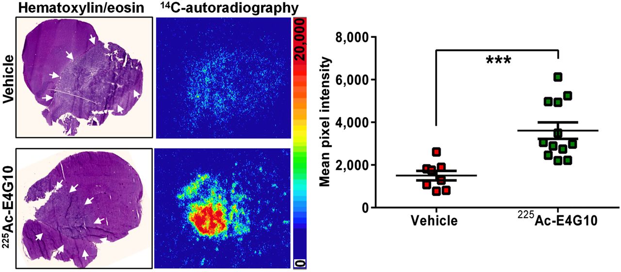
  • FIGURE 3.
    • Download figure
    • Open in new tab
    • Download powerpoint
    FIGURE 3.

    Dasatinib penetration of glioblastomas 10 d after 225Ac-E4G10 treatment. Representative axial whole-brain sections stained with hematoxylin and eosin are shown, with matching autoradiography of 14C-labeled dasatinib from mice treated with vehicle (n = 5) or 225Ac-E4G10 (n = 6). Arrows indicate tumor margins. Scale bar indicates relative intensities. Autoradiography data are quantified on right. Data are mean ± SEM of mean intensities of whole-tumor ROIs. ***P < 0.001.
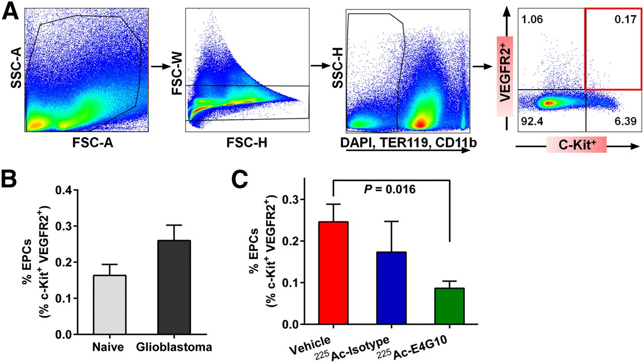
  • FIGURE 4.
    • Download figure
    • Open in new tab
    • Download powerpoint
    FIGURE 4.

    (A) Flow cytometry gating scheme to identify bone marrow EPCs in Ntva mice. After gating out debris (panel 1) and cell doublets based on forward scatter (panel 2), population of live (DAPI-negative) TER 119–negative and CD11b-negative cells is selected (third panel) and identified as VEGF receptor 2–positive, c-kit–positive EPCs (red square, panel 4). (B and C) Pretreatment (B) and posttreatment (C) comparisons of EPC population between Ntva mice and controls. Data are mean ± SEM. FSC = forward scatter; SSC = side scatter.

Additional Files

  • Figures
  • Supplemental Data

    Files in this Data Supplement:

    • Supplemental Data
PreviousNext
Back to top

In this issue

Journal of Nuclear Medicine: 57 (11)
Journal of Nuclear Medicine
Vol. 57, Issue 11
November 1, 2016
  • Table of Contents
  • Table of Contents (PDF)
  • About the Cover
  • Index by author
Print
Download PDF
Article Alerts
Sign In to Email Alerts with your Email Address
Email Article

Thank you for your interest in spreading the word on Journal of Nuclear Medicine.

NOTE: We only request your email address so that the person you are recommending the page to knows that you wanted them to see it, and that it is not junk mail. We do not capture any email address.

Enter multiple addresses on separate lines or separate them with commas.
Remodeling the Vascular Microenvironment of Glioblastoma with α-Particles
(Your Name) has sent you a message from Journal of Nuclear Medicine
(Your Name) thought you would like to see the Journal of Nuclear Medicine web site.
Citation Tools
Remodeling the Vascular Microenvironment of Glioblastoma with α-Particles
Katja Behling, William F. Maguire, Valentina Di Gialleonardo, Lukas E.M. Heeb, Iman F. Hassan, Darren R. Veach, Kayvan R. Keshari, Philip H. Gutin, David A. Scheinberg, Michael R. McDevitt
Journal of Nuclear Medicine Nov 2016, 57 (11) 1771-1777; DOI: 10.2967/jnumed.116.173559

Citation Manager Formats

  • BibTeX
  • Bookends
  • EasyBib
  • EndNote (tagged)
  • EndNote 8 (xml)
  • Medlars
  • Mendeley
  • Papers
  • RefWorks Tagged
  • Ref Manager
  • RIS
  • Zotero
Share
Remodeling the Vascular Microenvironment of Glioblastoma with α-Particles
Katja Behling, William F. Maguire, Valentina Di Gialleonardo, Lukas E.M. Heeb, Iman F. Hassan, Darren R. Veach, Kayvan R. Keshari, Philip H. Gutin, David A. Scheinberg, Michael R. McDevitt
Journal of Nuclear Medicine Nov 2016, 57 (11) 1771-1777; DOI: 10.2967/jnumed.116.173559
Twitter logo Facebook logo LinkedIn logo Mendeley logo
  • Tweet Widget
  • Facebook Like
  • Google Plus One
Bookmark this article

Jump to section

  • Article
    • Abstract
    • MATERIALS AND METHODS
    • RESULTS
    • DISCUSSION
    • CONCLUSION
    • DISCLOSURE
    • Acknowledgments
    • Footnotes
    • REFERENCES
  • Figures & Data
  • Supplemental
  • Info & Metrics
  • PDF

Related Articles

  • This Month in JNM
  • PubMed
  • Google Scholar

Cited By...

  • Leveraging Bioorthogonal Click Chemistry to Improve 225Ac-Radioimmunotherapy of Pancreatic Ductal Adenocarcinoma
  • {alpha}-Emitters for Radiotherapy: From Basic Radiochemistry to Clinical Studies--Part 2
  • {alpha}-Emitters for Radiotherapy: From Basic Radiochemistry to Clinical Studies--Part 1
  • Google Scholar

More in this TOC Section

  • Tumor-Specific Binding of Radiolabeled PEGylated GIRLRG Peptide: A Novel Agent for Targeting Cancers
  • PET/MRI of Hypoxic Atherosclerosis Using 64Cu-ATSM in a Rabbit Model
  • Tumor Uptake of Anti-CD20 Fabs Depends on Tumor Perfusion
Show more Basic Science Investigations

Similar Articles

Keywords

  • Glioblastoma
  • vascular endothelium
  • pericytes
  • radioimmunotherapy
  • actinium-225
  • 225Ac
SNMMI

© 2025 SNMMI

Powered by HighWire