
Main menu

  • Home
  • Content
    • Current
    • Ahead of print
    • Past Issues
    • JNM Supplement
    • SNMMI Annual Meeting Abstracts
    • Continuing Education
    • JNM Podcasts
  • Subscriptions
    • Subscribers
    • Institutional and Non-member
    • Rates
    • Journal Claims
    • Corporate & Special Sales
  • Authors
    • Submit to JNM
    • Information for Authors
    • Assignment of Copyright
    • AQARA requirements
  • Info
    • Reviewers
    • Permissions
    • Advertisers
  • About
    • About Us
    • Editorial Board
    • Contact Information
  • More
    • Alerts
    • Feedback
    • Help
    • SNMMI Journals
  • SNMMI
    • JNM
    • JNMT
    • SNMMI Journals
    • SNMMI

User menu

  • Subscribe
  • My alerts
  • Log in
  • My Cart

Search

  • Advanced search
Journal of Nuclear Medicine
  • SNMMI
    • JNM
    • JNMT
    • SNMMI Journals
    • SNMMI
  • Subscribe
  • My alerts
  • Log in
  • My Cart
Journal of Nuclear Medicine

Advanced Search

  • Home
  • Content
    • Current
    • Ahead of print
    • Past Issues
    • JNM Supplement
    • SNMMI Annual Meeting Abstracts
    • Continuing Education
    • JNM Podcasts
  • Subscriptions
    • Subscribers
    • Institutional and Non-member
    • Rates
    • Journal Claims
    • Corporate & Special Sales
  • Authors
    • Submit to JNM
    • Information for Authors
    • Assignment of Copyright
    • AQARA requirements
  • Info
    • Reviewers
    • Permissions
    • Advertisers
  • About
    • About Us
    • Editorial Board
    • Contact Information
  • More
    • Alerts
    • Feedback
    • Help
    • SNMMI Journals
  • View or Listen to JNM Podcast
  • Visit JNM on Facebook
  • Join JNM on LinkedIn
  • Follow JNM on Twitter
  • Subscribe to our RSS feeds
Research ArticleFocus on Molecular Imaging

Optical Imaging of Ionizing Radiation from Clinical Sources

Travis M. Shaffer, Charles Michael Drain and Jan Grimm
Journal of Nuclear Medicine November 2016, 57 (11) 1661-1666; DOI: https://doi.org/10.2967/jnumed.116.178624
Travis M. Shaffer
1Molecular Pharmacology Program, Memorial Sloan Kettering Cancer Center, New York, New York
2Department of Radiology, Memorial Sloan Kettering Cancer Center, New York, New York
3Department of Chemistry, Hunter College of City University of New York, New York, New York
4Department of Chemistry, Graduate Center of City University of New York, New York, New York
  • Find this author on Google Scholar
  • Find this author on PubMed
  • Search for this author on this site
Charles Michael Drain
3Department of Chemistry, Hunter College of City University of New York, New York, New York
4Department of Chemistry, Graduate Center of City University of New York, New York, New York
  • Find this author on Google Scholar
  • Find this author on PubMed
  • Search for this author on this site
Jan Grimm
1Molecular Pharmacology Program, Memorial Sloan Kettering Cancer Center, New York, New York
2Department of Radiology, Memorial Sloan Kettering Cancer Center, New York, New York
5Department of Pharmacology, Weill Cornell Medical College, New York, New York; and
6Department of Radiology, Weill Cornell Medical College, New York, New York
  • Find this author on Google Scholar
  • Find this author on PubMed
  • Search for this author on this site
  • Article
  • Figures & Data
  • Info & Metrics
  • PDF
Loading

Article Figures & Data

Figures

  • FIGURE 1.
    • Download figure
    • Open in new tab
    • Download powerpoint
    FIGURE 1.

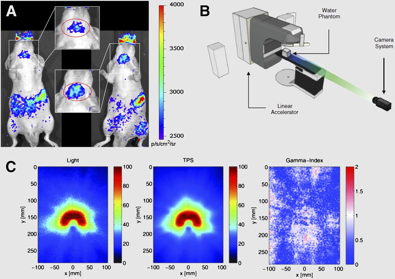
    Optical imaging of γ-emitters and photon beams. (A) Radioluminescence image of 99mTc-pertechnetate distribution in Nu/ν mice showing salivary, thyroid, and stomach uptake. (B) Linear accelerator phantom study to image Cerenkov light from photon beam interactions with water. Distance of camera to water phantom is 3 m. (C) Two-dimensional projections of experimentally imaged 3-dimensional (3D) Cerenkov light volume (left) and nonlinearly summed 3D dose matrix from TPS (center) are shown for the VMAT treatment plan. Resulting γ-index map is shown with a 3%/3-mm dose difference and DTA criterion (right). DTA = distance-to-agreement; TPS = treatment planning system; VMAT = volumetric-modulated arc therapy. (A and B–C adapted with permission of (6) and (9), respectively.)

  • FIGURE 2.
    • Download figure
    • Open in new tab
    • Download powerpoint
    FIGURE 2.

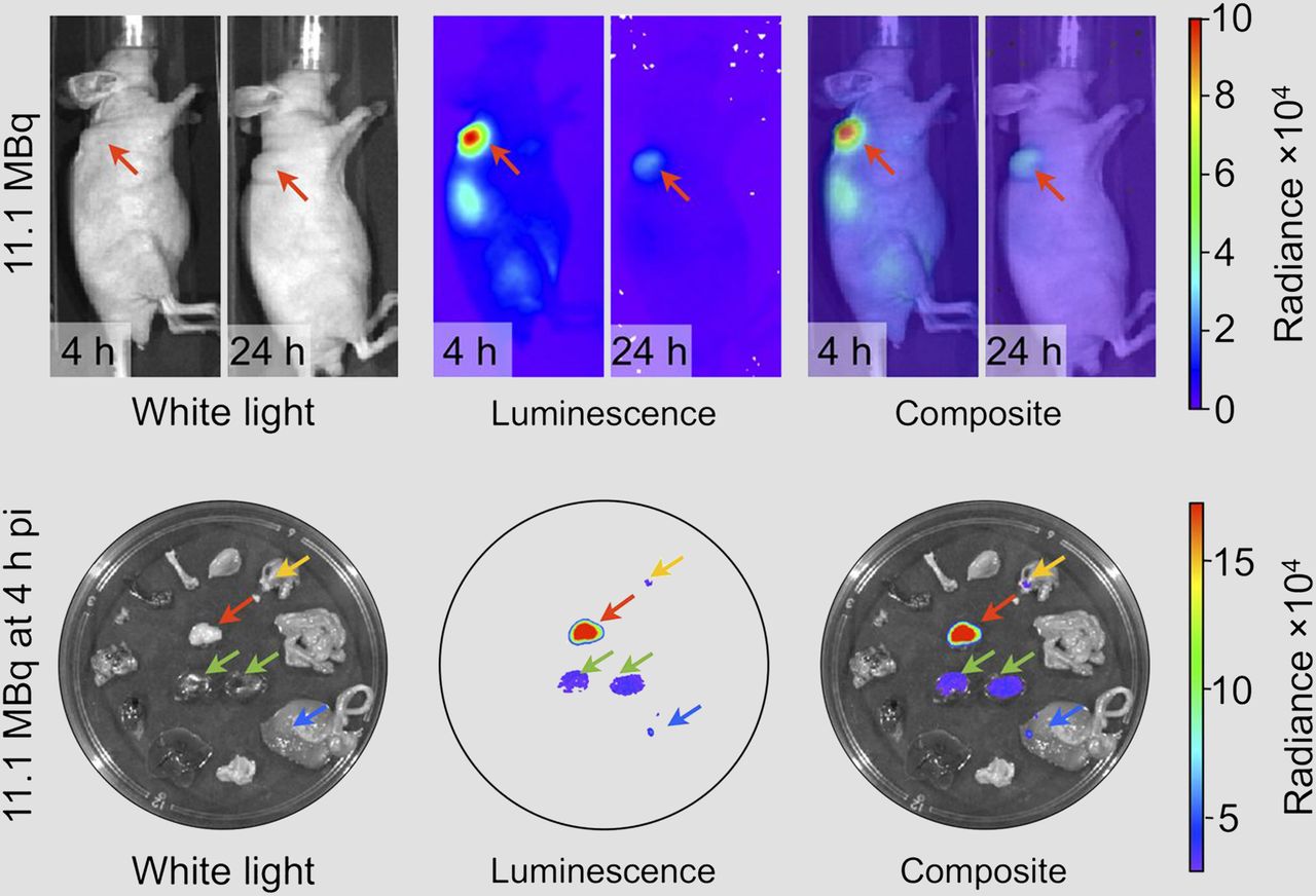
    Cerenkov imaging of therapeutic radiotracers. (Top) CLI of 90Y-DOTA-AR in mice with PC-3 prostate cancer tumors. Tumors (red arrow) show high radiance at 4 h after injection (pi) with partial clearance at 24 h after injection. (Bottom) Ex vivo images at 4 h after injection, with tumor clearly delineated (red arrow). Green arrows = kidneys; yellow arrow = stomach; blue arrow = large intestine. (Adapted with permission of (17).)

  • FIGURE 3.
    • Download figure
    • Open in new tab
    • Download powerpoint
    FIGURE 3.

    Optical imaging of charged particle beams. (A) Various interactions that may occur during charged particle beam therapy, including primary Cerenkov production, secondary Cerenkov, and various scattering and absorption processes. (B) Imaging of a patient undergoing postlumpectomy radiation therapy (inset, plan) using time domain–gated imaging system, which is synchronized to delivered radiation pulses. (C) Cerenkov images of each patient and corresponding white-light images (left). Edge-enhanced Cerenkov images for each patient (right). (Adapted with permission of (28), © Institute of Physics and Engineering in Medicine.)

  • FIGURE 4.
    • Download figure
    • Open in new tab
    • Download powerpoint
    FIGURE 4.

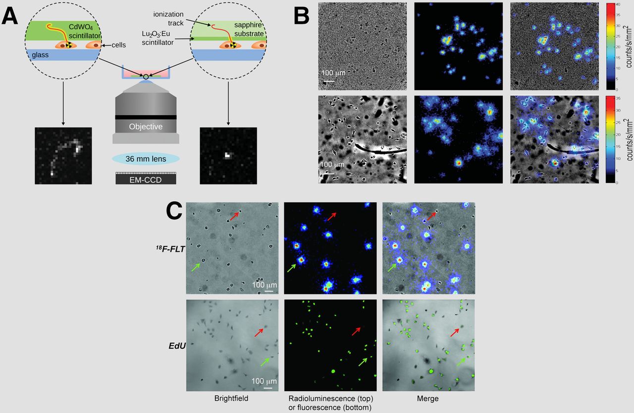
    Radioluminescence microscopy. (A) Choice of scintillator matters. Scheme of radioluminescence microscopy setup comparing 500-μm CdWO4 scintillator (left) and 10-μm Lu2O3:Eu scintillator (right). Because of thinness of Lu2O3:Eu scintillator, ionization tracks are shorter than with thicker CdWO4 scintillator. (B) Comparison of Lu2O3:Eu (top) and CdWO4 (bottom) scintillators with 18F-FDG, showing bright-field, radioluminescence, and overlaid images. (C) MDA-MB-231 cells imaged using 18F-FLT and radioluminescence microscopy (top) or 5-ethynyl-2′-deoxyuridine (EdU) and fluorescence microscopy (bottom). Red and green arrows indicate cells with negative and positive signals, respectively. (A–B and C adapted with permission of (36) and (37), respectively.)

PreviousNext
Back to top

In this issue

Journal of Nuclear Medicine: 57 (11)
Journal of Nuclear Medicine
Vol. 57, Issue 11
November 1, 2016
  • Table of Contents
  • Table of Contents (PDF)
  • About the Cover
  • Index by author
Print
Download PDF
Article Alerts
Sign In to Email Alerts with your Email Address
Email Article

Thank you for your interest in spreading the word on Journal of Nuclear Medicine.

NOTE: We only request your email address so that the person you are recommending the page to knows that you wanted them to see it, and that it is not junk mail. We do not capture any email address.

Enter multiple addresses on separate lines or separate them with commas.
Optical Imaging of Ionizing Radiation from Clinical Sources
(Your Name) has sent you a message from Journal of Nuclear Medicine
(Your Name) thought you would like to see the Journal of Nuclear Medicine web site.
Citation Tools
Optical Imaging of Ionizing Radiation from Clinical Sources
Travis M. Shaffer, Charles Michael Drain, Jan Grimm
Journal of Nuclear Medicine Nov 2016, 57 (11) 1661-1666; DOI: 10.2967/jnumed.116.178624

Citation Manager Formats

  • BibTeX
  • Bookends
  • EasyBib
  • EndNote (tagged)
  • EndNote 8 (xml)
  • Medlars
  • Mendeley
  • Papers
  • RefWorks Tagged
  • Ref Manager
  • RIS
  • Zotero
Share
Optical Imaging of Ionizing Radiation from Clinical Sources
Travis M. Shaffer, Charles Michael Drain, Jan Grimm
Journal of Nuclear Medicine Nov 2016, 57 (11) 1661-1666; DOI: 10.2967/jnumed.116.178624
Twitter logo Facebook logo LinkedIn logo Mendeley logo
  • Tweet Widget
  • Facebook Like
  • Google Plus One
Bookmark this article

Jump to section

  • Article
    • Abstract
    • OPTICAL IMAGING OF γ-EMITTING RADIOTRACERS
    • OPTICAL IMAGING OF CHARGED PARTICLES
    • NEW MICROSCOPY OPPORTUNITIES
    • CONCLUSION
    • DISCLOSURE
    • Footnotes
    • REFERENCES
  • Figures & Data
  • Info & Metrics
  • PDF

Related Articles

  • This Month in JNM
  • PubMed
  • Google Scholar

Cited By...

  • Cerenkov Radiation-Induced Photoimmunotherapy with 18F-FDG
  • Google Scholar

More in this TOC Section

  • From Genome to Phenome: Opportunities and Challenges of Molecular Imaging
  • Imaging the Activity of Efflux Transporters at the Blood–Brain Barrier in Neurologic Diseases: Radiotracer Selection Criteria
  • Molecular Imaging of Pulmonary Fibrosis
Show more Focus on Molecular Imaging

Similar Articles

Keywords

  • ionizing radiation
  • imaging
  • optical
  • dosimetry
  • radiation therapy
  • Cerenkov
SNMMI

© 2025 SNMMI

Powered by HighWire