


Article Figures & Data
Figures
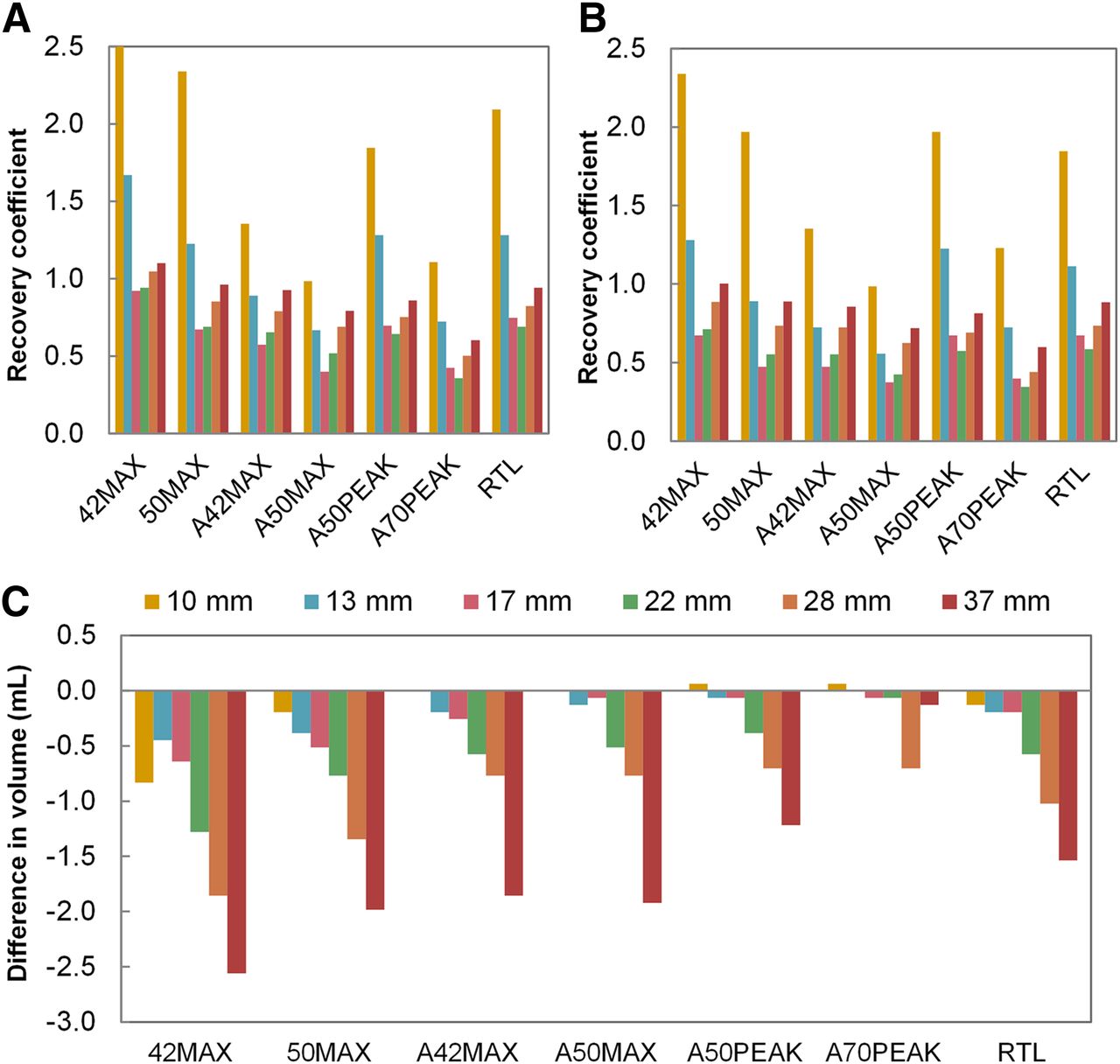
- FIGURE 1.
Volume recovery coefficients for non–PSF PVC images (A) and PSF PVC images (B) per VOI method and sphere size, and differences in PET-based volumes between non–PSF PVC images and PSF PVC images (C). Negative volume differences indicate smaller volumes for PSF reconstructed images than for non–PSF reconstructed images. Key indicates sphere diameters. Ten-millimeter sphere delineated with 42% maximal had recovery coefficient of 3.9 in non–PSF reconstructed images. 42MAX = 42% of maximal voxel value; 50MAX = 50% of maximal voxel value; A42MAX = 42% of maximal voxel value adapted for local background uptake; A50MAX = 50% of maximal voxel value adapted for local background uptake; A50PEAK = 50% of peak voxel value adapted for local background uptake; A70PEAK = 70% of peak voxel value adapted for local background uptake; RTL = relative threshold level.
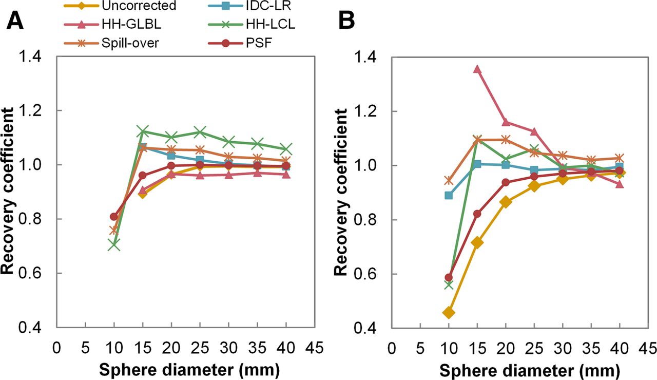
- FIGURE 2.
Activity concentration recovery coefficients as function of sphere diameter for all PVC methods, and uncorrected data, with their optimal PET-based VOI method (Table 1) for spheres in mediastinum (A) and lung (B). Missing values are due to delineation failure. HH-GLBL = global-background–adapted PVC; IDC-LR = iterative deconvolution Lucy–Richardson PVC; HH-LCL = local-background–adapted PVC.
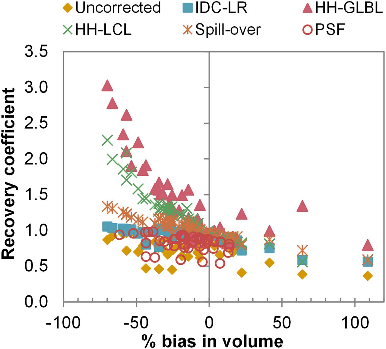
- FIGURE 3.
Activity concentration recovery coefficients as function of volumetric bias. Shown are results for all VOI methods for spheres in lung (noise-free images). HH-LCL = local-background–adapted PVC; IDC-LR = iterative deconvolution Lucy–Richardson PVC; HH-GLBL = global-background–adapted PVC.
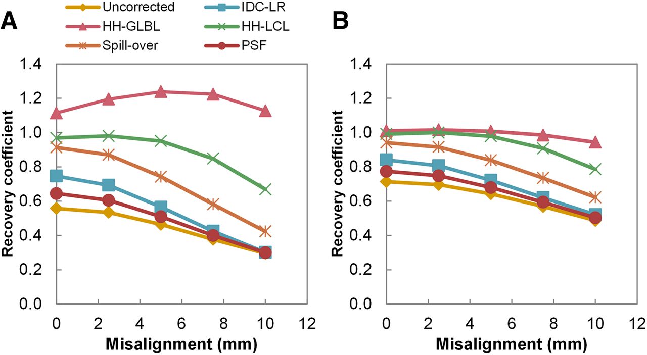
- FIGURE 4.
Activity concentration recovery coefficients as function of misalignment of true VOI. Shown are results from 15-mm (A) and 25-mm (B) spheres in lung (noise-free images). HH-LCL = local-background–adapted PVC; IDC-LR = iterative deconvolution Lucy–Richardson PVC; HH-GLBL = global-background–adapted PVC.
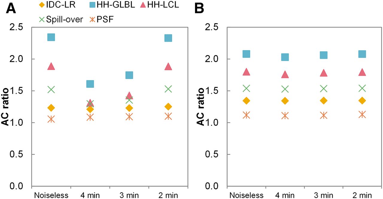
- FIGURE 5.
Activity concentration ratios as function of simulated acquisition time (thus, noise level). Shown are results from background-adapted 50% peak for 20-mm sphere (corresponding to median volumes of 18F-FDG and 18F-fluoromethylcholine PET cohorts delineated with background-adapted 50% peak) in mediastinum (A) and lung (B), respectively. AC = activity concentration; IDC-LR = iterative deconvolution Lucy–Richardson PVC; HH-GLBL = global-background–adapted PVC; HH-LCL = local-background–adapted PVC.
- FIGURE 6.
SDs of recovery coefficients for all combinations of PVC method. Shown are results for 20-mm spheres in lung (corresponding to median volumes of 18F-FDG and 18F-fluoromethylcholine PET cohorts delineated with background-adapted 50% peak). y-axis is scaled for visual interpretation; SD of global-background–adapted PVC using background-adapted 70% peak was 0.049. IDC-LR = iterative deconvolution Lucy–Richardson PVC; HH-GLBL = global-background–adapted PVC; HH-LCL = local-background–adapted PVC; 42MAX = 42% of maximal voxel value; 50MAX = 50% of maximal voxel value; A42MAX = 42% of maximal voxel value adapted for local background uptake; A50MAX = 50% of maximal voxel value adapted for local background uptake; A50PEAK = 50% of peak voxel value adapted for local background uptake; A70PEAK = 70% of peak voxel value adapted for local background uptake; RTL = relative threshold level.
- FIGURE 7.
ICCs of SUVmean (A) and TLG (B) for all combinations of PVC method. Shown are results for 18F-FDG PET cohort. Error bars represent 95% confidence intervals. Similar results were obtained for 18F-fluoromethylcholine PET cohort (Supplemental Fig. 3). IDC-LR = iterative deconvolution Lucy–Richardson PVC; HH-GLBL = global-background–adapted PVC; HH-LCL = local-background–adapted PVC; 42MAX = 42% of maximal voxel value; 50MAX = 50% of maximal voxel value; A42MAX = 42% of maximal voxel value adapted for local background uptake; A50MAX = 50% of maximal voxel value adapted for local background uptake; A50PEAK = 50% of peak voxel value adapted for local background uptake; A70PEAK = 70% of peak voxel value adapted for local background uptake; RTL = relative threshold level.


Tables
Location Uncorrected IDC-LR HH-GLBL HH-LCL Spillover PSF Mediastinum A70PEAK (97 ± 4.1) A50MAX (102 ± 2.7) 50MAX (96 ± 2.4) RTL (109 ± 2.6) RTL (104 ± 2.0) A70PEAK (99 ± 1.5) Lung A70PEAK (90 ± 9.8) A42MAX (99 ± 0.9) 42MAX (109 ± 15.8) 50MAX (103 ± 4.7) A42MAX (105 ± 3.3) A70PEAK (94 ± 6.0) IDC-LR = iterative deconvolution Lucy–Richardson PVC; HH-GLBL = global-background–adapted PVC; HH-LCL = local-background–adapted PVC; A70PEAK = 70% of peak voxel value adapted for local background uptake; A50MAX = 50% of maximal voxel value adapted for local background uptake; 50MAX = 50% of maximal voxel value; RTL = relative threshold level; A42MAX = 42% of maximal voxel value adapted for local background uptake; 42MAX = 42% of maximal voxel value.
Data are for noise-free simulated images. Mean accuracy (percentage ± SD) of spheres ≥ 15 mm is shown in parentheses.
Characteristic 18F-FDG PET (28) 18F-fluoromethylcholine PET (29) Type of cancer NSCLC Metastatic prostate cancer (4 castration-resistant cases) No. of patients 11 12 No. of lesions 70 67 Mean age ± SD (y) 60 ± 7 64 ± 8 Sex 7 male, 4 female 12 male Lesion location 16 intrapulmonary, 54 extrapulmonary 44 bone metastases, 23 lymph node metastases Median volume (mL) Non-PSF 3.94 (IQR, 10.85) 5.76 (IQR, 8.64) PSF 3.90 (IQR, 20.10) 5.28 (IQR, 7.92) IQR = interquartile range.
Data are median volumes as determined with background-adapted 50% peak, being the most accurate VOI method as determined in phantom experiment, on baseline.
Supplemental Data
Files in this Data Supplement:


Jump to section
Related Articles
Cited By...
- Lymph Node Staging with a Combined Protocol of 18F-FDG PET/MRI and Sentinel Node SPECT/CT: A Prospective Study in Patients with FIGO I/II Cervical Carcinoma
- Automated Segmentation of Baseline Metabolic Total Tumor Burden in Diffuse Large B-Cell Lymphoma: Which Method Is Most Successful? A Study on Behalf of the PETRA Consortium