
Main menu

  • Home
  • Content
    • Current
    • Ahead of print
    • Past Issues
    • JNM Supplement
    • SNMMI Annual Meeting Abstracts
    • Continuing Education
    • JNM Podcasts
  • Subscriptions
    • Subscribers
    • Institutional and Non-member
    • Rates
    • Journal Claims
    • Corporate & Special Sales
  • Authors
    • Submit to JNM
    • Information for Authors
    • Assignment of Copyright
    • AQARA requirements
  • Info
    • Reviewers
    • Permissions
    • Advertisers
  • About
    • About Us
    • Editorial Board
    • Contact Information
  • More
    • Alerts
    • Feedback
    • Help
    • SNMMI Journals
  • SNMMI
    • JNM
    • JNMT
    • SNMMI Journals
    • SNMMI

User menu

  • Subscribe
  • My alerts
  • Log in
  • My Cart

Search

  • Advanced search
Journal of Nuclear Medicine
  • SNMMI
    • JNM
    • JNMT
    • SNMMI Journals
    • SNMMI
  • Subscribe
  • My alerts
  • Log in
  • My Cart
Journal of Nuclear Medicine

Advanced Search

  • Home
  • Content
    • Current
    • Ahead of print
    • Past Issues
    • JNM Supplement
    • SNMMI Annual Meeting Abstracts
    • Continuing Education
    • JNM Podcasts
  • Subscriptions
    • Subscribers
    • Institutional and Non-member
    • Rates
    • Journal Claims
    • Corporate & Special Sales
  • Authors
    • Submit to JNM
    • Information for Authors
    • Assignment of Copyright
    • AQARA requirements
  • Info
    • Reviewers
    • Permissions
    • Advertisers
  • About
    • About Us
    • Editorial Board
    • Contact Information
  • More
    • Alerts
    • Feedback
    • Help
    • SNMMI Journals
  • View or Listen to JNM Podcast
  • Visit JNM on Facebook
  • Join JNM on LinkedIn
  • Follow JNM on Twitter
  • Subscribe to our RSS feeds
Research ArticleBasic Science Investigations

Modulation of Abnormal Metabolic Brain Networks by Experimental Therapies in a Nonhuman Primate Model of Parkinson Disease: An Application to Human Retinal Pigment Epithelial Cell Implantation

Shichun Peng, Yilong Ma, Joseph Flores, Michael Cornfeldt, Branka Mitrovic, David Eidelberg and Doris J. Doudet
Journal of Nuclear Medicine October 2016, 57 (10) 1591-1598; DOI: https://doi.org/10.2967/jnumed.115.161513
Shichun Peng
1Center for Neurosciences, The Feinstein Institute for Medical Research, Manhasset, New York, New York
  • Find this author on Google Scholar
  • Find this author on PubMed
  • Search for this author on this site
Yilong Ma
1Center for Neurosciences, The Feinstein Institute for Medical Research, Manhasset, New York, New York
  • Find this author on Google Scholar
  • Find this author on PubMed
  • Search for this author on this site
Joseph Flores
2Department of Neurology, University of British Columbia, Vancouver, British Columbia, Canada
  • Find this author on Google Scholar
  • Find this author on PubMed
  • Search for this author on this site
Michael Cornfeldt
3Titan Pharmaceuticals Inc., Somerville, New Jersey; and
  • Find this author on Google Scholar
  • Find this author on PubMed
  • Search for this author on this site
Branka Mitrovic
4Bayer HealthCare Pharmaceuticals Inc., Richmond, California
  • Find this author on Google Scholar
  • Find this author on PubMed
  • Search for this author on this site
David Eidelberg
1Center for Neurosciences, The Feinstein Institute for Medical Research, Manhasset, New York, New York
  • Find this author on Google Scholar
  • Find this author on PubMed
  • Search for this author on this site
Doris J. Doudet
2Department of Neurology, University of British Columbia, Vancouver, British Columbia, Canada
  • Find this author on Google Scholar
  • Find this author on PubMed
  • Search for this author on this site
  • Article
  • Figures & Data
  • Supplemental
  • Info & Metrics
  • PDF
Loading

Article Figures & Data

Figures

  • Tables
  • Additional Files
  • FIGURE 1.
    • Download figure
    • Open in new tab
    • Download powerpoint
    FIGURE 1.

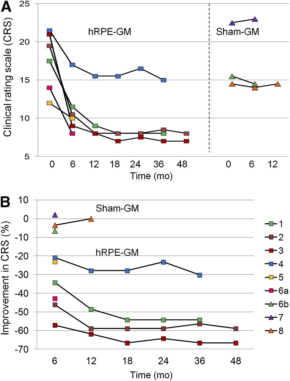
    Clinical improvement in individual macaques after cell-based therapy. hRPE-implanted animals showed motor recovery from 6 mo to 4 y after unilateral implantation but continued to express mild to moderate bradykinesia and hypokinesia. Maximal benefit was achieved within 1 y and remained stable afterward. Sham-implanted animals showed no clinical responses. Animal 6 was transplanted sequentially with GM and hRPE-GM in 2 different hemispheres. Animals 1–4 received fetal cells used in the successful phase I trial (15), and animals 5–6a received neonatal cells used in the failed phase II trial (22). ■ = hRPE-GM; ▲ = GM.

  • FIGURE 2.
    • Download figure
    • Open in new tab
    • Download powerpoint
    FIGURE 2.

    Mean images of relative glucose metabolism in healthy and parkinsonian macaques acquired using Siemens HRRT scanner. This high-resolution PET scanner provides superior image quality for revealing distinct regional differences in cortical and subcortical metabolism among normal, MPTP, and hRPE- and sham-implanted hemispheres. Each image represents brain 18F-FDG scans averaged over hemispheres in individual animal group spatially normalized to macaque PET brain template (23).
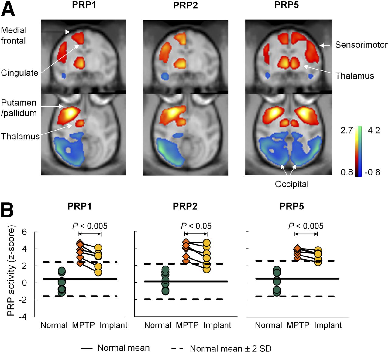
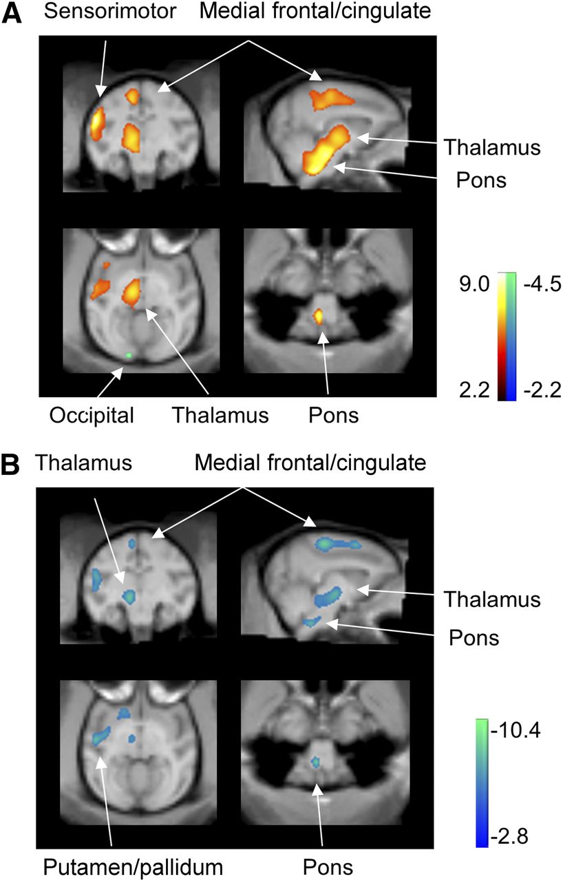
  • FIGURE 3.
    • Download figure
    • Open in new tab
    • Download powerpoint
    FIGURE 3.

    Modulation of abnormal metabolic brain networks in MPTP-induced experimental parkinsonism by hRPE cell transplantation therapy. (A) PRPs identified on a hemispheric (PRPs 1 and 2) and whole-brain (PRP 5) basis using 18F-FDG PET images in parkinsonian and age-matched healthy macaques (10). All PRPs shared analogous topographies with increased (red to yellow) and decreased (blue to green) metabolic activity in subcortical and cortical regions. (B) Network activity in individual hemispheres or brains was elevated (P < 0.00005) in the 7 untreated MPTP hemispheres compared with the 8 normal controls but declined consistently (P < 0.05) in the 6 contralateral MPTP hemispheres after hRPE cell implantation. The patterns are overlaid on macaque MRI brain template (23).

  • FIGURE 4.
    • Download figure
    • Open in new tab
    • Download powerpoint
    FIGURE 4.

    Modulation of abnormal regional metabolism in MPTP-induced experimental parkinsonism by hRPE cell transplantation therapy. (A) Metabolism in the 7 untreated MPTP hemispheres increased (red to yellow) in a set of subcortical and cortical motor regions relative to the 8 normal controls. (B) Metabolism in the 6 hRPE-implanted hemispheres decreased (blue to green) in the same set of subcortical and cortical motor regions compared with the 6 untreated MPTP hemispheres. SPM t maps of unpaired and paired comparisons are displayed at lower threshold (P = 0.025) for better visualization on macaque MRI brain template (23).
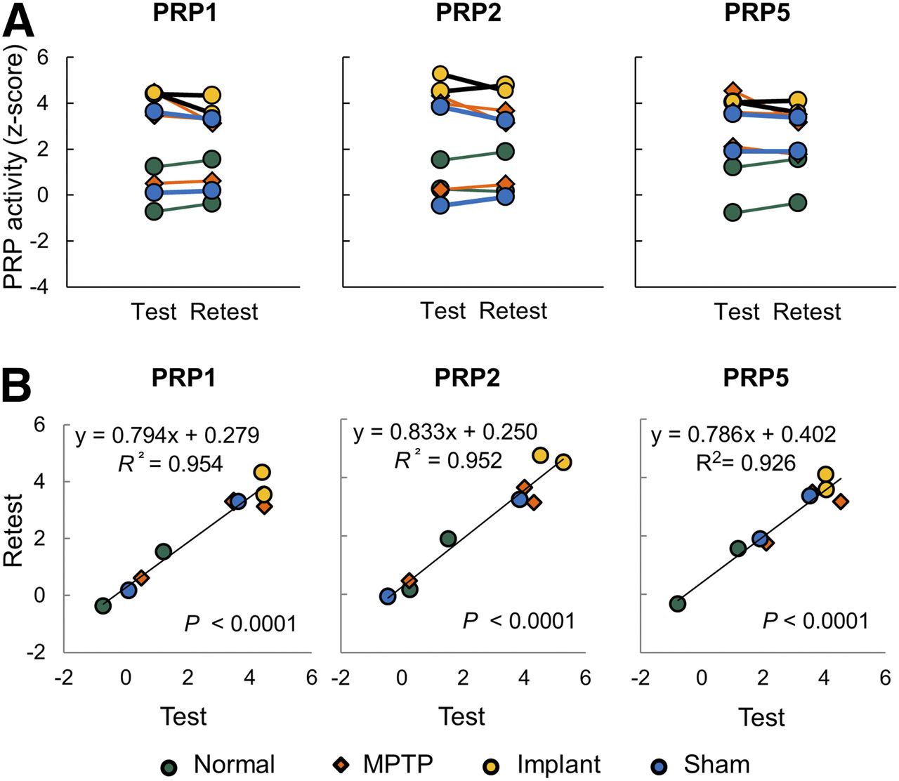
  • FIGURE 5.
    • Download figure
    • Open in new tab
    • Download powerpoint
    FIGURE 5.

    Test–retest reproducibility in PRP network activity. (A) Network scores in individual hemispheres or brains were highly reproducible (A: P > 0.29) and correlated (B: R2 > 0.92; P < 0.00005) between 9 test and retest scans in 4 subgroups of 7 macaques. Network scores were computed on hemispheric (PRPs 1–2) and whole-brain (PRP 5) basis, respectively.

Tables

  • Figures
  • Additional Files
    • View popup
    TABLE 1

    Parkinsonian Macaque Characteristics and Imaging-Related Parameters

    IDAge (y)Weight (kg)Motor ratingDose (MBq)Glucose (mmol/L)Condition, leftCondition, right
    1229102814.1hRPE*MPTP
    213881483.7hRPE*MPTP
    3131081853.3hRPE*MPTP
    42010142113.7hRPE*MPTP
    5912102443.6MPTPhRPE†
    614781854.1hRPE†GM
    787231853.5GMMPTP
    8910142224.5MPTPGM
    • ↵* Fetal cells.

    • ↵† Neonatal cells.

    • Data are provided for macaques undergoing 18F-FDG PET after unilateral striatal implantation of hRPE cells or gelatin carriers only (GM) in left or right striatum.

    • View popup
    TABLE 2

    Changes in Metabolic Network Activity Under Different Experimental Conditions

    Summary of analysisPRP1PRP2PRP3PRP4PRP5
    Pattern derivation
     Eigenvalue (%)42.943.327.827.248.2
     Control 10.00 ± 0.410.00 ± 0.450.00 ± 0.360.00 ± 0.330.00 ± 0.41
     MPTP 17.67 ± 1.377.91 ± 1.563.11 ± 0.252.66 ± 0.257.78 ± 1.41
     MPTP 1 vs. control 1*0.00370.00310.000030.000080.004
    Pattern validation
     Control 20.46 ± 0.360.09 ± 0.360.22 ± 0.320.36 ± 0.280.51 ± 0.37
     MPTP 23.80 ± 0.333.88 ± 0.323.04 ± 0.222.64 ± 0.213.64 ± 0.16
     MPTP 2 vs. control 2*0.000010.0000030.000010.000030.00002
    Implant effect
     Implant2.85 ± 0.433.10 ± 0.482.62 ± 0.391.92 ± 0.353.21 ± 0.21
     Implant vs. MPTP 2 (change %)−24.6 ± 5.9−21.6 ± 5.9−17.6 ± 7.2−26.8 ± 11.5−10.9 ± 2.3
     Implant vs. MPTP 2†0.00250.0110.0360.0330.0039
     Implant vs. control 2*0.0010.00030.00050.0040.00008
    Test–retest effect
     Test vs. retest†0.2920.3420.5770.5050.351
     R2 (Pearson correlation)0.9540.9520.9530.9560.926
     P0.0000060.0000070.0000070.0000050.00003
    • ↵* P values: unpaired Student t tests.

    • ↵† P values: paired Student t tests.

    • Eigenvalue for each PRP derivation is given as percentage of total subject × voxel variance (10). Subject scores are presented as mean ± SE for animals used to identify each PRP and to assess effects of implantation.

    • View popup
    TABLE 3

    Brain Regions with Significant Metabolic Changes Before and After hRPE Cell Implantation in Parkinsonian Macaques

    Metabolic increase* (MPTP > normal)Metabolic decrease† (implant < MPTP)
    Brain regionXYZZmaxSize (mm3)XYZZmaxSize (mm3)
    Medial frontal/cingulate86283.337646362.9208
    Insula/SMC/putamen3016184.91304268183.0744
    Frontal/SMA2824184.52626163.4160
    Thalamus61482.9187261462.8320
    Pons40−143.940−143.3184
    • ↵* Unpaired t test: P < 0.005 uncorrected and survived at false-discovery rate–corrected P < 0.05.

    • ↵† Paired t test: P < 0.005 uncorrected and did not survive at false-discovery rate–corrected P < 0.05.

    • SMC = sensorimotor cortex; SMA = supplementary motor area.

Additional Files

  • Figures
  • Tables
  • Supplemental Data

    Files in this Data Supplement:

    • Supplemental Data
PreviousNext
Back to top

In this issue

Journal of Nuclear Medicine: 57 (10)
Journal of Nuclear Medicine
Vol. 57, Issue 10
October 1, 2016
  • Table of Contents
  • Table of Contents (PDF)
  • About the Cover
  • Index by author
Print
Download PDF
Article Alerts
Sign In to Email Alerts with your Email Address
Email Article

Thank you for your interest in spreading the word on Journal of Nuclear Medicine.

NOTE: We only request your email address so that the person you are recommending the page to knows that you wanted them to see it, and that it is not junk mail. We do not capture any email address.

Enter multiple addresses on separate lines or separate them with commas.
Modulation of Abnormal Metabolic Brain Networks by Experimental Therapies in a Nonhuman Primate Model of Parkinson Disease: An Application to Human Retinal Pigment Epithelial Cell Implantation
(Your Name) has sent you a message from Journal of Nuclear Medicine
(Your Name) thought you would like to see the Journal of Nuclear Medicine web site.
Citation Tools
Modulation of Abnormal Metabolic Brain Networks by Experimental Therapies in a Nonhuman Primate Model of Parkinson Disease: An Application to Human Retinal Pigment Epithelial Cell Implantation
Shichun Peng, Yilong Ma, Joseph Flores, Michael Cornfeldt, Branka Mitrovic, David Eidelberg, Doris J. Doudet
Journal of Nuclear Medicine Oct 2016, 57 (10) 1591-1598; DOI: 10.2967/jnumed.115.161513

Citation Manager Formats

  • BibTeX
  • Bookends
  • EasyBib
  • EndNote (tagged)
  • EndNote 8 (xml)
  • Medlars
  • Mendeley
  • Papers
  • RefWorks Tagged
  • Ref Manager
  • RIS
  • Zotero
Share
Modulation of Abnormal Metabolic Brain Networks by Experimental Therapies in a Nonhuman Primate Model of Parkinson Disease: An Application to Human Retinal Pigment Epithelial Cell Implantation
Shichun Peng, Yilong Ma, Joseph Flores, Michael Cornfeldt, Branka Mitrovic, David Eidelberg, Doris J. Doudet
Journal of Nuclear Medicine Oct 2016, 57 (10) 1591-1598; DOI: 10.2967/jnumed.115.161513
Twitter logo Facebook logo LinkedIn logo Mendeley logo
  • Tweet Widget
  • Facebook Like
  • Google Plus One
Bookmark this article

Jump to section

  • Article
    • Abstract
    • MATERIALS AND METHODS
    • RESULTS
    • DISCUSSION
    • CONCLUSION
    • DISCLOSURE
    • Acknowledgments
    • Footnotes
    • REFERENCES
  • Figures & Data
  • Supplemental
  • Info & Metrics
  • PDF

Related Articles

  • Reverse Translation in Parkinson Disease
  • This Month in JNM
  • PubMed
  • Google Scholar

Cited By...

  • Dynamic 18F-FPCIT PET: Quantification of Parkinson Disease Metabolic Networks and Nigrostriatal Dopaminergic Dysfunction in a Single Imaging Session
  • Induced Cognitive Impairments Reversed by Grafts of Neural Precursors: a Longitudinal Study in a Macaque Model of Parkinsons Disease
  • Reverse Translation in Parkinson Disease
  • Google Scholar

More in this TOC Section

  • Design and Fabrication of Kidney Phantoms for Internal Radiation Dosimetry Using 3D Printing Technology
  • Synthesis and Biologic Evaluation of Novel 18F-Labeled Probes Targeting Prostate-Specific Membrane Antigen for PET of Prostate Cancer
  • Tumor-Specific Binding of Radiolabeled PEGylated GIRLRG Peptide: A Novel Agent for Targeting Cancers
Show more Basic Science Investigations

Similar Articles

Keywords

  • parkinsonism
  • primate model
  • FDG PET
  • network analysis
  • dopamine cell transplantation
  • retinal pigment epithelial cell
SNMMI

© 2025 SNMMI

Powered by HighWire