


Article Figures & Data
Figures
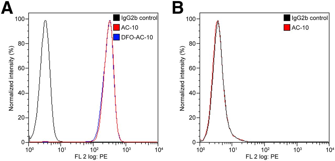
- FIGURE 1.
(A) Flow cytometry data showing specific binding of AC-10 and DFO-AC-10 to CD30-positive Karpas 299 cells. (B) Control data showing that AC-10 antibody does not bind to CD30-negative A-431 cells. FL = ▪▪▪; PE = phycoerythrin.
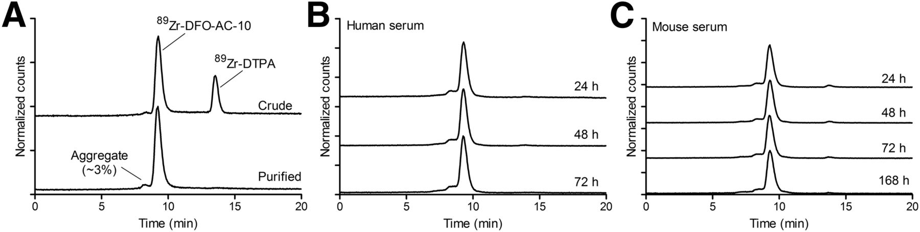
- FIGURE 2.
(A) Size-exclusion radio–high-performance liquid chromatograms showing radiochemical purity of formulated sample of 89Zr-DFO-AC-10. (B and C) Size-exclusion radio–high-performance liquid chromatogram data showing radiochemical stability of 89Zr-DFO-AC-10 after incubation at 37°C in human and mouse sera. DTPA = diethylenetriaminepentaacetic acid.
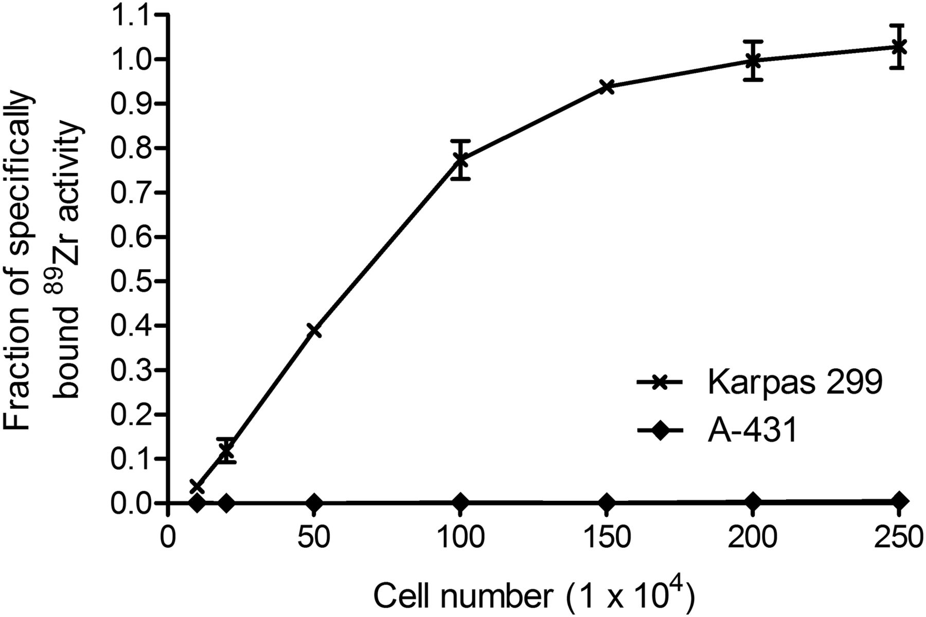
- FIGURE 3.
Cellular uptake data showing saturation of specific binding of 89Zr-DFO-AC-10 to CD30-positive Karpas 299 cells and lack of binding to CD30-negative A-431 cells.
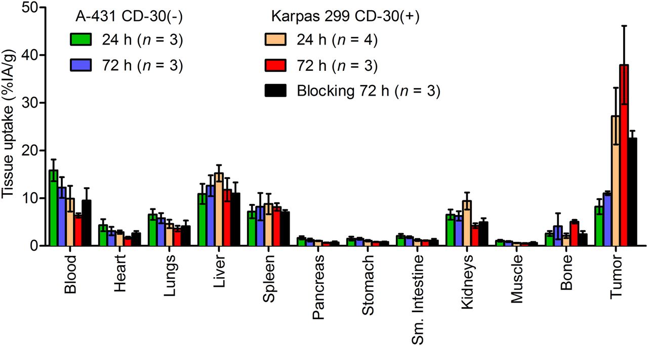
- FIGURE 4.
Biodistribution data showing accumulation and retention of 89Zr-DFO-AC-10 radioactivity in various tissues vs. time in mice bearing either A-431 or Karpas 299 tumors.
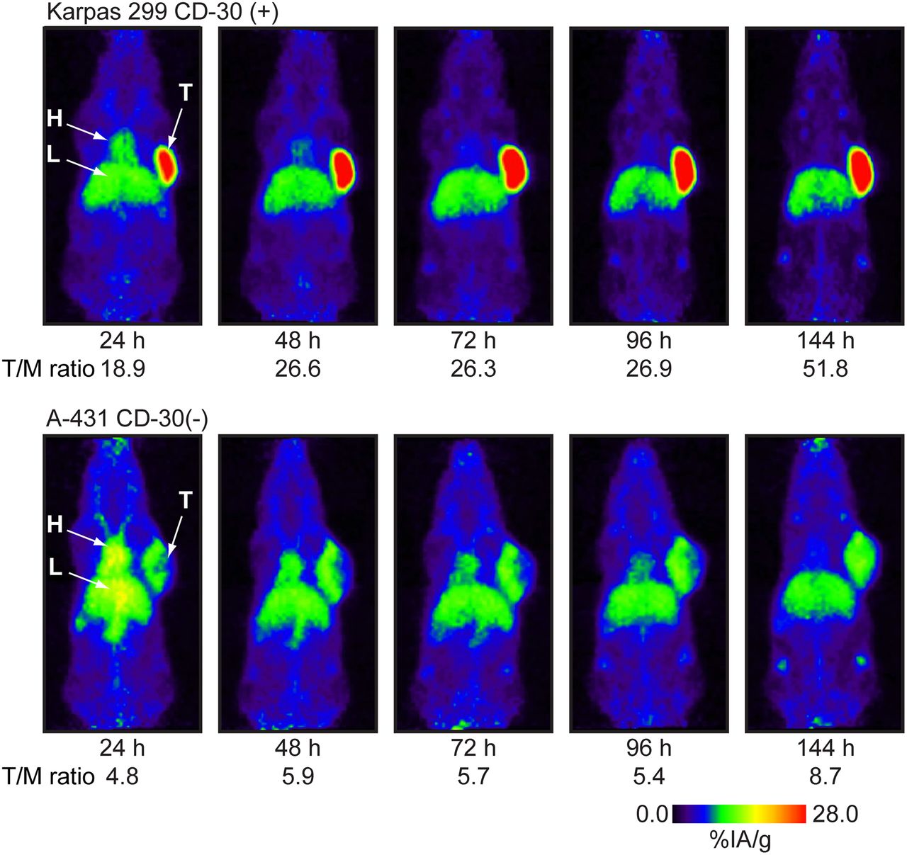
- FIGURE 5.
Longitudinal maximum-intensity-projection PET images showing distribution of 89Zr-DFO-AC-10 in Karpas 299 and A-431 tumor-bearing mice 24–144 h after administration. Mean tumor-to-muscle (T/M) ratios derived from volume-of-interest analysis of PET images are given. In A-431 model, liver and tumor time–activity curves overlap. T/M = tumor-to-muscle ratio.
- FIGURE 6.
Time–activity curves derived by VOI analysis of immuno-PET/CT images showing mean %IA/g tissue uptake vs. time for 89Zr-DFO-AC-10 radiotracer accumulation in mice bearing either Karpas 299 (A) or A-431 (B) tumors.
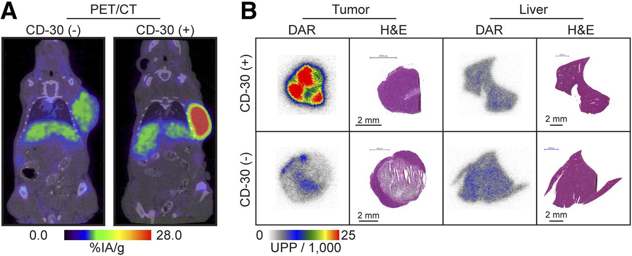
- FIGURE 7.
(A) PET/CT images of 2 representative mice bearing CD30-negative A-431 and CD30-positive Karpas 299 tumors at 144 h after injection. (B) Digital autoradiography and histology data showing accumulation and intratissue distribution of 89Zr-DFO-AC-10 in tumor and liver for both CD30-positive and CD30-negative models. H&E = hematoxylin and eosin. DAR = digital autoradiography; UPP = ▪▪▪.


Tables
A-431 CD30-negative Karpas 299 CD30-positive Site 24 h (n = 3) 72 h (n = 3) 24 h (n = 4) 72 h (n = 3) 72 h, blocking* (n = 3) Blood 15.83 ± 2.27 12.23 ± 2.18 9.89 ± 2.70 6.38 ± 0.46 9.49 ± 2.60 Heart 4.35 ± 1.27 3.11 ± 0.88 2.88 ± 0.36 1.71 ± 0.22 2.64 ± 0.47 Lung 6.58 ± 1.15 5.81 ± 1.07 4.60 ± 0.87 3.63 ± 0.59 4.12 ± 1.20 Liver 10.90 ± 2.10 12.61 ± 2.20 15.22 ± 1.71 11.78 ± 2.43 11.00 ± 2.32 Spleen 7.20 ± 1.40 8.21 ± 2.87 8.78 ± 2.14 8.15 ± 0.79 7.07 ± 0.46 Pancreas 1.68 ± 0.34 1.24 ± 0.30 1.05 ± 0.07 0.66 ± 0.06 0.74 ± 0.23 Stomach 1.52 ± 0.40 1.49 ± 0.20 1.09 ± 0.17 0.83 ± 0.05 0.83 ± 0.08 Small intestine 2.09 ± 0.44 1.82 ± 0.20 1.23 ± 0.20 1.12 ± 0.07 1.12 ± 0.36 Kidney 6.55 ± 1.08 6.28 ± 0.99 9.41 ± 1.78 4.22 ± 0.51 4.95 ± 0.86 Muscle 1.08 ± 0.19 0.91 ± 0.10 0.62 ± 0.06 0.49 ± 0.10 0.62 ± 0.20 Bone 2.58 ± 0.53 4.11 ± 2.73 2.12 ± 0.48 5.06 ± 0.38 2.46 ± 0.65 Tumor 8.23 ± 1.58 11.04 ± 0.40 27.21 ± 5.96 37.92 ± 8.22 22.54 ± 1.58 Tumor/blood 0.52 ± 0.12 0.90 ± 0.16 2.75 ± 0.96 5.95 ± 1.36 2.37 ± 0.67 Tumor/liver 0.76 ± 0.21 0.88 ± 0.16 1.79 ± 0.44 3.22 ± 0.96 2.05 ± 0.45 Tumor/kidney 1.14 ± 0.31 1.34 ± 0.47 2.89 ± 0.84 9.00 ± 2.23 4.56 ± 0.85 Tumor/spleen 1.26 ± 0.32 1.76 ± 0.28 3.10 ± 1.01 4.65 ± 1.10 3.19 ± 0.31 Tumor/muscle 7.63 ± 2.00 12.09 ± 1.45 43.95 ± 10.67 77.24 ± 22.92 36.21 ± 11.85 Tumor/bone 3.20 ± 0.90 2.68 ± 1.79 12.82 ± 4.03 7.49 ± 1.72 9.18 ± 2.51 ↵* With 500-μg mAb blocking.
Data are mean %IA/g ± SD. Errors for tumor-to-tissue ratios are geometric mean of SD.
Supplemental Data
Files in this Data Supplement:


