
Main menu

  • Home
  • Content
    • Current
    • Ahead of print
    • Past Issues
    • JNM Supplement
    • SNMMI Annual Meeting Abstracts
    • Continuing Education
    • JNM Podcasts
  • Subscriptions
    • Subscribers
    • Institutional and Non-member
    • Rates
    • Journal Claims
    • Corporate & Special Sales
  • Authors
    • Submit to JNM
    • Information for Authors
    • Assignment of Copyright
    • AQARA requirements
  • Info
    • Reviewers
    • Permissions
    • Advertisers
  • About
    • About Us
    • Editorial Board
    • Contact Information
  • More
    • Alerts
    • Feedback
    • Help
    • SNMMI Journals
  • SNMMI
    • JNM
    • JNMT
    • SNMMI Journals
    • SNMMI

User menu

  • Subscribe
  • My alerts
  • Log in
  • My Cart

Search

  • Advanced search
Journal of Nuclear Medicine
  • SNMMI
    • JNM
    • JNMT
    • SNMMI Journals
    • SNMMI
  • Subscribe
  • My alerts
  • Log in
  • My Cart
Journal of Nuclear Medicine

Advanced Search

  • Home
  • Content
    • Current
    • Ahead of print
    • Past Issues
    • JNM Supplement
    • SNMMI Annual Meeting Abstracts
    • Continuing Education
    • JNM Podcasts
  • Subscriptions
    • Subscribers
    • Institutional and Non-member
    • Rates
    • Journal Claims
    • Corporate & Special Sales
  • Authors
    • Submit to JNM
    • Information for Authors
    • Assignment of Copyright
    • AQARA requirements
  • Info
    • Reviewers
    • Permissions
    • Advertisers
  • About
    • About Us
    • Editorial Board
    • Contact Information
  • More
    • Alerts
    • Feedback
    • Help
    • SNMMI Journals
  • View or Listen to JNM Podcast
  • Visit JNM on Facebook
  • Join JNM on LinkedIn
  • Follow JNM on Twitter
  • Subscribe to our RSS feeds
Research ArticleClinical Investigations

68Ga-NOTA-Aca-BBN(7–14) PET/CT in Healthy Volunteers and Glioma Patients

Jingjing Zhang, Deling Li, Lixin Lang, Zhaohui Zhu, Ling Wang, Peilin Wu, Gang Niu, Fang Li and Xiaoyuan Chen
Journal of Nuclear Medicine January 2016, 57 (1) 9-14; DOI: https://doi.org/10.2967/jnumed.115.165316
Jingjing Zhang
1Department of Nuclear Medicine, Peking Union Medical College Hospital, Chinese Academy of Medical Sciences and Peking Union Medical College, Beijing, China
2Laboratory of Molecular Imaging and Nanomedicine, National Institute of Biomedical Imaging and Bioengineering, National Institutes of Health, Bethesda, Maryland; and
  • Find this author on Google Scholar
  • Find this author on PubMed
  • Search for this author on this site
Deling Li
3Department of Neurosurgery, Beijing Tiantan Hospital, Capital Medical University, Beijing, China
  • Find this author on Google Scholar
  • Find this author on PubMed
  • Search for this author on this site
Lixin Lang
2Laboratory of Molecular Imaging and Nanomedicine, National Institute of Biomedical Imaging and Bioengineering, National Institutes of Health, Bethesda, Maryland; and
  • Find this author on Google Scholar
  • Find this author on PubMed
  • Search for this author on this site
Zhaohui Zhu
1Department of Nuclear Medicine, Peking Union Medical College Hospital, Chinese Academy of Medical Sciences and Peking Union Medical College, Beijing, China
  • Find this author on Google Scholar
  • Find this author on PubMed
  • Search for this author on this site
Ling Wang
1Department of Nuclear Medicine, Peking Union Medical College Hospital, Chinese Academy of Medical Sciences and Peking Union Medical College, Beijing, China
  • Find this author on Google Scholar
  • Find this author on PubMed
  • Search for this author on this site
Peilin Wu
1Department of Nuclear Medicine, Peking Union Medical College Hospital, Chinese Academy of Medical Sciences and Peking Union Medical College, Beijing, China
  • Find this author on Google Scholar
  • Find this author on PubMed
  • Search for this author on this site
Gang Niu
2Laboratory of Molecular Imaging and Nanomedicine, National Institute of Biomedical Imaging and Bioengineering, National Institutes of Health, Bethesda, Maryland; and
  • Find this author on Google Scholar
  • Find this author on PubMed
  • Search for this author on this site
Fang Li
1Department of Nuclear Medicine, Peking Union Medical College Hospital, Chinese Academy of Medical Sciences and Peking Union Medical College, Beijing, China
  • Find this author on Google Scholar
  • Find this author on PubMed
  • Search for this author on this site
Xiaoyuan Chen
2Laboratory of Molecular Imaging and Nanomedicine, National Institute of Biomedical Imaging and Bioengineering, National Institutes of Health, Bethesda, Maryland; and
  • Find this author on Google Scholar
  • Find this author on PubMed
  • Search for this author on this site
  • Article
  • Figures & Data
  • Info & Metrics
  • PDF
Loading

Article Figures & Data

Figures

  • Tables
  • FIGURE 1.
    • Download figure
    • Open in new tab
    • Download powerpoint
    FIGURE 1.

    (A) Schematic structure of 68Ga-BBN. (B) Representative maximum-intensity-projection torso images of healthy volunteer at different time points after intravenous injection of 68Ga-BBN. Main regions with prominent 68Ga-BBN uptake are pancreas, kidneys, urinary ducts, and bladder.

  • FIGURE 2.
    • Download figure
    • Open in new tab
    • Download powerpoint
    FIGURE 2.

    Representative 68Ga-BBN PET/CT images (left), MR images (middle), and immunohistochemically stained tissue samples (right) of patients with gliomas of different grades. (A) A 5-y-old girl with space-occupying lesion in cerebellar lobe diagnosed as pilocytic astrocytoma, WHO grade I. Lesion had SUVmax of 2.35 and SUVmean of 1.31, showed enhancement on T1-weighted MRI, and stained positively for GRPR. (B) A 4-y-old girl with multiple space-occupying lesions diagnosed as WHO grade II astrocytoma. Lesions had SUVmax of 2.05 and SUVmean of 1.45, were identified by contrast-enhanced T1-weighted MRI, and stained positively for GRPR. (C) A 14-y-old boy with space-occupying lesion in thalamic connections of temporal lobe diagnosed as WHO grade III anaplastic oligoastrocytoma. Lesion had SUVmax of 1.98 and SUVmean of 1.08, showed enhancement on T1-weighted MRI, and stained positively for GRPR. (D) A 44-y-old man with space-occupying lesion in left temporal lobe diagnosed as WHO grade IV glioblastoma multiforme. Lesion had SUVmax of 1.95 and SUVmean of 0.98, showed enhancement on T2-weighted MRI, and stained positively for GRPR.
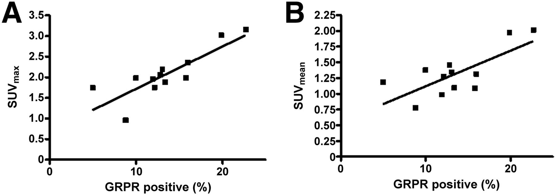
  • FIGURE 3.
    • Download figure
    • Open in new tab
    • Download powerpoint
    FIGURE 3.

    Correlation of SUVmax (A) and SUVmean (B) as determined by 68Ga-BBN PET with GRPR expression level as determined by immunohistochemical staining (r2 = 0.71, P < 0.001, for SUVmax and r2 = 0.54, P < 0.01, for SUVmean).

Tables

  • Figures
    • View popup
    TABLE 1

    Patient Demographics

    Patient no.Age (y)SexWHO gradeHistologyLocationMRI findings
    112MIGangliogliomaThird ventricleHeterogeneous, intense
    239MIIIAnaplastic oligoastrocytomaRight frontalHeterogeneous, intense
    314MIIIAnaplastic oligoastrocytomaThalamus, temporal lobeHeterogeneous, intense
    44FIIAstrocytomaSeller areaHeterogeneous, intense
    55MIIPilocytic astrocytoma with necrosisSeller areaHeterogeneous, moderate
    65FIPilocytic astrocytomaCerebellar lobeHeterogeneous, moderate
    733FIPilocytic astrocytomaSeller areaHeterogeneous, intense
    834MIIIAnaplastic oligoastrocytomaFrontal lobeHeterogeneous, moderate
    937MIVGlioblastoma multiformeFrontal lobeHeterogeneous, intense
    1034FIIOligoastrocytomaLeft frontalNot applicable
    1112MIPilocytic astrocytomaSeller areaHomogeneous, intense
    1244MIVGlioblastoma multiformeLeft temporal lobeHomogeneous, intense
    • View popup
    TABLE 2

    Biodistribution of 68Ga-BBN in Healthy Volunteers

    Time (min)
    Organ151015304560
    Brain0.23 ± 0.050.18 ± 0.050.18 ± 0.050.15 ± 0.060.15 ± 0.060.13 ± 0.050.15 ± 0.06
    Heart3.50 ± 0.592.88 ± 0.432.30 ± 0.231.85 ± 0.211.60 ± 0.181.43 ± 0.221.25 ± 0.24
    Lung1.13 ± 0.260.81 ± 0.060.70 ± 0.080.60 ± 0.180.50 ± 0.080.40 ± 0.080.43 ± 0.10
    Liver2.68 ± 0.322.18 ± 0.781.58 ± 0.211.55 ± 0.371.43 ± 0.261.33 ± 0.261.25 ± 0.17
    Spleen2.84 ± 0.302.18 ± 0.741.65 ± 0.381.53 ± 0.431.58 ± 0.361.50 ± 0.351.30 ± 0.35
    Pancreas13.59 ± 2.2919.79 ± 2.3220.05 ± 2.1120.06 ± 4.1917.15 ± 3.0013.10 ± 0.1412.70 ± 2.00
    Kidneys7.73 ± 1.036.83 ± 1.267.88 ± 3.594.88 ± 0.623.83 ± 0.633.15 ± 0.772.70 ± 0.54
    Bladder2.58 ± 1.4442.67 ± 5.5774.86 ± 5.5180.57 ± 5.7180.30 ± 6.5575.95 ± 7.2371.63 ± 5.96
    Red marrow0.50 ± 0.450.80 ± 0.240.90 ± 0.290.85 ± 0.240.78 ± 0.240.80 ± 0.410.73 ± 0.26
    Muscle0.18 ± 0.050.48 ± 0.050.53 ± 0.100.53 ± 0.100.45 ± 0.060.45 ± 0.060.40 ± 0.08
    • Data are mean SUV ± SD (n = 4).

    • View popup
    TABLE 3

    Estimated Absorbed 68Ga-BBN Dose in Healthy Volunteers

    mSv/MBq
    OrganMeanSD
    Adrenals0.000210.00004
    Brain0.000010.00001
    Breasts0.000040.00002
    Gallbladder wall0.000530.00013
    Lower large intestine wall0.005660.00121
    Small intestine0.002280.00057
    Stomach wall0.000320.00001
    Upper large intestine wall0.001740.00044
    Heart wall0.000170.00001
    Kidneys0.000580.00013
    Liver0.000320.00006
    Lungs0.000090.00003
    Muscle0.001390.00025
    Ovaries0.005300.00124
    Pancreas0.001050.00015
    Red marrow0.000980.00022
    Osteogenic cells0.000580.00011
    Skin0.000500.00001
    Spleen0.000320.00007
    Testes0.01000—
    Thymus0.001590.00177
    Thyroid0.000040.00001
    Urinary bladder wall0.307000.35559
    Uterus0.01310—
    Total body0.001500.00029
    Effective dose equivalent0.033500.00790
    Effective dose0.027600.00660
PreviousNext
Back to top

In this issue

Journal of Nuclear Medicine: 57 (1)
Journal of Nuclear Medicine
Vol. 57, Issue 1
January 1, 2016
  • Table of Contents
  • Table of Contents (PDF)
  • About the Cover
  • Index by author
Print
Download PDF
Article Alerts
Sign In to Email Alerts with your Email Address
Email Article

Thank you for your interest in spreading the word on Journal of Nuclear Medicine.

NOTE: We only request your email address so that the person you are recommending the page to knows that you wanted them to see it, and that it is not junk mail. We do not capture any email address.

Enter multiple addresses on separate lines or separate them with commas.
68Ga-NOTA-Aca-BBN(7–14) PET/CT in Healthy Volunteers and Glioma Patients
(Your Name) has sent you a message from Journal of Nuclear Medicine
(Your Name) thought you would like to see the Journal of Nuclear Medicine web site.
Citation Tools
68Ga-NOTA-Aca-BBN(7–14) PET/CT in Healthy Volunteers and Glioma Patients
Jingjing Zhang, Deling Li, Lixin Lang, Zhaohui Zhu, Ling Wang, Peilin Wu, Gang Niu, Fang Li, Xiaoyuan Chen
Journal of Nuclear Medicine Jan 2016, 57 (1) 9-14; DOI: 10.2967/jnumed.115.165316

Citation Manager Formats

  • BibTeX
  • Bookends
  • EasyBib
  • EndNote (tagged)
  • EndNote 8 (xml)
  • Medlars
  • Mendeley
  • Papers
  • RefWorks Tagged
  • Ref Manager
  • RIS
  • Zotero
Share
68Ga-NOTA-Aca-BBN(7–14) PET/CT in Healthy Volunteers and Glioma Patients
Jingjing Zhang, Deling Li, Lixin Lang, Zhaohui Zhu, Ling Wang, Peilin Wu, Gang Niu, Fang Li, Xiaoyuan Chen
Journal of Nuclear Medicine Jan 2016, 57 (1) 9-14; DOI: 10.2967/jnumed.115.165316
Twitter logo Facebook logo LinkedIn logo Mendeley logo
  • Tweet Widget
  • Facebook Like
  • Google Plus One
Bookmark this article

Jump to section

  • Article
    • Abstract
    • MATERIALS AND METHODS
    • RESULTS
    • DISCUSSION
    • CONCLUSION
    • DISCLOSURE
    • Footnotes
    • REFERENCES
  • Figures & Data
  • Info & Metrics
  • PDF

Related Articles

  • This Month in JNM
  • PubMed
  • Google Scholar

Cited By...

  • Head-to-Head Comparison of [68Ga]Ga-NOTA-RM26 and [18F]FDG PET/CT in Patients with Gastrointestinal Stromal Tumors: A Prospective Study
  • PET Using a GRPR Antagonist 68Ga-RM26 in Healthy Volunteers and Prostate Cancer Patients
  • Glu-Ureido-Based Inhibitors of Prostate-Specific Membrane Antigen: Lessons Learned During the Development of a Novel Class of Low-Molecular-Weight Theranostic Radiotracers
  • Clinical Translation of a Dual Integrin {alpha}v{beta}3- and Gastrin-Releasing Peptide Receptor-Targeting PET Radiotracer, 68Ga-BBN-RGD
  • Google Scholar

More in this TOC Section

  • Feasibility of Ultra-Low-Activity 18F-FDG PET/CT Imaging Using a Long–Axial-Field-of-View PET/CT System
  • Cardiac Presynaptic Sympathetic Nervous Function Evaluated by Cardiac PET in Patients with Chronotropic Incompetence Without Heart Failure
  • Validation and Evaluation of a Vendor-Provided Head Motion Correction Algorithm on the uMI Panorama PET/CT System
Show more Clinical Investigations

Similar Articles

Keywords

  • BBN peptide
  • gastrin-releasing peptide receptor (GRPR)
  • 68Ga-BBN
  • PET/CT
  • glioma
SNMMI

© 2025 SNMMI

Powered by HighWire