


Article Figures & Data
Figures
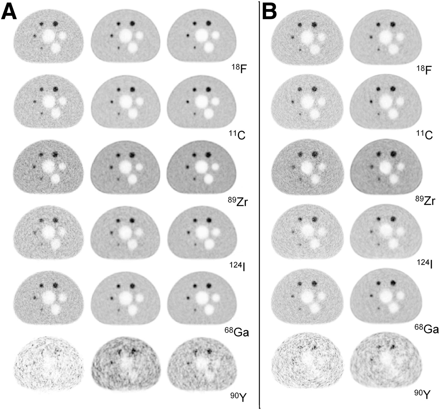
- FIGURE 1.
PET images reconstructed for all isotopes. (A) Images obtained on mCT using 3D OP-OSEM (left), 3D OP-OSEM+PSF (center), and 3D OP-OSEM+PSF+TOF (right). (B) Images obtained on mMR using 3D OP-OSEM (left) and 3D OP-OSEM+PSF (right). All images have 2-mm pixel size and 2-mm postreconstruction gaussian filter applied.
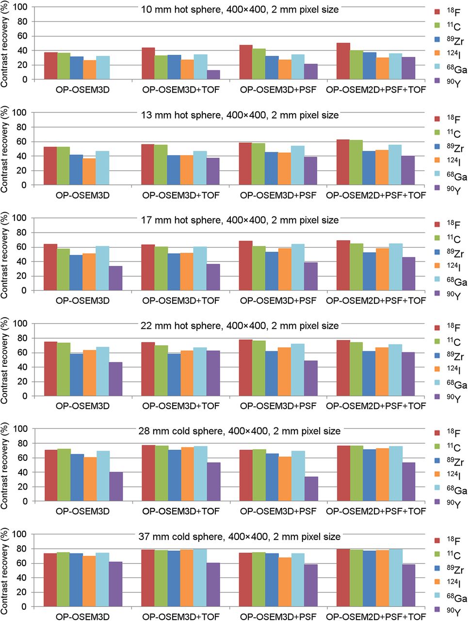
- FIGURE 2.
mCT contrast recovery for all isotopes. 90Y data were omitted from plot when sphere was not visible in image.
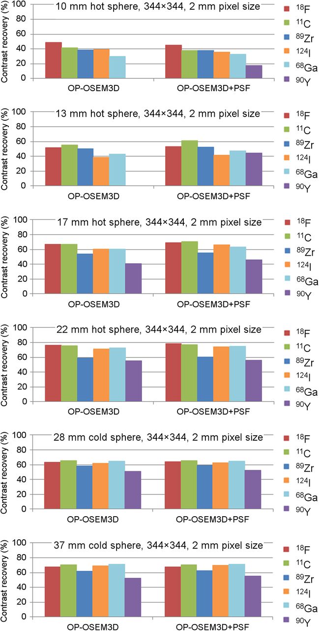
- FIGURE 3.
mMR contrast recovery for all isotopes. 90Y data were omitted from plot when sphere was not visible in image.
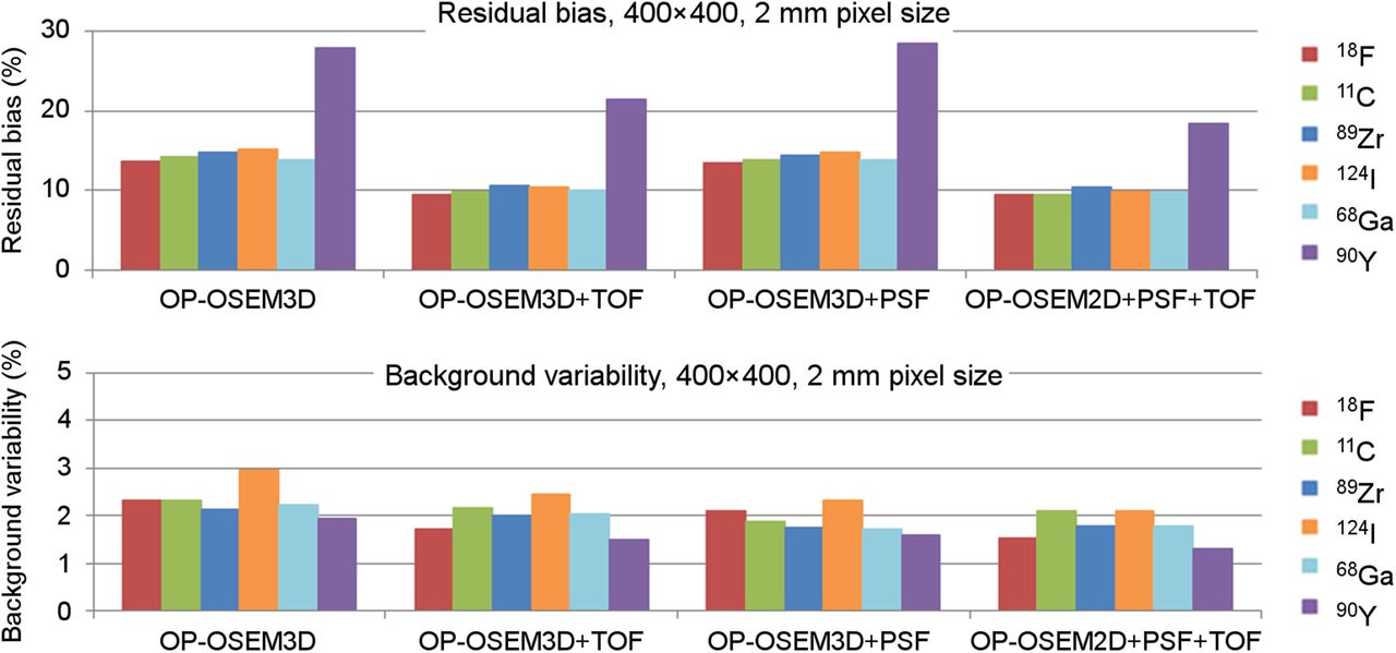
- FIGURE 4.
mCT residual bias in lung insert and background variability. For background variability, 90Y data have been normalized to 200-million-true-count scan.
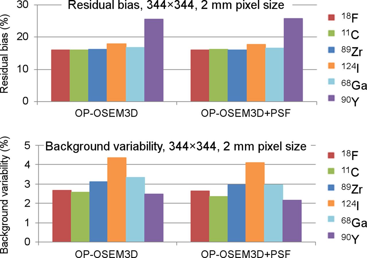
- FIGURE 5.
mMR residual bias in lung insert (top) and background variability (bottom). For background variability, 90Y data have been normalized to 200-million-true-count scan.
- FIGURE 6.
mCT (top) and mMR (bottom) FWHM measurements (2-mm pixel size) obtained with FBP and no postreconstruction filter. Error bars are 2 times SD of repeated measurements.
- FIGURE 7.
mCT (top) and mMR (bottom) FWHM-to-FWTM ratios (2-mm pixel size) obtained with FBP and no postreconstruction filter.


Tables
Method mCT* mMR* FBP Backprojection Filtered backprojection FBP+TOF Backprojection+TOF — 3D OP-OSEM Iterative (3 iterations, 24 subsets) 3D iterative (3 iterations, 21 subsets) 3D OP-OSEM+TOF Iterative+TOF (2 iterations, 21 subsets) — 3D OP-OSEM+PSF TrueX (3 iterations, 24 subsets) HD PET (3 iterations, 21 subsets) 3D OP-OSEM+PSF+TOF TrueX+TOF (2 iterations, 21 subsets) — ↵* Reconstruction label in user interface.
Supplemental Data
Files in this Data Supplement:


