
Main menu

  • Home
  • Content
    • Current
    • Ahead of print
    • Past Issues
    • JNM Supplement
    • SNMMI Annual Meeting Abstracts
    • Continuing Education
    • JNM Podcasts
  • Subscriptions
    • Subscribers
    • Institutional and Non-member
    • Rates
    • Journal Claims
    • Corporate & Special Sales
  • Authors
    • Submit to JNM
    • Information for Authors
    • Assignment of Copyright
    • AQARA requirements
  • Info
    • Reviewers
    • Permissions
    • Advertisers
  • About
    • About Us
    • Editorial Board
    • Contact Information
  • More
    • Alerts
    • Feedback
    • Help
    • SNMMI Journals
  • SNMMI
    • JNM
    • JNMT
    • SNMMI Journals
    • SNMMI

User menu

  • Subscribe
  • My alerts
  • Log in
  • My Cart

Search

  • Advanced search
Journal of Nuclear Medicine
  • SNMMI
    • JNM
    • JNMT
    • SNMMI Journals
    • SNMMI
  • Subscribe
  • My alerts
  • Log in
  • My Cart
Journal of Nuclear Medicine

Advanced Search

  • Home
  • Content
    • Current
    • Ahead of print
    • Past Issues
    • JNM Supplement
    • SNMMI Annual Meeting Abstracts
    • Continuing Education
    • JNM Podcasts
  • Subscriptions
    • Subscribers
    • Institutional and Non-member
    • Rates
    • Journal Claims
    • Corporate & Special Sales
  • Authors
    • Submit to JNM
    • Information for Authors
    • Assignment of Copyright
    • AQARA requirements
  • Info
    • Reviewers
    • Permissions
    • Advertisers
  • About
    • About Us
    • Editorial Board
    • Contact Information
  • More
    • Alerts
    • Feedback
    • Help
    • SNMMI Journals
  • View or Listen to JNM Podcast
  • Visit JNM on Facebook
  • Join JNM on LinkedIn
  • Follow JNM on Twitter
  • Subscribe to our RSS feeds
Research ArticleBasic Science Investigations

In Vivo Cancer Dual-Targeting and Dual-Modality Imaging with Functionalized Quantum Dots

Kongzhen Hu, Hongliang Wang, Ganghua Tang, Tingting Huang, Xiaolan Tang, Xiang Liang, Shaobo Yao and Dahong Nie
Journal of Nuclear Medicine August 2015, 56 (8) 1278-1284; DOI: https://doi.org/10.2967/jnumed.115.158873
Kongzhen Hu
1PET-CT Center, Department of Nuclear Medicine, the First Affiliated Hospital, Sun Yat-Sen University, Guangzhou, China; and
  • Find this author on Google Scholar
  • Find this author on PubMed
  • Search for this author on this site
Hongliang Wang
1PET-CT Center, Department of Nuclear Medicine, the First Affiliated Hospital, Sun Yat-Sen University, Guangzhou, China; and
  • Find this author on Google Scholar
  • Find this author on PubMed
  • Search for this author on this site
Ganghua Tang
1PET-CT Center, Department of Nuclear Medicine, the First Affiliated Hospital, Sun Yat-Sen University, Guangzhou, China; and
  • Find this author on Google Scholar
  • Find this author on PubMed
  • Search for this author on this site
Tingting Huang
1PET-CT Center, Department of Nuclear Medicine, the First Affiliated Hospital, Sun Yat-Sen University, Guangzhou, China; and
  • Find this author on Google Scholar
  • Find this author on PubMed
  • Search for this author on this site
Xiaolan Tang
2College of Science, Southern China Agricultural University, Guangzhou, China
  • Find this author on Google Scholar
  • Find this author on PubMed
  • Search for this author on this site
Xiang Liang
1PET-CT Center, Department of Nuclear Medicine, the First Affiliated Hospital, Sun Yat-Sen University, Guangzhou, China; and
  • Find this author on Google Scholar
  • Find this author on PubMed
  • Search for this author on this site
Shaobo Yao
1PET-CT Center, Department of Nuclear Medicine, the First Affiliated Hospital, Sun Yat-Sen University, Guangzhou, China; and
  • Find this author on Google Scholar
  • Find this author on PubMed
  • Search for this author on this site
Dahong Nie
1PET-CT Center, Department of Nuclear Medicine, the First Affiliated Hospital, Sun Yat-Sen University, Guangzhou, China; and
  • Find this author on Google Scholar
  • Find this author on PubMed
  • Search for this author on this site
  • Article
  • Figures & Data
  • Supplemental
  • Info & Metrics
  • PDF
Loading

Article Figures & Data

Figures

  • Tables
  • Additional Files
  • FIGURE 1.
    • Download figure
    • Open in new tab
    • Download powerpoint
    FIGURE 1.

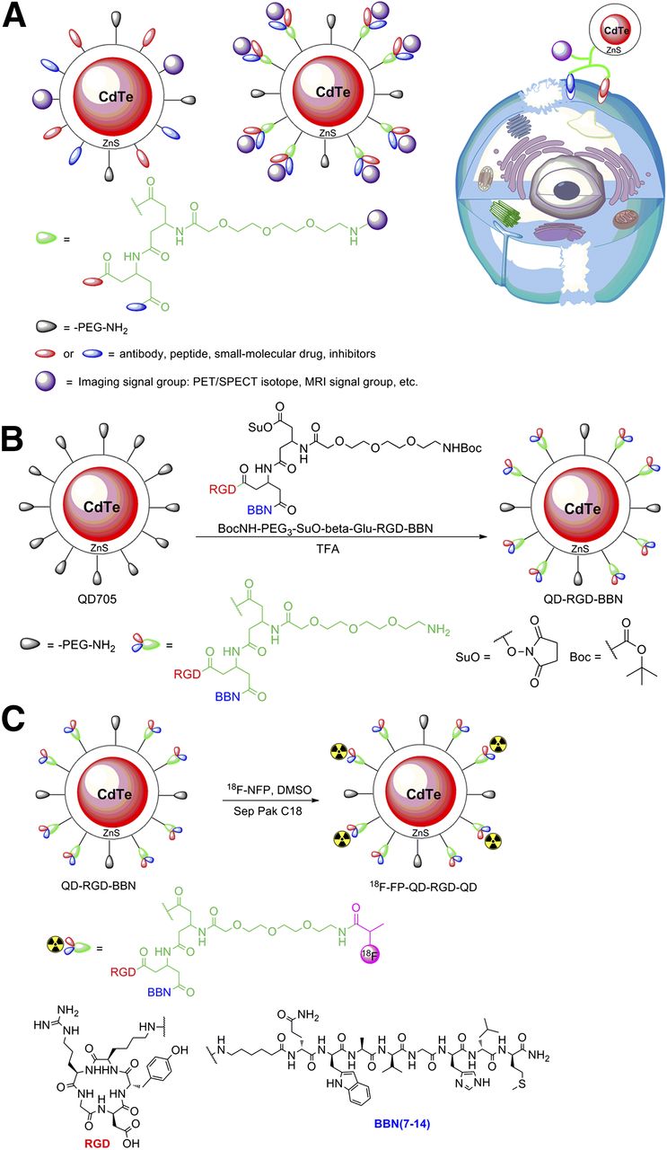
    (A) Schematic illustration of functionalized QD probe for in vivo cancer dual-targeting and dual-modality imaging: structure of multiplex modifying multifunctional QD probe (left), structure of single modifying multifunctional QD probe (middle), and strategy for enhancing synergistic binding of heterodimeric multifunctional QD probe (right). (B) Synthesis of QD-RGD-BBN. (C) Synthesis of dual receptor–targeting dual-modality PET probe 18F-FP-QD-RGD-BBN. CdTe = cadmium telluride; DMSO = dimethyl sulfoxide; TFA = trifluoroacetic acid.

  • FIGURE 2.
    • Download figure
    • Open in new tab
    • Download powerpoint
    FIGURE 2.

    Characterization of QD probes. (A) Dynamic light scattering data of QD705. (B) Dynamic light scattering data of QD-RGD-BBN. (C) Fluorescence spectra of QD-RGD-BBN and QD705 in aqueous solution. (D) Ultraviolet visible absorbance spectra of NH2-RGD-BBN, QD-RGD-BBN, and QD705.

  • FIGURE 3.
    • Download figure
    • Open in new tab
    • Download powerpoint
    FIGURE 3.

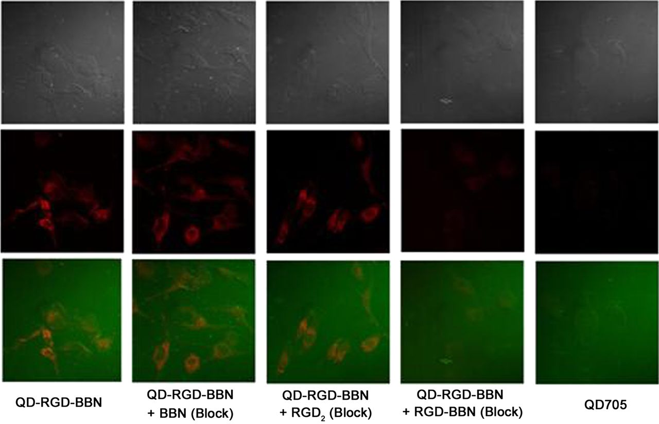
    In vitro staining of PC-3 cells (both integrin αvβ3 and GRPR high expression) using 1 nM QD-RGD-BBN (left-first column) and 1 nM QD705 (right-first column). Staining of PC-3 cells with 1 nM QD-RGD-BBN in presence of 2 μM BBN (QD-RGD-BBN + BBN Block), 2 μM RGD2 (QD-RGD-BBN + RGD2 Block), and mixture of 2 μM BBN and 2 μM RGD2 (QD-RGD-BBN + RGD-BBN Block) is also shown. Filter set: excitation, 420/40 nm; emission, 705/40 nm. Magnification, ×400, 0.5-s exposure. All fluorescence images were acquired under same condition and displayed under same scale.

  • FIGURE 4.
    • Download figure
    • Open in new tab
    • Download powerpoint
    FIGURE 4.

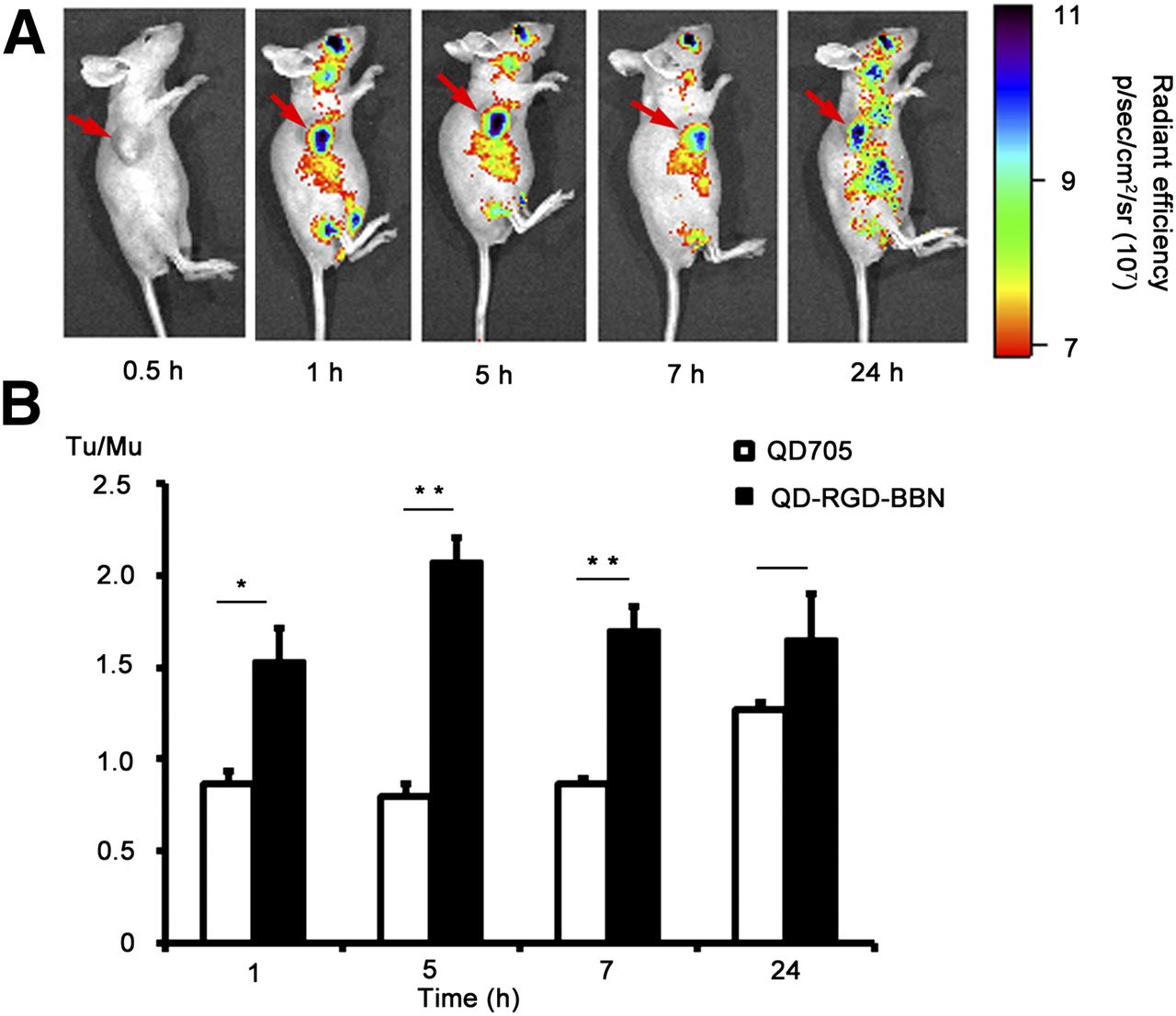
    (A) In vivo NIRF imaging of PC-3 tumor–bearing mice at 0.5, 1, 5, 7, and 24 h after injection of 200 pmol QD-RGD-BBN. Arrows indicate tumor. (B) Tumor-to-muscle uptake ratios (Tu/Mu) of mice injected with QD705 and QD-RGD-BBN. Data are mean ± SD. *P < 0.05, as compared with mice injected with QD705, 1-tailed paired Student t test (n = 3). **P < 0.01.
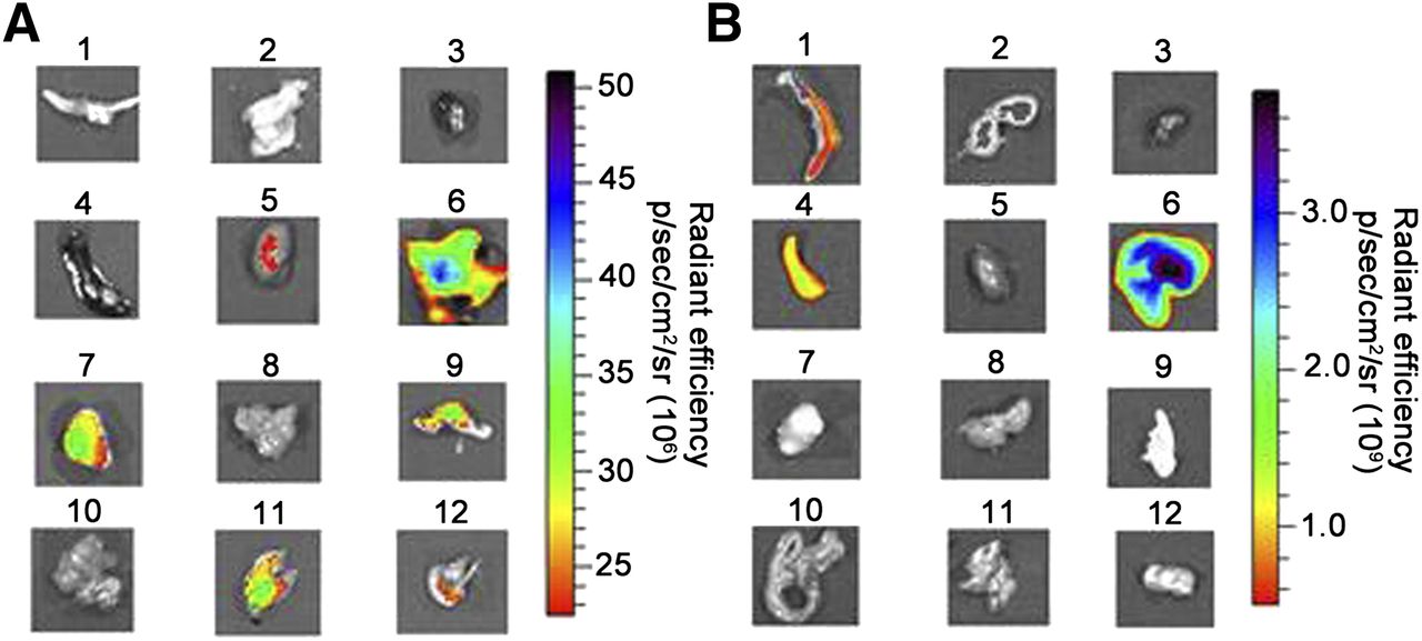
  • FIGURE 5.
    • Download figure
    • Open in new tab
    • Download powerpoint
    FIGURE 5.

    Ex vivo NIRF imaging. (A) NIRF image of harvested major organs at 5 h after injection of QD-RGD-BBN. (B) NIRF image of harvested major organs at 5 h after injection of QD705. 1 = bone; 2 = brain; 3 = heart; 4 =spleen; 5 = kidney; 6 = liver; 7 = tumor; 8 = muscle; 9 = skin; 10 = intestine; 11 = lung; 12 = stomach.

  • FIGURE 6.
    • Download figure
    • Open in new tab
    • Download powerpoint
    FIGURE 6.

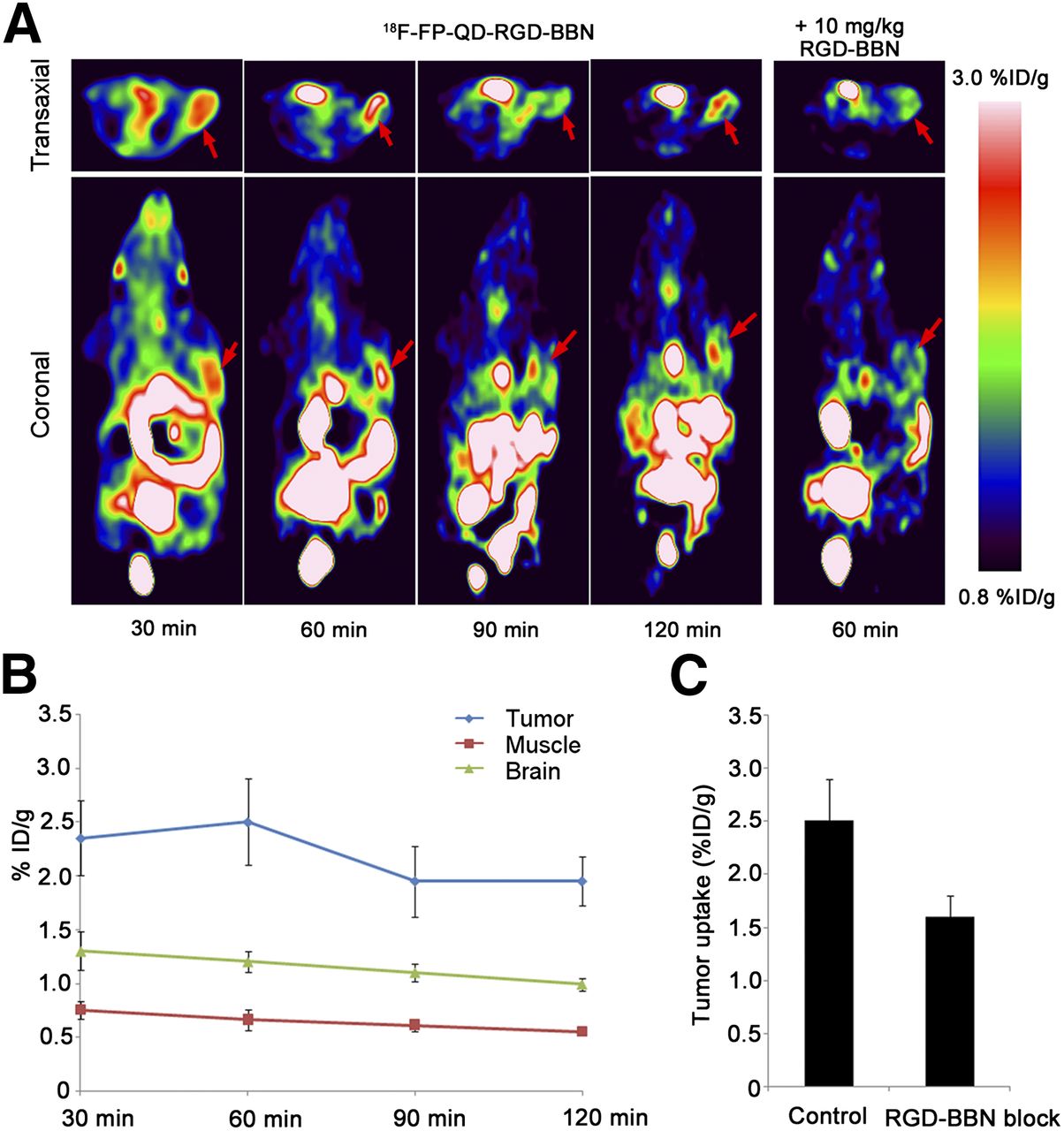
    PET imaging of PC-3 tumor–bearing mice with targeting dual-receptor PET/NIRF probe. (A) PET images of same mouse at 30, 60, 90, and 120 min after injection of 18F-FP-QD-RGD-BBN and in presence of blocking agent RGD-BBN. Red arrows indicate tumor. (B) Uptake of 18F-FP-QD-RGD-BBN in tumor, muscle, and brain over time, as quantified by ROI analysis of small-animal PET scans (n = 3 per group). (C) Tumor uptake of 18F-FP-QD-RGD-BBN at 60 min after injection in absence of blocking agent (control) and in presence of RGD-BBN, as quantified by ROI analysis of small-animal PET scans (n = 3 per group). %ID/g = percentage injected dose per gram.

Tables

  • Figures
  • Additional Files
    • View popup
    TABLE 1

    In Vivo Biodistribution of 18F-FP-QD-RGD-BBN in Normal Mice

    Organ5 min30 min60 min120 min
    Blood4.57 ± 0.963.01 ± 0.501.81 ± 0.111.73 ± 0.17
    Brain1.89 ± 0.331.86 ± 0.201.17 ± 0.071.04 ± 0.13
    Heart2.51 ± 0.441.98 ± 0.121.39 ± 0.251.36 ± 0.30
    Lung6.87 ± 2.563.49 ± 0.742.22 ± 0.421.50 ± 0.36
    Liver7.03 ± 0.693.46 ± 0.842.19 ± 0.141.98 ± 0.38
    Spleen1.83 ± 0.631.62 ± 0.331.04 ± 0.200.81 ± 0.15
    Pancreas2.73 ± 0.771.59 ± 0.430.88 ± 0.220.61 ± 0.14
    Kidneys10.8 ± 3.827.35 ± 1.692.60 ± 0.441.98 ± 0.34
    Intestine2.56 ± 0.633.53 ± 0.691.94 ± 0.561.56 ± 0.28
    Muscle2.20 ± 0.711.51 ± 0.380.97 ± 0.090.69 ± 0.15
    Stomach2.49 ± 0.681.61 ± 0.660.96 ± 0.240.88 ± 0.10
    Bone1.78 ± 0.611.72 ± 0.251.04 ± 0.240.92 ± 0.14
    • Data are average percentage injected dose per gram, mean ± SD (n = 4).

Additional Files

  • Figures
  • Tables
  • Supplemental Data

    Files in this Data Supplement:

    • Supplemental Data
PreviousNext
Back to top

In this issue

Journal of Nuclear Medicine: 56 (8)
Journal of Nuclear Medicine
Vol. 56, Issue 8
August 1, 2015
  • Table of Contents
  • Table of Contents (PDF)
  • About the Cover
  • Index by author
Print
Download PDF
Article Alerts
Sign In to Email Alerts with your Email Address
Email Article

Thank you for your interest in spreading the word on Journal of Nuclear Medicine.

NOTE: We only request your email address so that the person you are recommending the page to knows that you wanted them to see it, and that it is not junk mail. We do not capture any email address.

Enter multiple addresses on separate lines or separate them with commas.
In Vivo Cancer Dual-Targeting and Dual-Modality Imaging with Functionalized Quantum Dots
(Your Name) has sent you a message from Journal of Nuclear Medicine
(Your Name) thought you would like to see the Journal of Nuclear Medicine web site.
Citation Tools
In Vivo Cancer Dual-Targeting and Dual-Modality Imaging with Functionalized Quantum Dots
Kongzhen Hu, Hongliang Wang, Ganghua Tang, Tingting Huang, Xiaolan Tang, Xiang Liang, Shaobo Yao, Dahong Nie
Journal of Nuclear Medicine Aug 2015, 56 (8) 1278-1284; DOI: 10.2967/jnumed.115.158873

Citation Manager Formats

  • BibTeX
  • Bookends
  • EasyBib
  • EndNote (tagged)
  • EndNote 8 (xml)
  • Medlars
  • Mendeley
  • Papers
  • RefWorks Tagged
  • Ref Manager
  • RIS
  • Zotero
Share
In Vivo Cancer Dual-Targeting and Dual-Modality Imaging with Functionalized Quantum Dots
Kongzhen Hu, Hongliang Wang, Ganghua Tang, Tingting Huang, Xiaolan Tang, Xiang Liang, Shaobo Yao, Dahong Nie
Journal of Nuclear Medicine Aug 2015, 56 (8) 1278-1284; DOI: 10.2967/jnumed.115.158873
Twitter logo Facebook logo LinkedIn logo Mendeley logo
  • Tweet Widget
  • Facebook Like
  • Google Plus One
Bookmark this article

Jump to section

  • Article
    • Abstract
    • MATERIALS AND METHODS
    • RESULTS
    • DISCUSSION
    • CONCLUSION
    • DISCLOSURE
    • Acknowledgments
    • Footnotes
    • REFERENCES
  • Figures & Data
  • Supplemental
  • Info & Metrics
  • PDF

Related Articles

  • This Month in JNM
  • PubMed
  • Google Scholar

Cited By...

  • No citing articles found.
  • Google Scholar

More in this TOC Section

  • Tumor Uptake of Anti-CD20 Fabs Depends on Tumor Perfusion
  • How Sensitive Is the Upper Gastrointestinal Tract to 90Y Radioembolization? A Histologic and Dosimetric Analysis in a Porcine Model
  • 11C-Methionine PET of Myocardial Inflammation in a Rat Model of Experimental Autoimmune Myocarditis
Show more Basic Science Investigations

Similar Articles

Keywords

  • Quantum dots
  • fluorescence
  • PET
  • integrin αvβ3
  • GRPR
SNMMI

© 2025 SNMMI

Powered by HighWire