
Main menu

  • Home
  • Content
    • Current
    • Ahead of print
    • Past Issues
    • JNM Supplement
    • SNMMI Annual Meeting Abstracts
    • Continuing Education
    • JNM Podcasts
  • Subscriptions
    • Subscribers
    • Institutional and Non-member
    • Rates
    • Journal Claims
    • Corporate & Special Sales
  • Authors
    • Submit to JNM
    • Information for Authors
    • Assignment of Copyright
    • AQARA requirements
  • Info
    • Reviewers
    • Permissions
    • Advertisers
  • About
    • About Us
    • Editorial Board
    • Contact Information
  • More
    • Alerts
    • Feedback
    • Help
    • SNMMI Journals
  • SNMMI
    • JNM
    • JNMT
    • SNMMI Journals
    • SNMMI

User menu

  • Subscribe
  • My alerts
  • Log in
  • Log out
  • My Cart

Search

  • Advanced search
Journal of Nuclear Medicine
  • SNMMI
    • JNM
    • JNMT
    • SNMMI Journals
    • SNMMI
  • Subscribe
  • My alerts
  • Log in
  • Log out
  • My Cart
Journal of Nuclear Medicine

Advanced Search

  • Home
  • Content
    • Current
    • Ahead of print
    • Past Issues
    • JNM Supplement
    • SNMMI Annual Meeting Abstracts
    • Continuing Education
    • JNM Podcasts
  • Subscriptions
    • Subscribers
    • Institutional and Non-member
    • Rates
    • Journal Claims
    • Corporate & Special Sales
  • Authors
    • Submit to JNM
    • Information for Authors
    • Assignment of Copyright
    • AQARA requirements
  • Info
    • Reviewers
    • Permissions
    • Advertisers
  • About
    • About Us
    • Editorial Board
    • Contact Information
  • More
    • Alerts
    • Feedback
    • Help
    • SNMMI Journals
  • View or Listen to JNM Podcast
  • Visit JNM on Facebook
  • Join JNM on LinkedIn
  • Follow JNM on Twitter
  • Subscribe to our RSS feeds
Research ArticleClinical Investigations

Accuracy of 18F-FDG PET/CT, Multidetector CT, and MR Imaging in the Diagnosis of Pancreatic Cysts: A Prospective Single-Center Study

Saila Kauhanen, Irina Rinta-Kiikka, Jukka Kemppainen, Juha Grönroos, Sami Kajander, Marko Seppänen, Kalle Alanen, Risto Gullichsen, Pirjo Nuutila and Jari Ovaska
Journal of Nuclear Medicine August 2015, 56 (8) 1163-1168; DOI: https://doi.org/10.2967/jnumed.114.148940
Saila Kauhanen
1Division of Digestive Surgery and Urology, Turku University Hospital, Turku, Finland
2Turku PET Centre, Turku University Hospital, Turku, Finland
  • Find this author on Google Scholar
  • Find this author on PubMed
  • Search for this author on this site
Irina Rinta-Kiikka
3Department of Radiology, Tampere University Hospital, Tampere, Finland
  • Find this author on Google Scholar
  • Find this author on PubMed
  • Search for this author on this site
Jukka Kemppainen
2Turku PET Centre, Turku University Hospital, Turku, Finland
4Department of Clinical Physiology and Nuclear Medicine, Turku, Finland
  • Find this author on Google Scholar
  • Find this author on PubMed
  • Search for this author on this site
Juha Grönroos
1Division of Digestive Surgery and Urology, Turku University Hospital, Turku, Finland
  • Find this author on Google Scholar
  • Find this author on PubMed
  • Search for this author on this site
Sami Kajander
2Turku PET Centre, Turku University Hospital, Turku, Finland
  • Find this author on Google Scholar
  • Find this author on PubMed
  • Search for this author on this site
Marko Seppänen
2Turku PET Centre, Turku University Hospital, Turku, Finland
4Department of Clinical Physiology and Nuclear Medicine, Turku, Finland
  • Find this author on Google Scholar
  • Find this author on PubMed
  • Search for this author on this site
Kalle Alanen
5Department of Pathology, Turku University Hospital, Turku, Finland; and
  • Find this author on Google Scholar
  • Find this author on PubMed
  • Search for this author on this site
Risto Gullichsen
1Division of Digestive Surgery and Urology, Turku University Hospital, Turku, Finland
  • Find this author on Google Scholar
  • Find this author on PubMed
  • Search for this author on this site
Pirjo Nuutila
2Turku PET Centre, Turku University Hospital, Turku, Finland
6Department of Medicine, University of Turku, Turku, Finland
  • Find this author on Google Scholar
  • Find this author on PubMed
  • Search for this author on this site
Jari Ovaska
1Division of Digestive Surgery and Urology, Turku University Hospital, Turku, Finland
  • Find this author on Google Scholar
  • Find this author on PubMed
  • Search for this author on this site
  • Article
  • Figures & Data
  • Info & Metrics
  • PDF
Loading

Article Figures & Data

Figures

  • Tables
  • FIGURE 1.
    • Download figure
    • Open in new tab
    • Download powerpoint
    FIGURE 1.

    Characterization of 31 patients with pancreatic cysts. Twenty-two patients were operated on, and 9 patients were followed up. Histopathologic findings of operated patients are also shown. MCN = mucinous cystic neoplasia; MD = main duct; NET = neuroendocrine tumor; PC = pseudocysts; PDAC = pancreatic ductal adenocarcinoma; SCN = serous cystic neoplasia; SPT = solid pseudopapillary tumor. *One patient with severe dysplasia in resection margin. #Three-branch-duct IPMN. §One-branch-duct IPMN and 2 undifferentiated lesions.
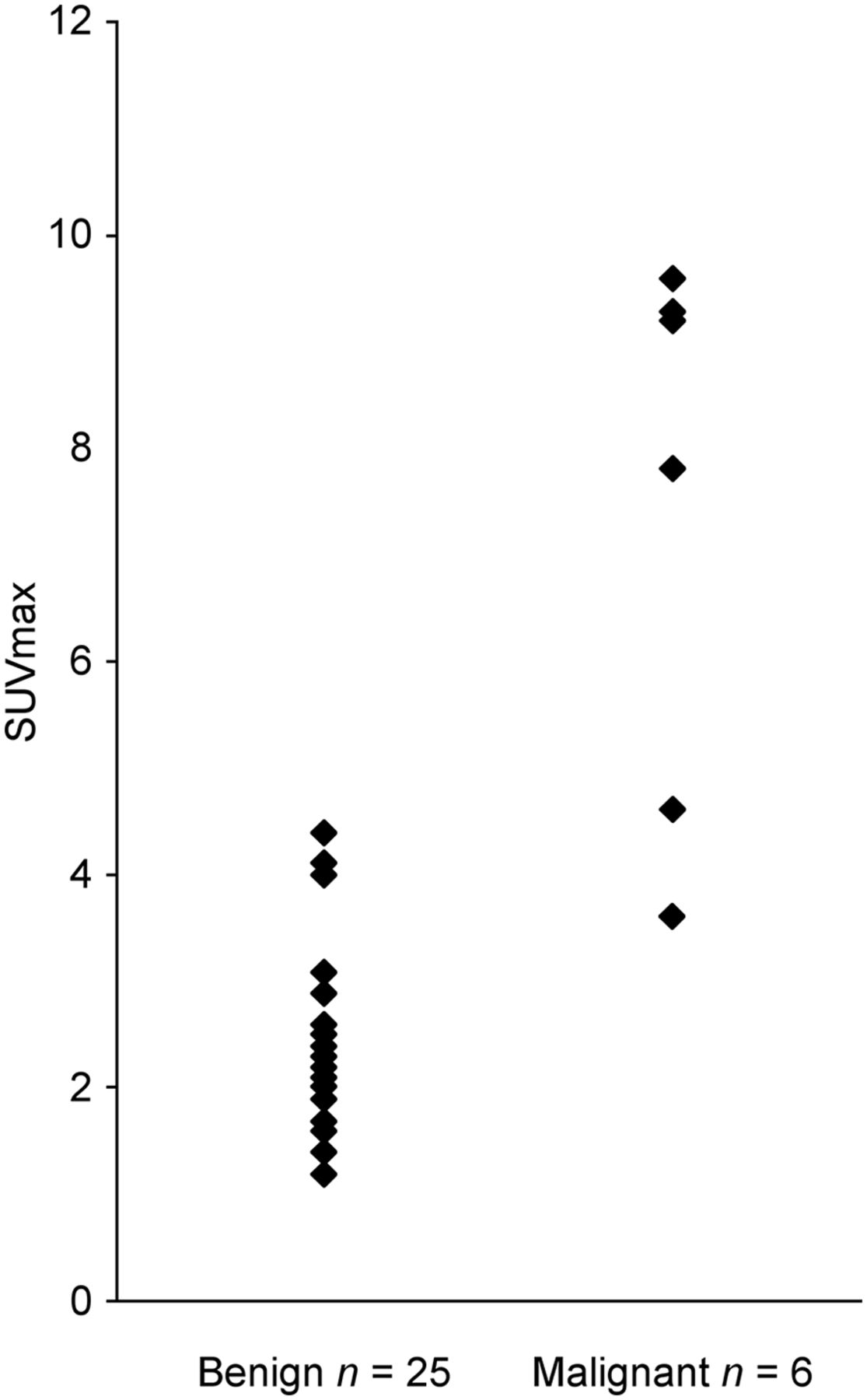
  • FIGURE 2.
    • Download figure
    • Open in new tab
    • Download powerpoint
    FIGURE 2.

    Distributions of SUVmax in benign and malignant cystic pancreatic lesions.
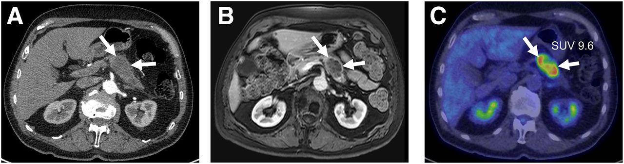
  • FIGURE 3.
    • Download figure
    • Open in new tab
    • Download powerpoint
    FIGURE 3.

    (A and B) Enhanced MDCT image (A) and enhanced T1-weighted MR image (B) of patient with lesion in tail of pancreas (arrows). Both MDCT and MR imaging had findings suggestive of malignancy. (C) Focal uptake was observed on 18F-FDG PET/CT (arrows). Histopathology confirmed adenocarcinoma.

  • FIGURE 4.
    • Download figure
    • Open in new tab
    • Download powerpoint
    FIGURE 4.

    46-y-old man with abdominal pain for 1 mo, significant weight loss, and jaundice. He had neither previous episodes of pancreatitis nor excess alcohol consumption. (A) MDCT showed thick-walled cystic mass enhancing with contrast in head of pancreas (arrows). ERCP showed biliary stricture, and stent was placed. Serum Ca19-9 activity was slightly elevated (51 kU/L). (B) T1-weighted enhanced MR imaging showed thick, irregular walls containing cystic lesion suspected of being malignant (arrows). (C) 18F-FDG PET/CT showed only diffuse uptake throughout pancreas. Patient underwent pancreaticoduodenectomy, and histopathology revealed pseudocyst (7 cm).
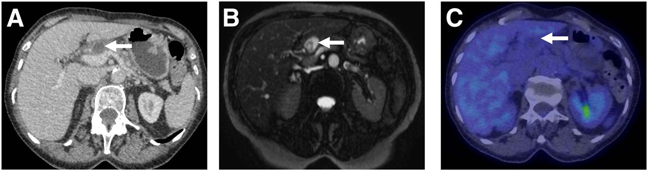
  • FIGURE 5.
    • Download figure
    • Open in new tab
    • Download powerpoint
    FIGURE 5.

    Corresponding MDCT (A), MR (B), and PET/CT (C) images of patient with 3-cm cystic lesion in head of pancreas (arrows). Both MDCT and MR imaging were suggestive of malignancy. No uptake was observed on 18F-FDG PET/CT. During 18 mo of follow-up, patient was asymptomatic.

Tables

  • Figures
    • View popup
    TABLE 1

    Patient Demographics

    ParameterTotal (n = 31)Malignant (n = 6)Benign (n = 25)P
    Mean age ± SD (y)57.1 ± 14.560.8 ± 16.356.2 ± 14.2NS
    Sex (F/M)19/123/316/9NS
    Body mass index (kg/m2)25.0 ± 4.123.9 ± 2.825.5 ± 4.3NS
    Use of alcohol (yes/no)2/290/62/23NS
    Smoking (yes/no)6/251/55/20NS
    History of pancreatitis (yes/no)6/250/62/23NS
    Symptoms (yes/no)19/125/114/11NS
    Blood chemistry
     Ca19-9 (kU/L)87 ± 263232 ± 49447 ± 156*NS
     Carcinoembryonic antigen (μg/L)2.6 ± 1.63.0 ± 2.32.5 ± 1.4NS
     Total bilirubin (μmol/L)11.9 ± 10.39.7 ± 2.612.8 ± 11.6NS
     Alkaline phosphatase (U/L)70 ± 3276 ± 5068 ± 28NS
     Alanine transferase (U/L)25 ± 1828 ± 2824 ± 15NS
     Amylase (U/L)69 ± 3153 ± 2474 ± 32NS
     γ-glutamyl transferase (U/L)45 ± 6242 ± 2846 ± 68NS
    Location
     Head15213NS
     Body514NS
     Tail826NS
     Whole pancreas312†NS
    Size† (cm)NS
     Mean ± SD5.3 ± 3.85.1 ± 4.04.8 ± 2.7
     Range1.3–182.5–181.3–10
    • ↵* One patient with benign mucinous cystic neoplasia had Ca19-9 of 775 kU/L.

    • ↵† Two patients with main-duct IPMN.

    • NS = not statistically significant.

    • Data are numbers of patients, unless otherwise indicated.

    • View popup
    TABLE 2

    Comparison of the 3 Modalities in Differential Diagnosis of Pancreatic Cysts

    Parameter18F-FDG PET/CTMDCTMR imaging18F-FDG PET/CT vs. MDCT18F-FDG PET/CT vs. MR imaging
    Sensitivity*10083830.3170.317
    Specificity*9276880.0460.564
    Accuracy9477870.0250.317
    Positive predictive value754563
    Negative predictive value1009596
    • ↵* In differentiating malignant from benign cystic lesion.

    • n = 31.

PreviousNext
Back to top

In this issue

Journal of Nuclear Medicine: 56 (8)
Journal of Nuclear Medicine
Vol. 56, Issue 8
August 1, 2015
  • Table of Contents
  • Table of Contents (PDF)
  • About the Cover
  • Index by author
Print
Download PDF
Article Alerts
Sign In to Email Alerts with your Email Address
Email Article

Thank you for your interest in spreading the word on Journal of Nuclear Medicine.

NOTE: We only request your email address so that the person you are recommending the page to knows that you wanted them to see it, and that it is not junk mail. We do not capture any email address.

Enter multiple addresses on separate lines or separate them with commas.
Accuracy of 18F-FDG PET/CT, Multidetector CT, and MR Imaging in the Diagnosis of Pancreatic Cysts: A Prospective Single-Center Study
(Your Name) has sent you a message from Journal of Nuclear Medicine
(Your Name) thought you would like to see the Journal of Nuclear Medicine web site.
Citation Tools
Accuracy of 18F-FDG PET/CT, Multidetector CT, and MR Imaging in the Diagnosis of Pancreatic Cysts: A Prospective Single-Center Study
Saila Kauhanen, Irina Rinta-Kiikka, Jukka Kemppainen, Juha Grönroos, Sami Kajander, Marko Seppänen, Kalle Alanen, Risto Gullichsen, Pirjo Nuutila, Jari Ovaska
Journal of Nuclear Medicine Aug 2015, 56 (8) 1163-1168; DOI: 10.2967/jnumed.114.148940

Citation Manager Formats

  • BibTeX
  • Bookends
  • EasyBib
  • EndNote (tagged)
  • EndNote 8 (xml)
  • Medlars
  • Mendeley
  • Papers
  • RefWorks Tagged
  • Ref Manager
  • RIS
  • Zotero
Share
Accuracy of 18F-FDG PET/CT, Multidetector CT, and MR Imaging in the Diagnosis of Pancreatic Cysts: A Prospective Single-Center Study
Saila Kauhanen, Irina Rinta-Kiikka, Jukka Kemppainen, Juha Grönroos, Sami Kajander, Marko Seppänen, Kalle Alanen, Risto Gullichsen, Pirjo Nuutila, Jari Ovaska
Journal of Nuclear Medicine Aug 2015, 56 (8) 1163-1168; DOI: 10.2967/jnumed.114.148940
Twitter logo Facebook logo LinkedIn logo Mendeley logo
  • Tweet Widget
  • Facebook Like
  • Google Plus One
Bookmark this article

Jump to section

  • Article
    • Abstract
    • MATERIALS AND METHODS
    • RESULTS
    • DISCUSSION
    • CONCLUSION
    • Acknowledgments
    • Footnotes
    • REFERENCES
  • Figures & Data
  • Info & Metrics
  • PDF

Related Articles

  • This Month in JNM
  • PubMed
  • Google Scholar

Cited By...

  • No citing articles found.
  • Google Scholar

More in this TOC Section

  • Feasibility of Ultra-Low-Activity 18F-FDG PET/CT Imaging Using a Long–Axial-Field-of-View PET/CT System
  • Cardiac Presynaptic Sympathetic Nervous Function Evaluated by Cardiac PET in Patients with Chronotropic Incompetence Without Heart Failure
  • Validation and Evaluation of a Vendor-Provided Head Motion Correction Algorithm on the uMI Panorama PET/CT System
Show more Clinical Investigations

Similar Articles

Keywords

  • pancreatic cyst
  • PET
  • diagnosis
SNMMI

© 2025 SNMMI

Powered by HighWire