
Main menu

  • Home
  • Content
    • Current
    • Ahead of print
    • Past Issues
    • JNM Supplement
    • SNMMI Annual Meeting Abstracts
    • Continuing Education
    • JNM Podcasts
  • Subscriptions
    • Subscribers
    • Institutional and Non-member
    • Rates
    • Journal Claims
    • Corporate & Special Sales
  • Authors
    • Submit to JNM
    • Information for Authors
    • Assignment of Copyright
    • AQARA requirements
  • Info
    • Reviewers
    • Permissions
    • Advertisers
  • About
    • About Us
    • Editorial Board
    • Contact Information
  • More
    • Alerts
    • Feedback
    • Help
    • SNMMI Journals
  • SNMMI
    • JNM
    • JNMT
    • SNMMI Journals
    • SNMMI

User menu

  • Subscribe
  • My alerts
  • Log in
  • Log out
  • My Cart

Search

  • Advanced search
Journal of Nuclear Medicine
  • SNMMI
    • JNM
    • JNMT
    • SNMMI Journals
    • SNMMI
  • Subscribe
  • My alerts
  • Log in
  • Log out
  • My Cart
Journal of Nuclear Medicine

Advanced Search

  • Home
  • Content
    • Current
    • Ahead of print
    • Past Issues
    • JNM Supplement
    • SNMMI Annual Meeting Abstracts
    • Continuing Education
    • JNM Podcasts
  • Subscriptions
    • Subscribers
    • Institutional and Non-member
    • Rates
    • Journal Claims
    • Corporate & Special Sales
  • Authors
    • Submit to JNM
    • Information for Authors
    • Assignment of Copyright
    • AQARA requirements
  • Info
    • Reviewers
    • Permissions
    • Advertisers
  • About
    • About Us
    • Editorial Board
    • Contact Information
  • More
    • Alerts
    • Feedback
    • Help
    • SNMMI Journals
  • View or Listen to JNM Podcast
  • Visit JNM on Facebook
  • Join JNM on LinkedIn
  • Follow JNM on Twitter
  • Subscribe to our RSS feeds
Research ArticleBasic Science Investigations

In Vivo Evaluation of 18F-SiFAlin–Modified TATE: A Potential Challenge for 68Ga-DOTATATE, the Clinical Gold Standard for Somatostatin Receptor Imaging with PET

Sabrina Niedermoser, Joshua Chin, Carmen Wängler, Alexey Kostikov, Vadim Bernard-Gauthier, Nils Vogler, Jean-Paul Soucy, Alexander J. McEwan, Ralf Schirrmacher and Björn Wängler
Journal of Nuclear Medicine July 2015, 56 (7) 1100-1105; DOI: https://doi.org/10.2967/jnumed.114.149583
Sabrina Niedermoser
1Division of Molecular Imaging and Radiochemistry, Department of Clinical Radiology and Nuclear Medicine, Medical Faculty Mannheim of Heidelberg University, Mannheim, Germany
  • Find this author on Google Scholar
  • Find this author on PubMed
  • Search for this author on this site
Joshua Chin
2McConnell Brain Imaging Centre, Montreal Neurological Institute, McGill University, Montreal, Canada
  • Find this author on Google Scholar
  • Find this author on PubMed
  • Search for this author on this site
Carmen Wängler
3Division of Biomedical Chemistry, Department of Clinical Radiology and Nuclear Medicine, Medical Faculty Mannheim of Heidelberg University, Mannheim, Germany
  • Find this author on Google Scholar
  • Find this author on PubMed
  • Search for this author on this site
Alexey Kostikov
2McConnell Brain Imaging Centre, Montreal Neurological Institute, McGill University, Montreal, Canada
  • Find this author on Google Scholar
  • Find this author on PubMed
  • Search for this author on this site
Vadim Bernard-Gauthier
2McConnell Brain Imaging Centre, Montreal Neurological Institute, McGill University, Montreal, Canada
4Division of Oncological Imaging, Department of Oncology, University of Alberta, Edmonton, AB, Canada
  • Find this author on Google Scholar
  • Find this author on PubMed
  • Search for this author on this site
Nils Vogler
5Division of Nuclear Medicine, Department of Clinical Radiology and Nuclear Medicine, Medical Faculty Mannheim of Heidelberg University, Mannheim, Germany; and
  • Find this author on Google Scholar
  • Find this author on PubMed
  • Search for this author on this site
Jean-Paul Soucy
2McConnell Brain Imaging Centre, Montreal Neurological Institute, McGill University, Montreal, Canada
6Concordia–PERFORM Centre, Concordia University, Montreal, Canada
  • Find this author on Google Scholar
  • Find this author on PubMed
  • Search for this author on this site
Alexander J. McEwan
4Division of Oncological Imaging, Department of Oncology, University of Alberta, Edmonton, AB, Canada
  • Find this author on Google Scholar
  • Find this author on PubMed
  • Search for this author on this site
Ralf Schirrmacher
2McConnell Brain Imaging Centre, Montreal Neurological Institute, McGill University, Montreal, Canada
4Division of Oncological Imaging, Department of Oncology, University of Alberta, Edmonton, AB, Canada
  • Find this author on Google Scholar
  • Find this author on PubMed
  • Search for this author on this site
Björn Wängler
1Division of Molecular Imaging and Radiochemistry, Department of Clinical Radiology and Nuclear Medicine, Medical Faculty Mannheim of Heidelberg University, Mannheim, Germany
  • Find this author on Google Scholar
  • Find this author on PubMed
  • Search for this author on this site
  • Article
  • Figures & Data
  • Supplemental
  • Info & Metrics
  • PDF
Loading

Article Figures & Data

Figures

  • Tables
  • Additional Files
  • FIGURE 1.
    • Download figure
    • Open in new tab
    • Download powerpoint
    FIGURE 1.

    Structures of building blocks SiFA and SiFAlin.
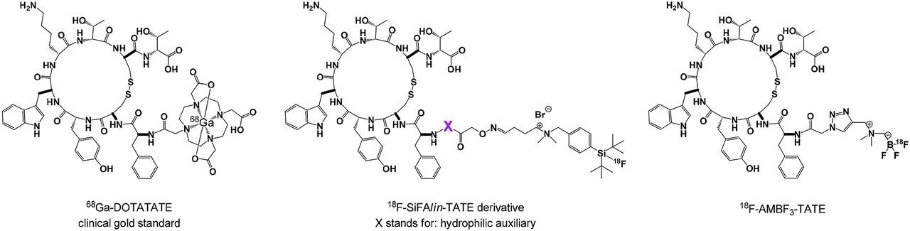
  • FIGURE 2.
    • Download figure
    • Open in new tab
    • Download powerpoint
    FIGURE 2.

    Structures of 68Ga-DOTATATE, 18F-SiFAlin analogs (X = site of introduction of hydrophilic auxiliaries for optimizing in vivo biodistribution properties), and 18F-trifluoroborate derivative 18F-AMBF3-TATE.
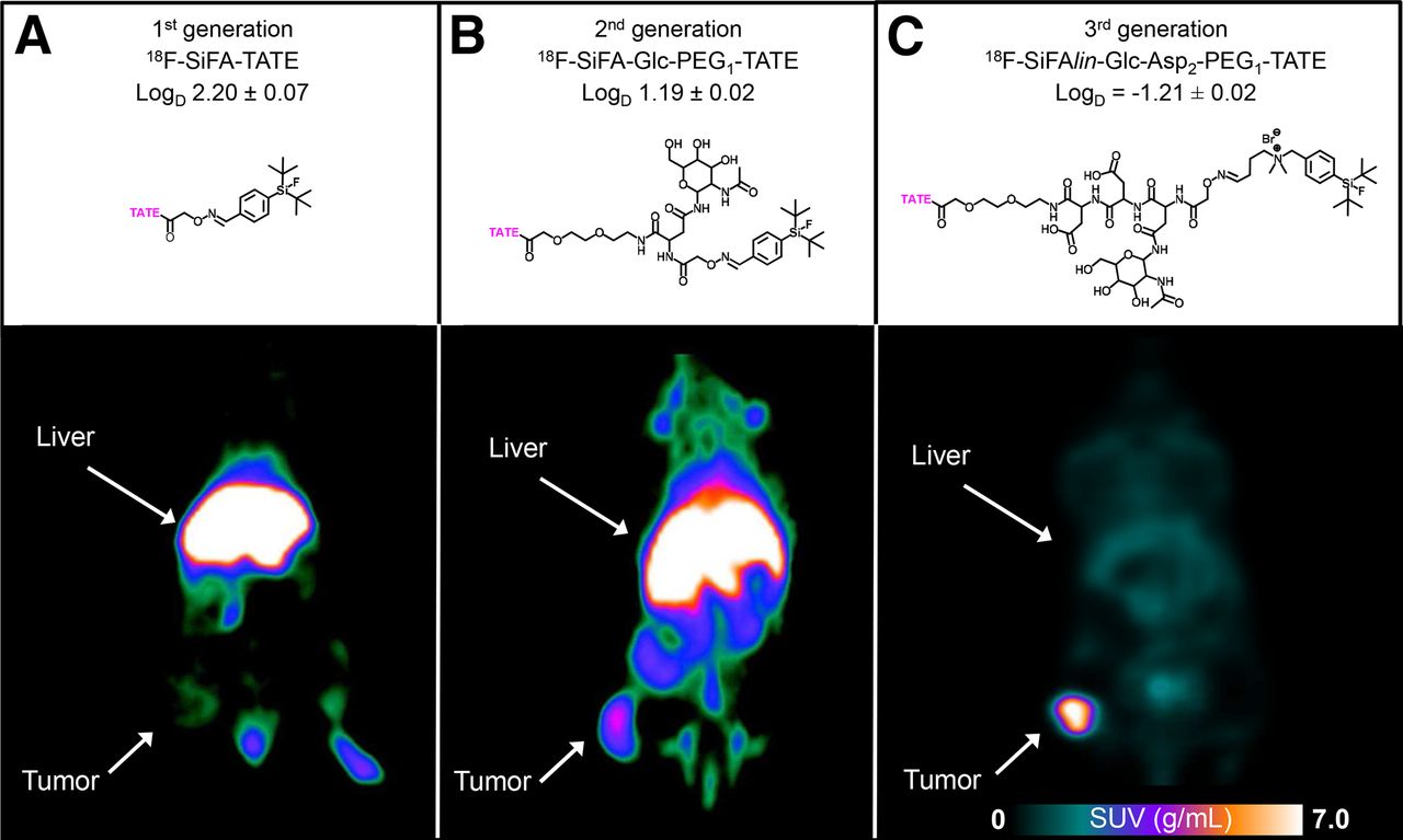
  • FIGURE 3.
    • Download figure
    • Open in new tab
    • Download powerpoint
    FIGURE 3.

    Small-animal PET images of 3 generations of SiFA-derivatized somatostatin analogs evaluated in AR42J tumor–bearing rodents. All images show coronal slices of the 50–90 min postinjection time frame. (A) 18F-SiFA-TATE (18F-1) in vivo images were obtained using a Philips Mosaic small-animal PET scanner. (B) 18F-SiFA-Glc-PEG1-TATE (18F-3) images were obtained with a Siemens Inveon small-animal PET-scanner. (C) Images of 18F-SiFAlin-Glc-Asp2-PEG1-TATE (18F-9) distribution were obtained using a Bruker Albira small-animal PET/SPECT/CT scanner, indicating both a renal clearance and high activity accumulation in the SSTR-positive tumor tissue.
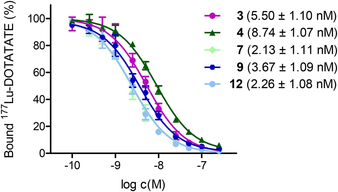
  • FIGURE 4.
    • Download figure
    • Open in new tab
    • Download powerpoint
    FIGURE 4.

    High tolerance of SSTR binding affinities of Tyr3-octreotate toward hydrophilic chemical modifications without loss of bioactive potency. Binding affinities of third-generation SiFA (green, PEG1: 4 and PEG5: 7) and SiFAlin (blue, PEG1: 9 and PEG5: 12) derivatives compared with second-generation (magenta, 3) were determined by competitive receptor binding affinity studies using AR42J cells and 177Lu-DOTATATE as competitor. After incubation for 60 min in buffer at ambient temperature and successive washing steps, both cell-bound and internalized activity were measured using a γ counter. All experiments were performed in triplicate (error bars, mean ± SD).
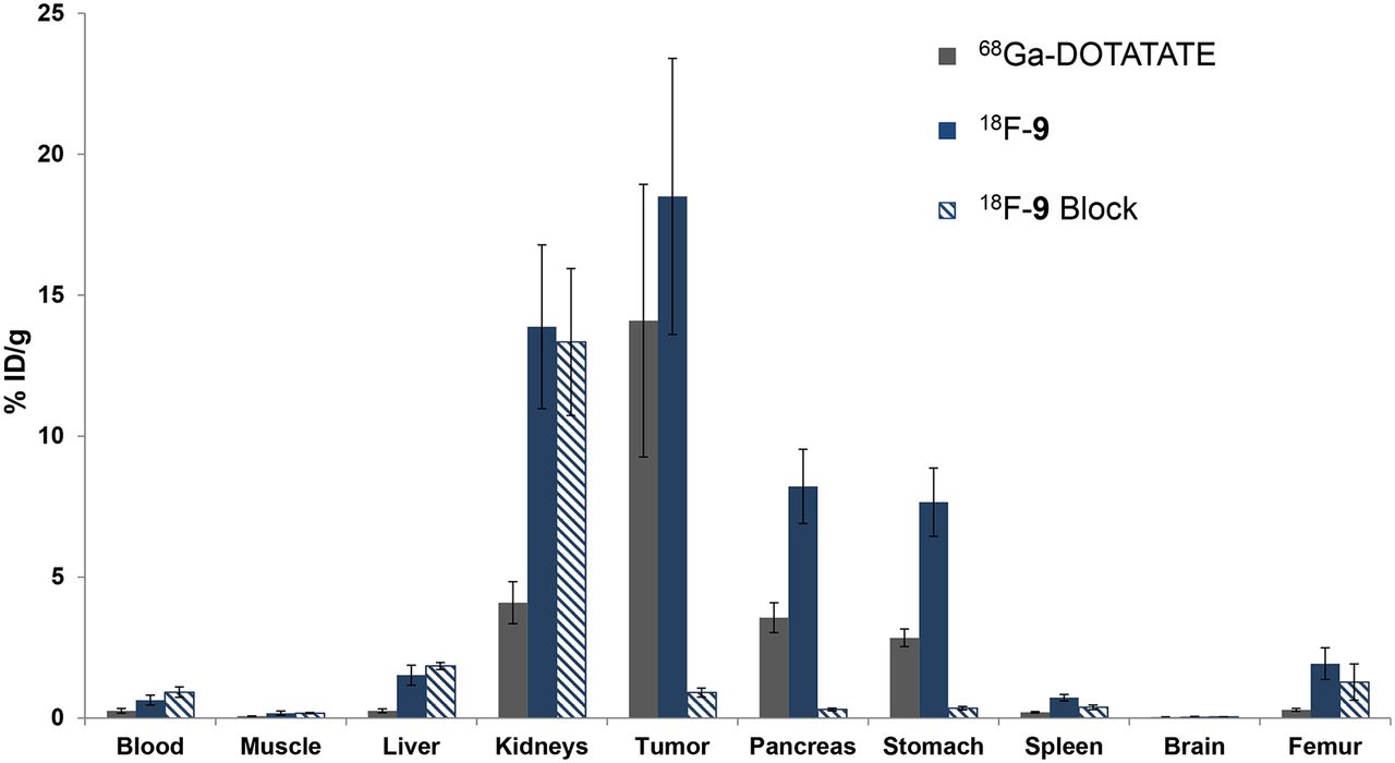
  • FIGURE 5.
    • Download figure
    • Open in new tab
    • Download powerpoint
    FIGURE 5.

    Results from ex vivo biodistribution studies comparing 68Ga-DOTATATE (n = 5) with 18F-9 (n = 10) in AR42J tumor–bearing mice at 60 min after injection. Values are given as %ID/g. Most promising 18F-SiFAlin-derivatized derivative 18F-9 demonstrates highly specific and even slightly higher tumor uptake than gold standard 68Ga-DOTATATE. For 18F-9, a blocking experiment using DOTATATE (200 μg/mouse; n = 5) was performed (dashed blue columns), showing specific binding of tracer to tumor and physiologically SSTR-positive tissues. 18F-SiFAlin derivatives enable tumor-to-background ratios comparable to 68Ga-DOTATATE.
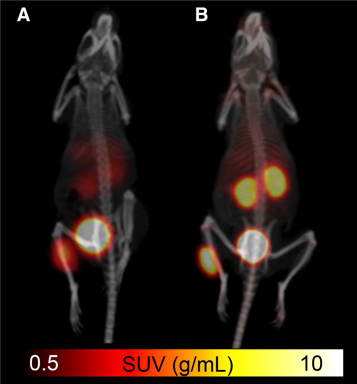
  • FIGURE 6.
    • Download figure
    • Open in new tab
    • Download powerpoint
    FIGURE 6.

    Comparative small-animal PET/CT imaging of SSTR-positive tumor-bearing mice using 68Ga-DOTATATE (A) and 18F-9 (B) with identical standardized uptake value (SUV) scale for both images. Tumor accumulation SUVs in last frame (80–90 min after injection; showing highest accumulation of both tracers) are 5.50 for 68Ga-DOTATATE and 7.80 for 18F-9.

Tables

  • Figures
  • Additional Files
    • View popup
    TABLE 1

    Tumor-to-Blood and Tumor-to-Muscle Ratios for Tested Newly Developed 18F-Labeled Peptides 18F-4, 18F-7, 18F-9, and 18F-12 Obtained from Ex Vivo Biodistribution at 90 Minutes after Injection

    Ratio18F-418F-918F-718F-12
    Tumor to blood4.30 ± 0.2657.58 ± 35.8912.31 ± 4.1974.42 ± 20.37
    Tumor to muscle33.31 ± 11.94211.05 ± 143.3894.12 ± 26.37256.49 ± 61.17

Additional Files

  • Figures
  • Tables
  • Supplemental Data

    Files in this Data Supplement:

    • Supplemental Data
PreviousNext
Back to top

In this issue

Journal of Nuclear Medicine: 56 (7)
Journal of Nuclear Medicine
Vol. 56, Issue 7
July 1, 2015
  • Table of Contents
  • Table of Contents (PDF)
  • About the Cover
  • Index by author
Print
Download PDF
Article Alerts
Sign In to Email Alerts with your Email Address
Email Article

Thank you for your interest in spreading the word on Journal of Nuclear Medicine.

NOTE: We only request your email address so that the person you are recommending the page to knows that you wanted them to see it, and that it is not junk mail. We do not capture any email address.

Enter multiple addresses on separate lines or separate them with commas.
In Vivo Evaluation of 18F-SiFAlin–Modified TATE: A Potential Challenge for 68Ga-DOTATATE, the Clinical Gold Standard for Somatostatin Receptor Imaging with PET
(Your Name) has sent you a message from Journal of Nuclear Medicine
(Your Name) thought you would like to see the Journal of Nuclear Medicine web site.
Citation Tools
In Vivo Evaluation of 18F-SiFAlin–Modified TATE: A Potential Challenge for 68Ga-DOTATATE, the Clinical Gold Standard for Somatostatin Receptor Imaging with PET
Sabrina Niedermoser, Joshua Chin, Carmen Wängler, Alexey Kostikov, Vadim Bernard-Gauthier, Nils Vogler, Jean-Paul Soucy, Alexander J. McEwan, Ralf Schirrmacher, Björn Wängler
Journal of Nuclear Medicine Jul 2015, 56 (7) 1100-1105; DOI: 10.2967/jnumed.114.149583

Citation Manager Formats

  • BibTeX
  • Bookends
  • EasyBib
  • EndNote (tagged)
  • EndNote 8 (xml)
  • Medlars
  • Mendeley
  • Papers
  • RefWorks Tagged
  • Ref Manager
  • RIS
  • Zotero
Share
In Vivo Evaluation of 18F-SiFAlin–Modified TATE: A Potential Challenge for 68Ga-DOTATATE, the Clinical Gold Standard for Somatostatin Receptor Imaging with PET
Sabrina Niedermoser, Joshua Chin, Carmen Wängler, Alexey Kostikov, Vadim Bernard-Gauthier, Nils Vogler, Jean-Paul Soucy, Alexander J. McEwan, Ralf Schirrmacher, Björn Wängler
Journal of Nuclear Medicine Jul 2015, 56 (7) 1100-1105; DOI: 10.2967/jnumed.114.149583
Twitter logo Facebook logo LinkedIn logo Mendeley logo
  • Tweet Widget
  • Facebook Like
  • Google Plus One
Bookmark this article

Jump to section

  • Article
    • Abstract
    • MATERIALS AND METHODS
    • RESULTS
    • DISCUSSION
    • CONCLUSION
    • DISCLOSURE
    • Acknowledgments
    • Footnotes
    • REFERENCES
  • Figures & Data
  • Supplemental
  • Info & Metrics
  • PDF

Related Articles

  • This Month in JNM
  • PubMed
  • Google Scholar

Cited By...

  • Radiohybrid Ligands: A Novel Tracer Concept Exemplified by 18F- or 68Ga-Labeled rhPSMA Inhibitors
  • Recent Advances in 18F Radiochemistry: A Focus on B-18F, Si-18F, Al-18F, and C-18F Radiofluorination via Spirocyclic Iodonium Ylides
  • Google Scholar

More in this TOC Section

  • 11C-Methionine PET of Myocardial Inflammation in a Rat Model of Experimental Autoimmune Myocarditis
  • Counting Rate Characteristics and Image Distortion in Preclinical PET Imaging During Radiopharmaceutical Therapy
  • Design and Fabrication of Kidney Phantoms for Internal Radiation Dosimetry Using 3D Printing Technology
Show more Basic Science Investigations

Similar Articles

Keywords

  • 68Ga-DOTATATE
  • PET
  • kitlike 18F labeling
  • 18F-SiFAlin-TATE
  • somatostatin receptor imaging
SNMMI

© 2025 SNMMI

Powered by HighWire